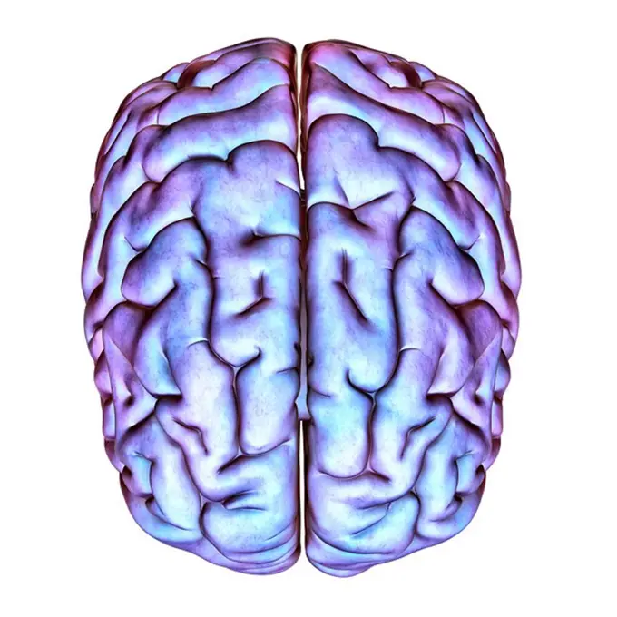
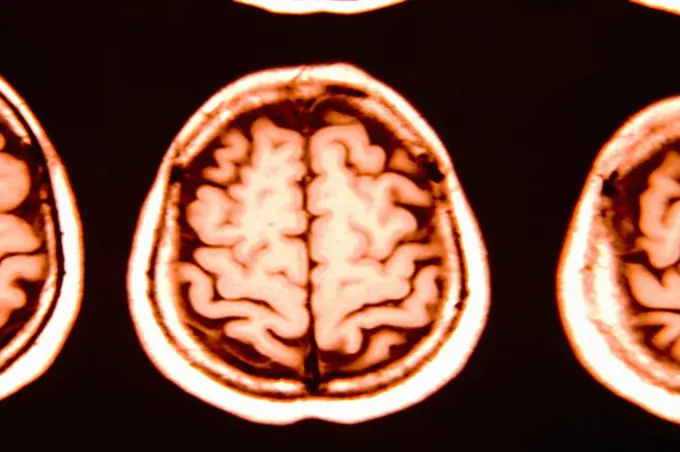
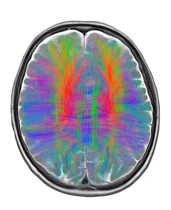
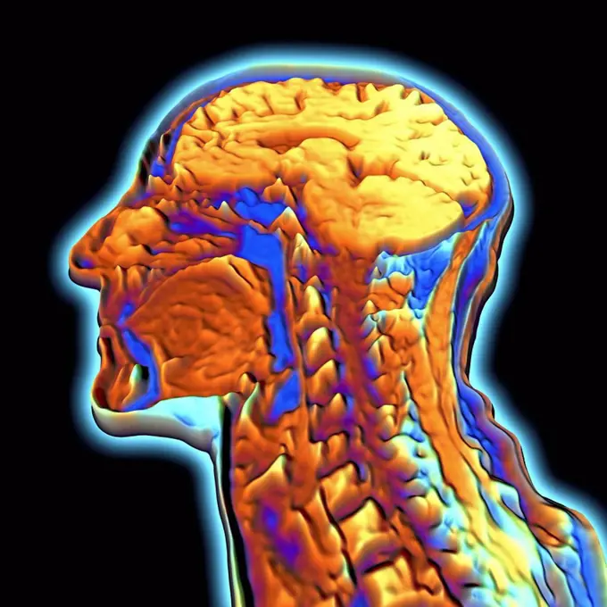
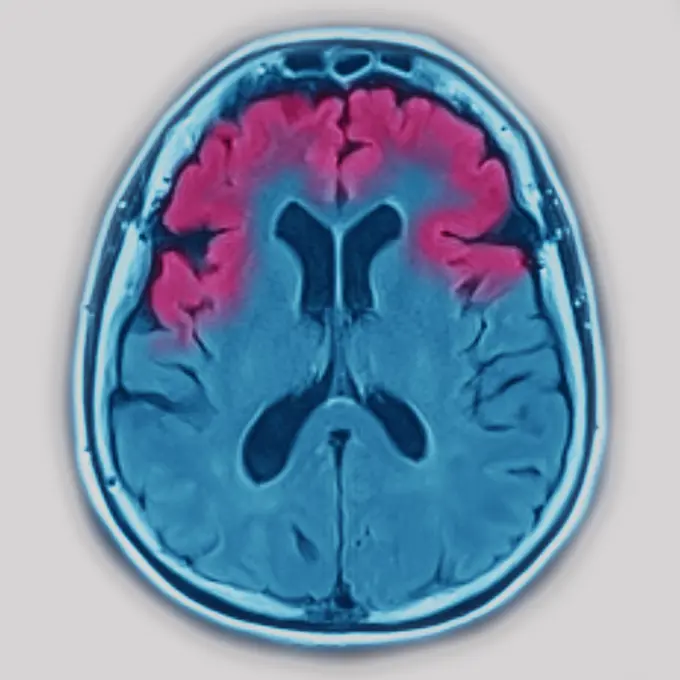
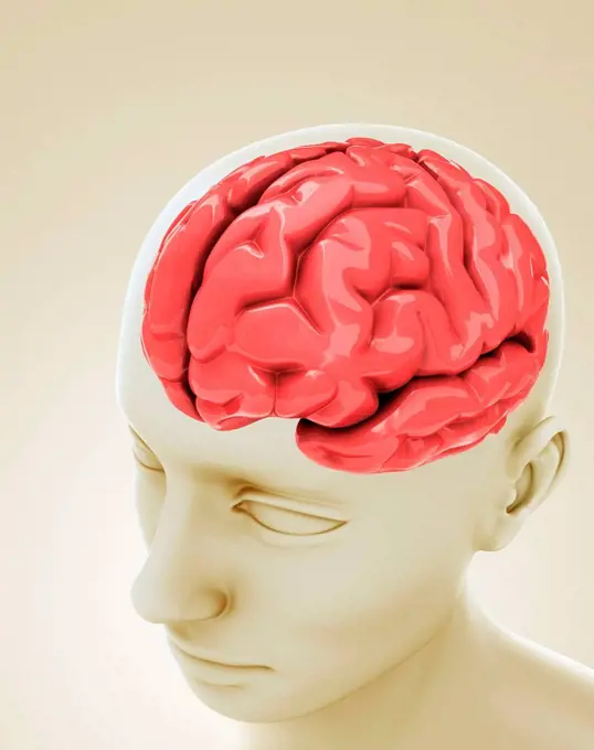
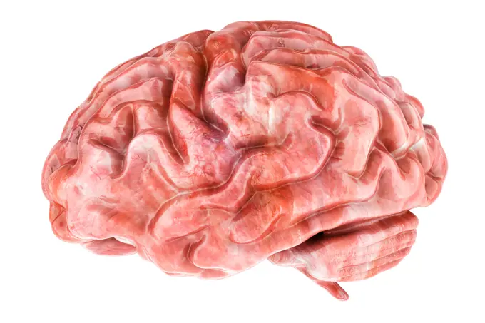
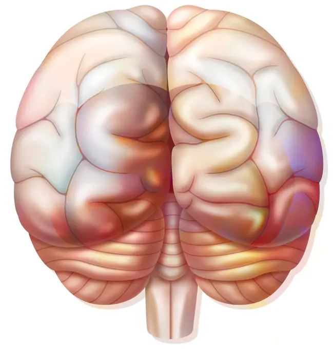
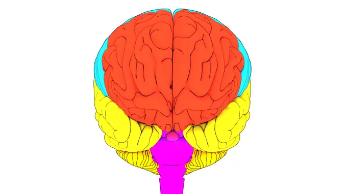
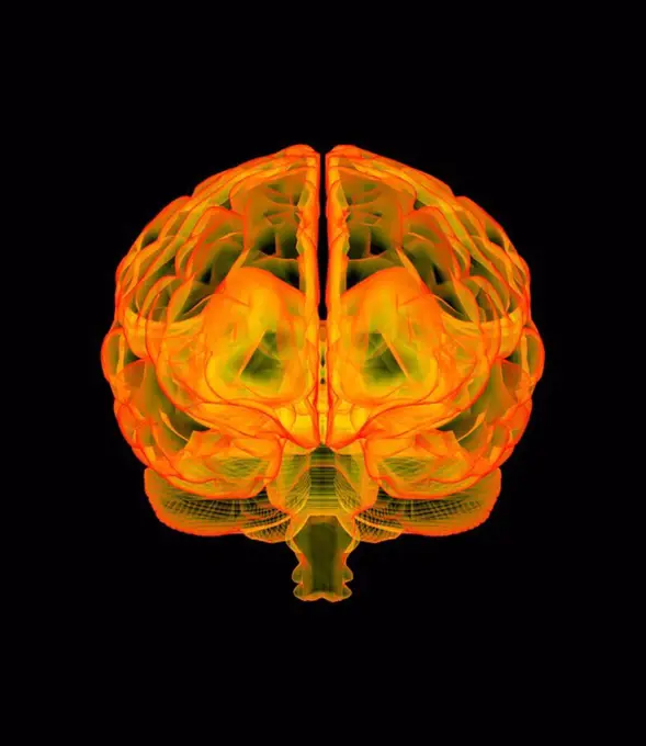
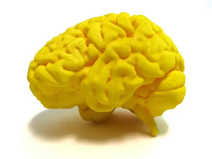
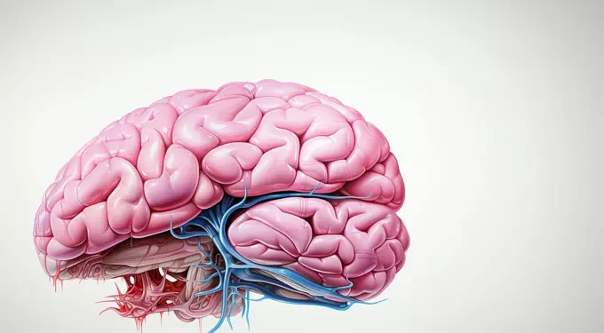
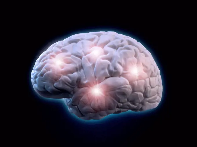
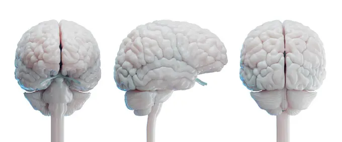
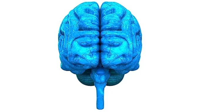
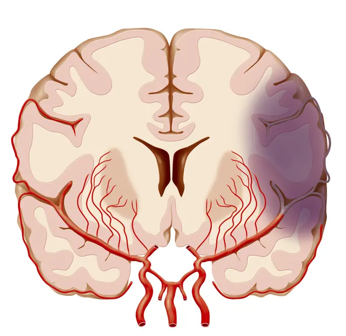
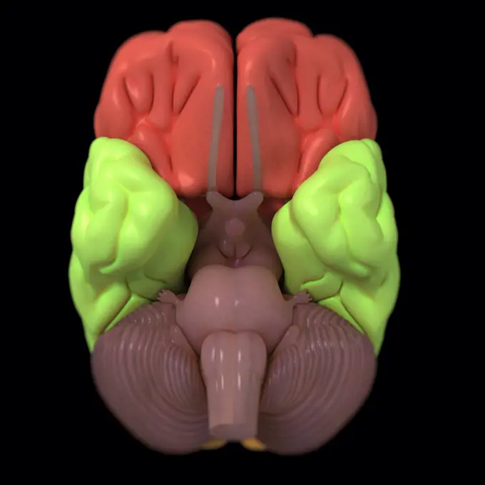
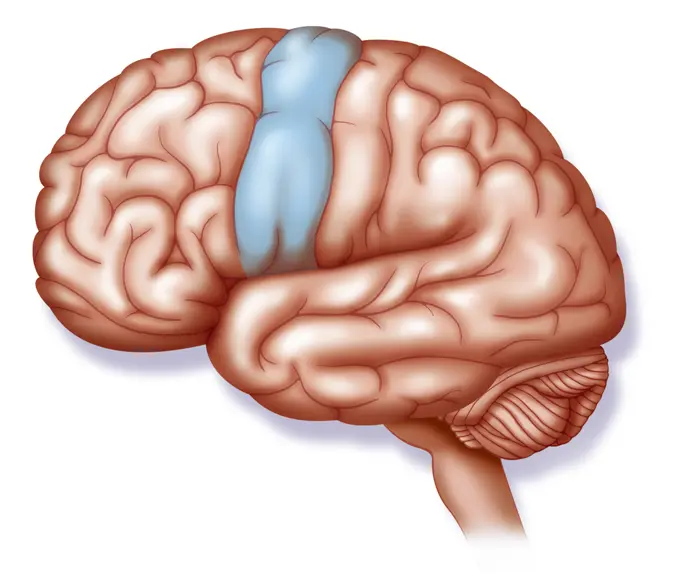
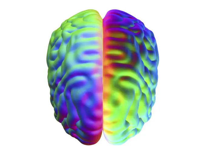
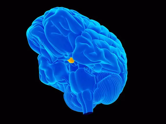
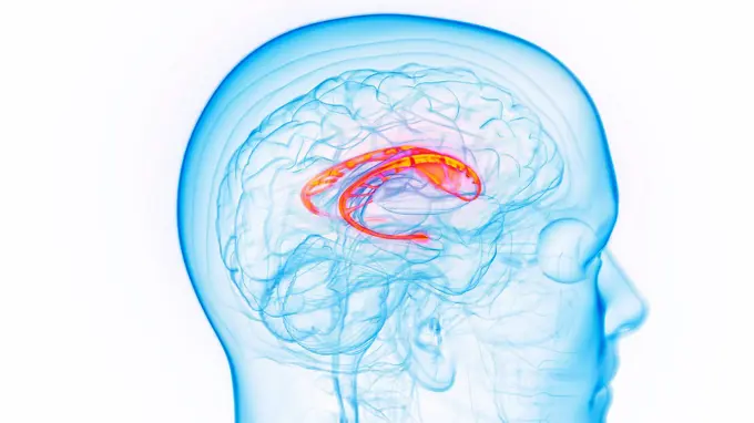
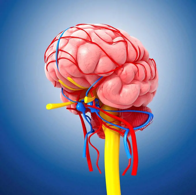
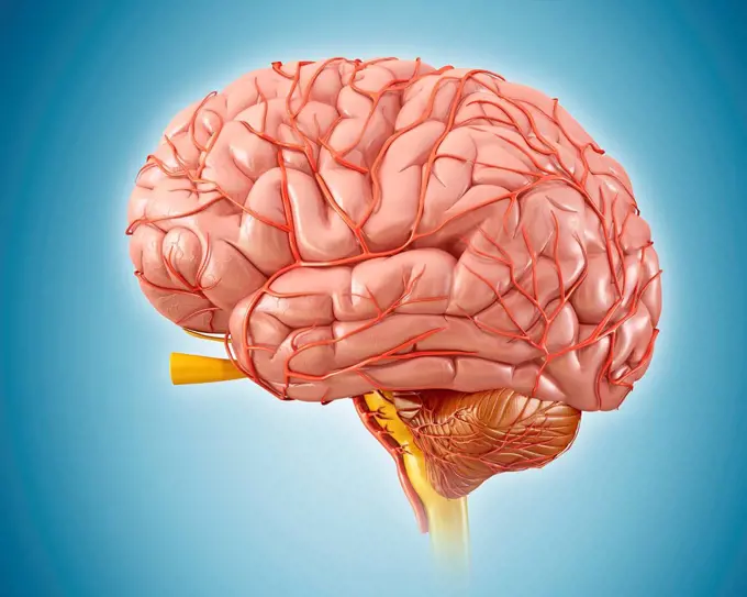
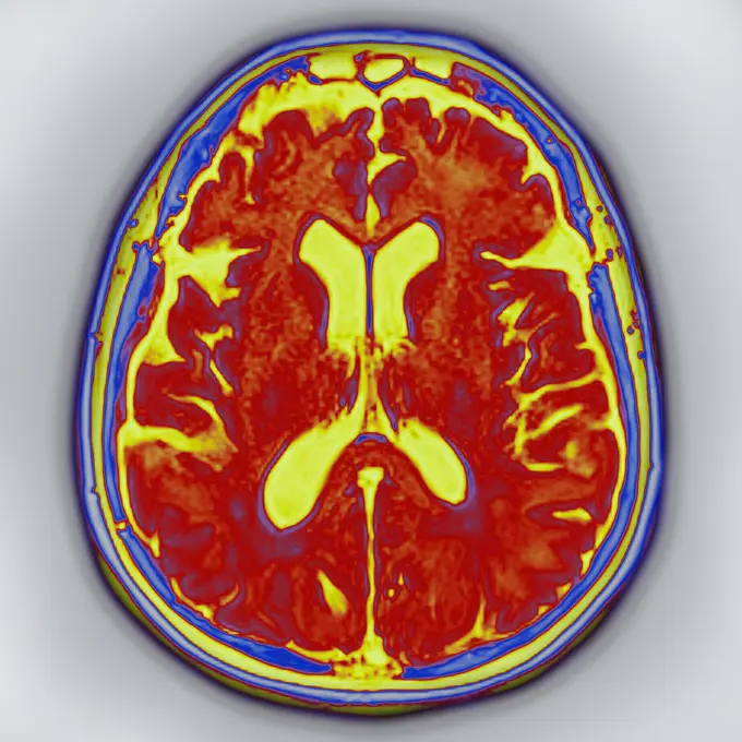
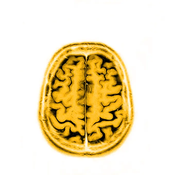
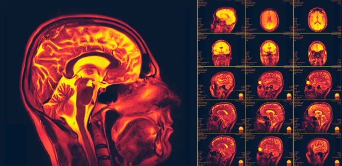
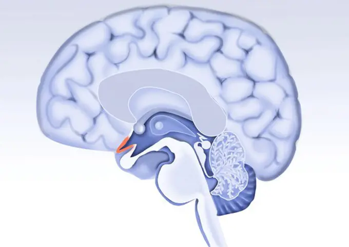
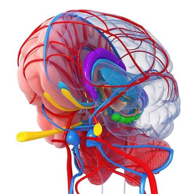
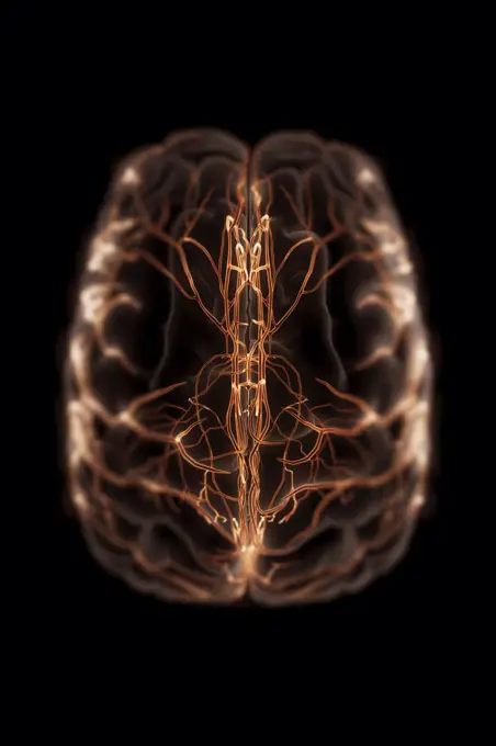
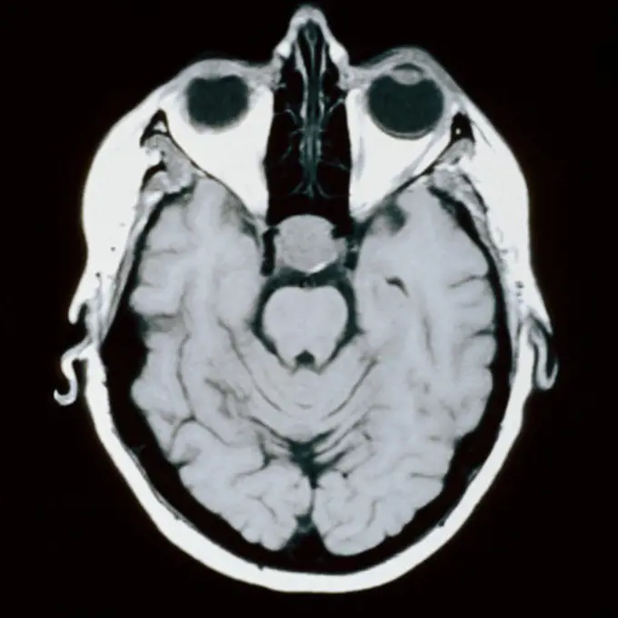
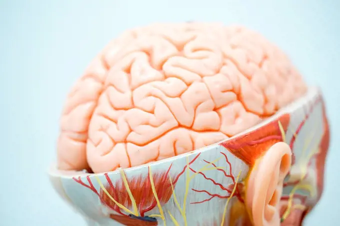
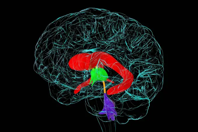
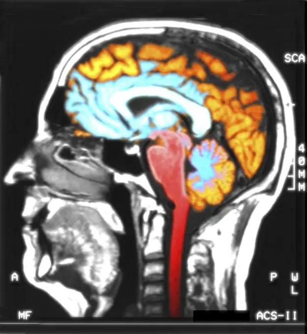
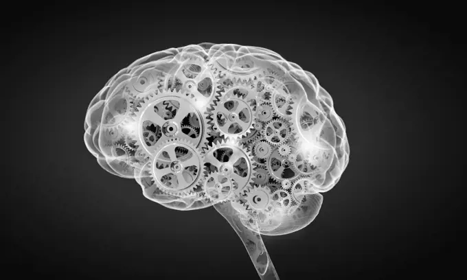
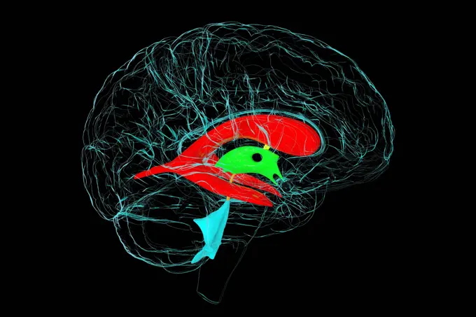
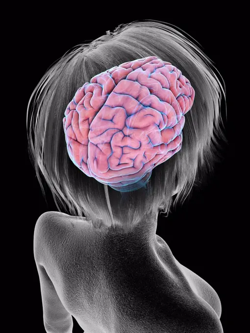
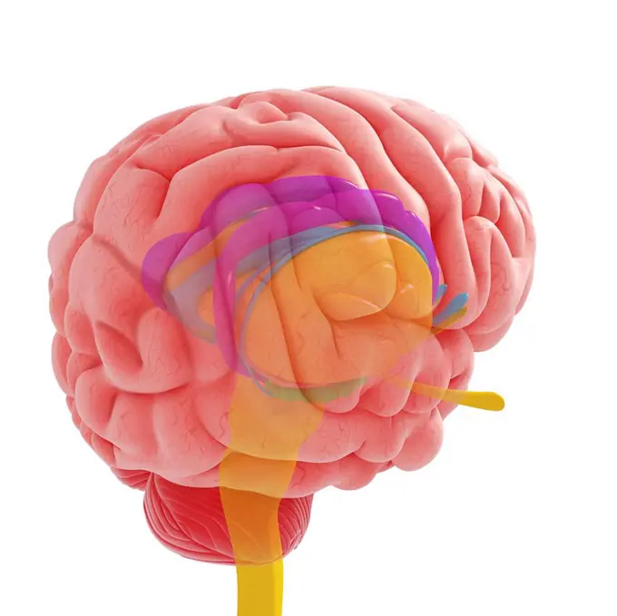
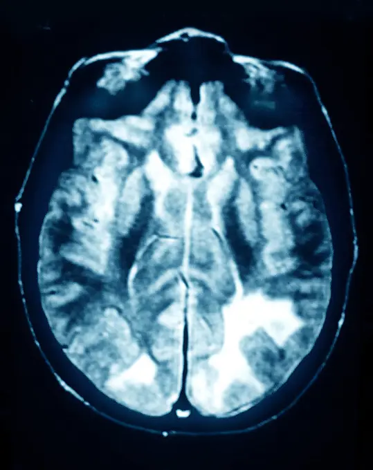
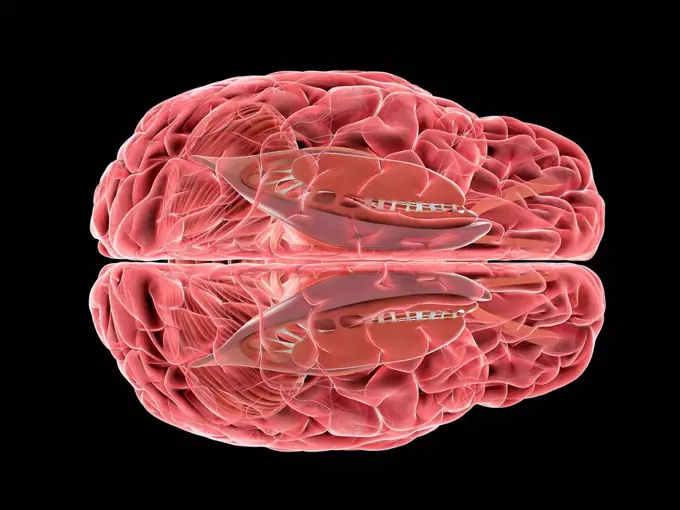
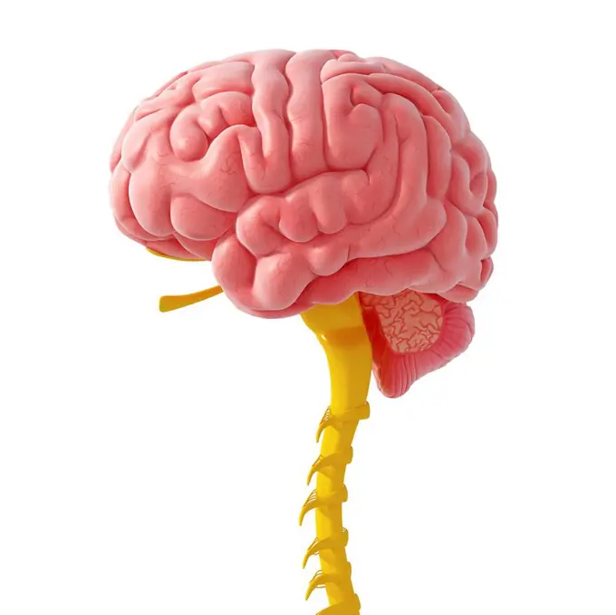
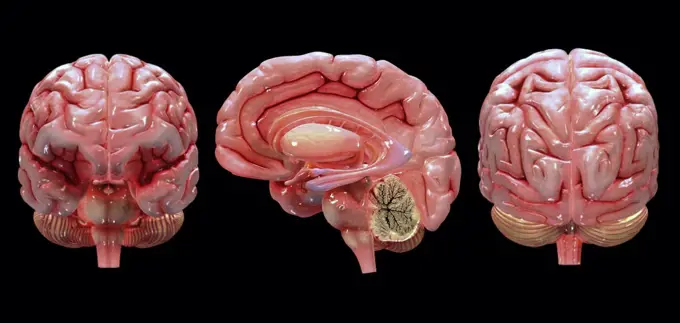
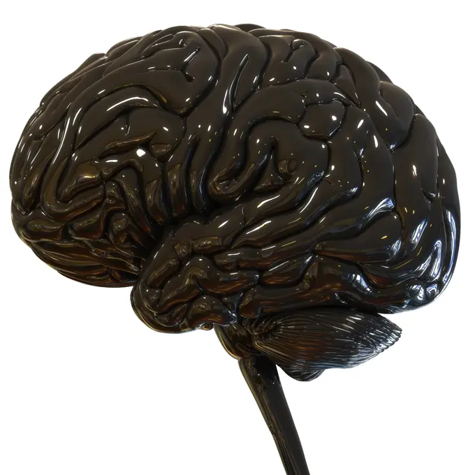
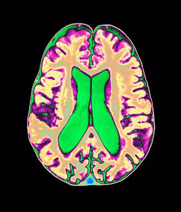
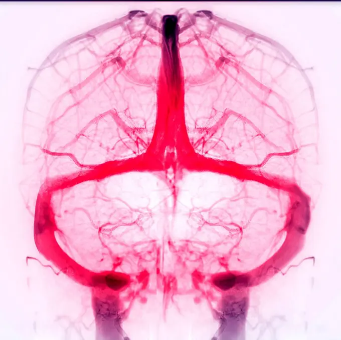
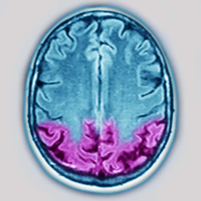
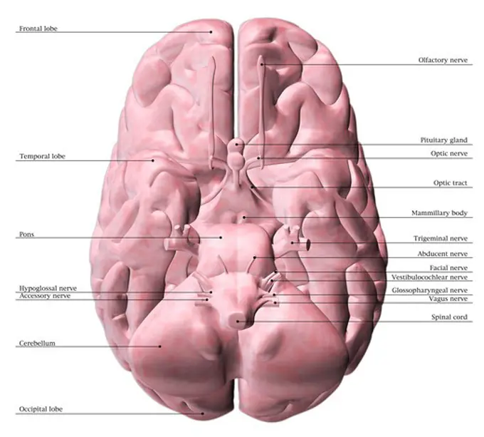
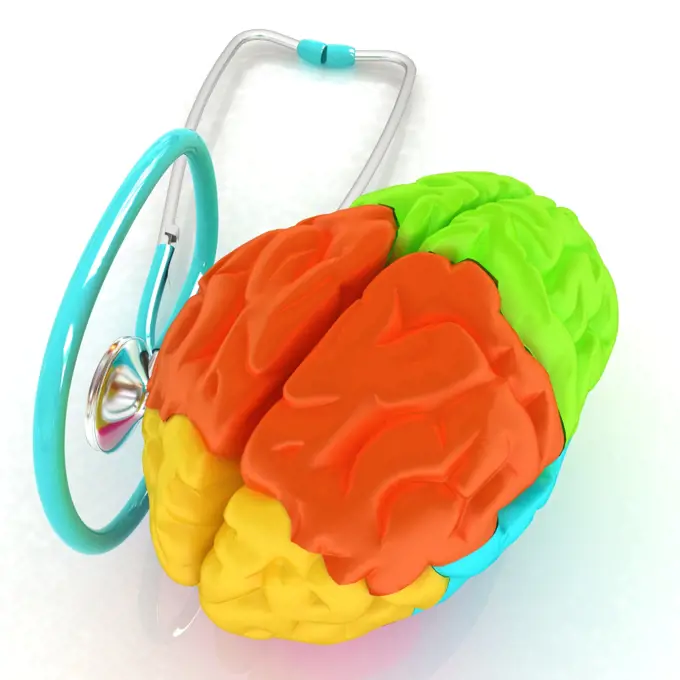
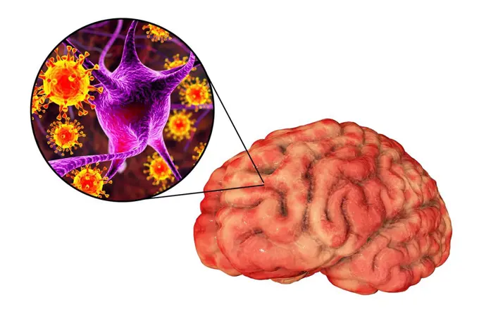
Fine ArtVintageFootageSubscriptionBoardsFree ResearchPricing
  • Fine Art
  • Vintage
  • Footage
  • Subscription
  • Boards
  • Free Research
  • Pricing

Human Brain Artwork

Vibrant computer-generated images of the human brain, illustrating various perspectives and highlighting intricate structures and electrical activity.

Human brain, artwork
248 assets in this story
1525-24964628
Scan
4128R-13682188
Computer artwork of a MRI top view of the brain showing white matter fibres. Coloured 3D diffusion spectral imaging (DSI) scan of the bundles of white...
4128R-28098
Brain scan. Computer enhanced Magnetic Resonance Imaging (MRI) scan through a human head, showing a healthy brain in side view. The face is seen in pr...
1899-53509519
Frontal section of the brain with white and gray matter.
1899-53511275
Cerebral atrophy - Bilateral cortical and subcortical temporal atrophy (frontotemporal degeneration responsible for neurodegenerative diseases including Alzheimer's disease, Lewy body disease, cognitive disorders, and attention disorders. Radial section of the brain, visualized by an MRI in radial section.
4128R-30628
Human brain, computer artwork.
6188-56067053
Human Brain, side view. 3D rendering Human Brain, side view. 3D rendering isolated on white background Copyright: xZoonar.com/alexlmxx 21216685
824-63164380
Brain with occipital, parietal, temporal lobes, cerebellum.
4128R-22396
Human brain, computer artwork.
4128R-15516201
Illustration of a red glowing glass brain
6188-60962725
Central Organ of Human Nervous System Brain Anatomy 3D Illustration Concept of Central Organ of Human Nervous System Brain Anatomy Copyright: xZoonar.com/magicminex 21747971
4128R-652
Human brain, artwork.
824-66065396
Normal Human Brain
5507-48578836
Colorful human brain on white background. Left right human brain concept. Creative part and logic part with social and business doodle watercolor design copy space. Concept human thinking
824-63186542
Representation of the brain in case of dyslexia. The dark blue area represents face recognition, the green area represents object recognition, and the turquoise blue area represents shape recognition. The red area (absent here) in the left parietal lobe corresponds to the area stimulated during word recognition.
4128R-12577579
Human brain, computer illustration.
4128R-14637807
Human brain arteries, illustration
4128-30415619
Brain, illustration
1848-77403600
3D Illustration Concept of Central Organ of Human Nervous System Brain Anatomy
4128R-14637814
Human brain olfactory nerve, illustration
4128-30416566
Brain, illustration
4128R-13387122
Illustration of human brain anatomy.
824-63206067
Frontal cross-sectionllustration of a transient ischemic attack (purple area) in the brain. The transient ischemic attack (mini CVA), caused by a thrombus, a clot blocking the small blood vessel, preventing blood from flowing in the vessel.
4269-24705
Brain. Sagital MRI (Magnetic Resonance Imaging) scan of a normal brain.
4128R-25971
human brains, computer artwork.
4286-39891
CT scan showing a cross section of a human head.
824-129455
Localised malignant astrocytoma in the left hemisphere of the brain. Transversal MRI brain scan.
4128R-12577581
Human brain with a microchip, computer illustration.
4378-1676
A model of the human brain, isolating different lobes with color.
824-63181813
Location of the motor cortex (areas 4 and 6) from an external view of the brain.
6188-60013147
3D rendering of a human brain isolated in studio background 3D rendering of a human brain isolated in studio background. Copyright: xZoonar.com/HugoxKurkx 13914979
4128R-13620451
Brain vascular system and blood supply. Artwork showing the brain with arteries (red) and veins (blue).
4128R-12922546
Human brain, illustration
4128R-13620462
Brain vascular system and blood supply. Artwork showing the brain with arteries (red) and veins (blue).
4128R-13245014
Human brain, illustration.
4128R-13371078
Human brain, illustration.
4128R-13387263
Illustration of human brain anatomy.
4128-30421705
Caudate nucleus, illustration
4128R-28933
Brain anatomy, computer artwork.
4128R-13586400
Illustration of the human brain.
824-63224455
Cerebral atrophy - Bilateral cortical and subcortical temporal atrophy (frontotemporal degeneration responsible for neurodegenerative diseases including Alzheimer's disease, Lewy body disease, cognitive disorders, and attention disorders. Radial section of the brain , visualized by an MRI in radial section.
824-63128401
Brain, mri
4128R-11542177
Human brain, computer illustration.
1525R-177051
6188-60048724
Magnetic resonance imaging of the brain. MRI scan Magnetic resonance imaging of the brain with no visible abnormalities. MRI in different views Copyright: xZoonar.com/MaximxxPavlovx 13221419
4252-4448
Brain, optic chiasm
4128R-24248
Brain anatomy, computer artwork.
4378-3606
Superior view of an anatomical model of the brain and its blood supply.
4380-320
CT Scan Showing Cross-Section of Human Head
4128R-15516211
Illustration of a brain and syringes
4128R-19171
Nervous system, computer artwork.
4128-17810492
Human brain anatomy model
4128-30422570
Ventricular system of the brain, illustration
1574R-09827
MRI scan of a human head
4128-30421639
Human brain, illustration
4128-V58573971
6188-62240789
Colorfull human brain Colorfull human brain Copyright: xZoonar.com/SergiixSvyerchkovx 5957325
1525-23221923
Mechanism inside human brain. Illustration of human brain with cogwheel mechanisms
4128-30422563
Ventricular system of the brain, illustration
4128R-15516681
Illustration of an old woman's brain
4128R-24058
Brain anatomy, computer artwork.
824-63175243
MELAS syndrome, Mitochondrial Encephalomyopathy, Lactic Acidosis, and Stroke-like episodes. Axial CT scan of the brain.
4128R-11289184
Human brain, computer artwork.
4128R-22272
Brain and circular network diagram, depicting neural connections in the brain.
4128-111579472
Human brain, computer illustration.
1525-19891230
Glass human brain with question mark within
4128R-29526
Central nervous system, computer artwork.
824-63227300
This is a pseudo-colored image of a high resolution gradient echo MRI of a fixed cerebral hemisphere of a person with multiple sclerosis.
1525-56234402
Normal Visual Pathway with Eye Movement 3D rendering. Normal Visual Pathway with Eye Movement
4269-39213
Coloured magnetic resonance imaging (MRI) scan and CT of an axial section through the brain of patient, showing the damage (red) caused 12 hours after a cerebral infarction.
4239-18641529
3D rendering of human brain.
824-63211579
Posterior cortical atrophy (Bensons syndrome) seen on a radial CT-scan.
1439R-1071388
Brain
6188-62318356
Central Organ of Human Nervous System Brain Anatomy 3D Illustration Concept of Central Organ of Human Nervous System Brain Anatomy Copyright: xZoonar.com/magicminex 21748087
4128R-13371247
Human brain, illustration.
1525-23187664
Human mind. Science image with human brain on white background
4128R-4527
Coloured MRI scan of organophosphate brain damage
4239-18641131
Superior view of human brain.
4128-30419900
Cerebral veins, MRV scan
1525-23369581
Human mind. Science image with human brain on white background
4128R-28106
Brain scan. False-colour magnetic resonance imaging (MRI) scan of a human head containing a healthy brain, seen in horizontal view. At upper frame, th...
4128R-13928409
Computer enhanced false-colour Magnetic Resonance Images (MRI) of two mid-sagittal and one axial (cross sectional) sections through the head of a norm...
824-63198874
Cerebral atrophy. Parietal cortico-sub-cortical atrophies (aftereffects of left, frontoparietal parenchymal ischemia), seen on a frontal cross-section brain CT scan.
1525-26716913
Human skull. Red X-ray image isolated on black background. Human skull. Red X-ray image on black background
824-63128375
Bilateral lesions of the caudate heads, the putamens, and the internal posterior region of the two thalami (image on the right).
824-63224445
Cerebral atrophy - Posterior cortical atrophy (posterior cortical degeneration responsible for agnosia, apraxia, aphasia, Gerstmann syndrome, acalculia, indistinction, agraphia, Balint syndrome, optic ataxia, psychic paralysis. Radial section of the brain, visualized by a cross-sectional MRI radial.
4128R-12663
Arteries of the brain. 3D computed tomography CT scan of normal arteries in the brain of a 35 year old.
4239-18641780
Medical diagram illustrating how the left side of the brain works compared to the right side of the brain.
1660R-41554
brain with a straw in it
4297-1671
Anatomical illustration of the underside of the human brain
4128-111452963
Human brain, illustration
1525-23339154
stethoscope and brain. 3d illustration
4128R-13410259
Human brain anatomical, illustration
4128R-15290615
Viral encephalitis, illustration
4128-28970834
Healthy human brain, MRI scan
4128R-4249
Coloured MRI brain scan showing a meningioma
4128-30421636
Human brain, illustration
4128-V58573967
1899-61461954
Anterior view of the brain with the two cerebral hemispheres, the cerebellum and the beginning of the brainstem
4378-3071
Superior view of an anatomical model of the brain and its blood supply.
PREVIOUS
of 3
NEXT
2401 S. Ervay, Suite 206
Dallas, Texas 75215
United States
Browse
CategoriesGalleriesFree Research
For Creators
License Your ContentSuperStock For StudentsFrame of Mind BlogContributor PortalNew Image SpotlightLicense AI Imagery
Resources
About UsLicensingTerms & ConditionsAPI AccessPrivacyPricing
Contact
+1 866 236 0087help@superstock.com Contact form
©2026 SuperStock. All Rights Reserved. -A
This site is protected by reCAPTCHA and Google Privacy Policy and Terms of Service apply.