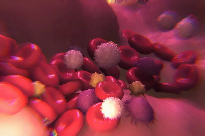
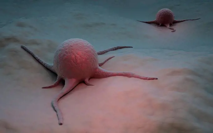
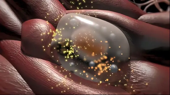
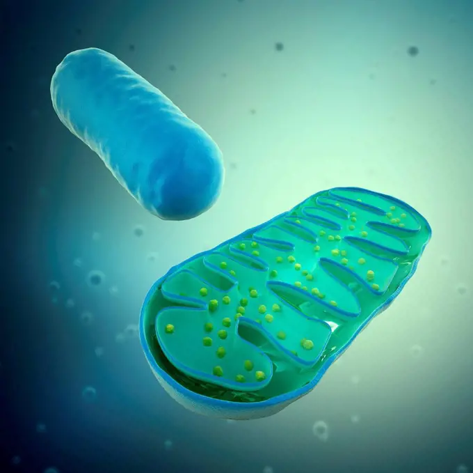
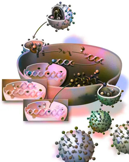
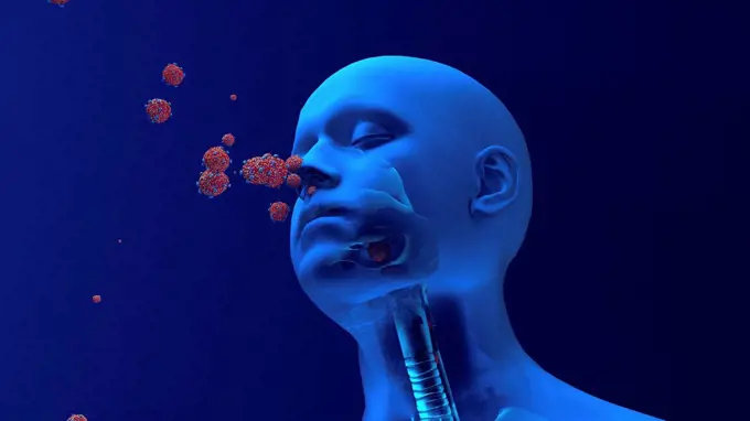
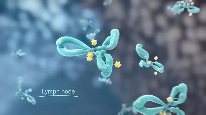
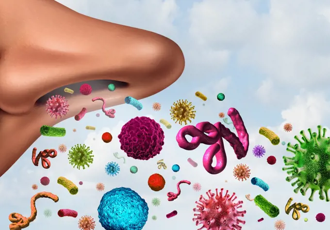
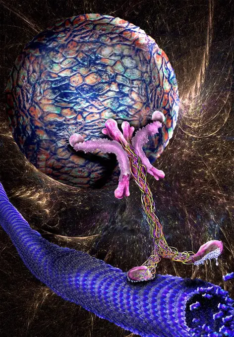
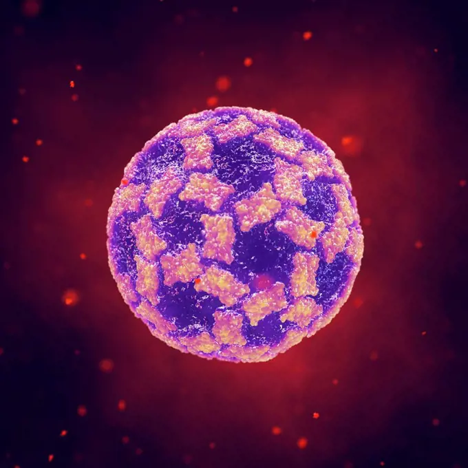
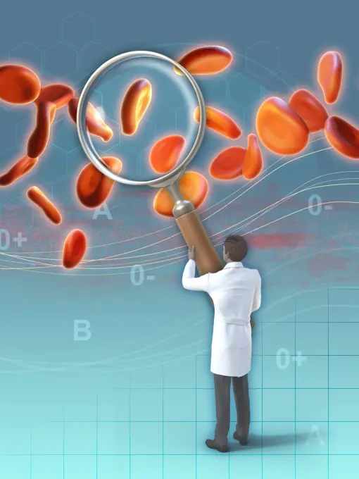
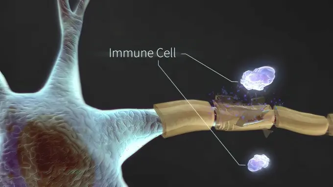
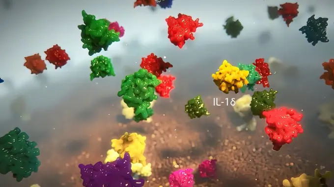
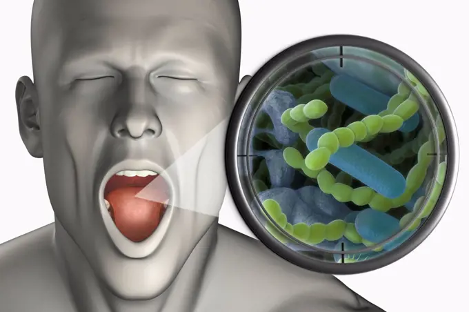
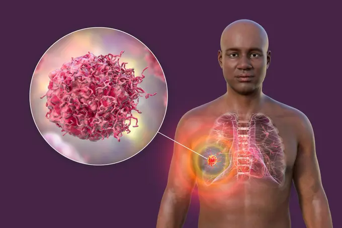
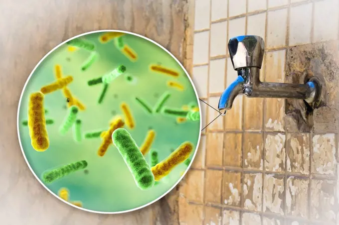
Fine ArtVintageFootageSubscriptionBoardsFree ResearchPricing
  • Fine Art
  • Vintage
  • Footage
  • Subscription
  • Boards
  • Free Research
  • Pricing

3D Biological Cell Structures

Detailed 3D illustrations depicting cellular structures like lysosomes and immune cells, showcasing vibrant colors and intricate designs related to lung immunity.

Group of red and white blood cells circulating in the bloodstream.
358 assets in this story
824-63189629
Illustration of the penetration and duplication of the aids virus in a CD4 host cell. The virus penetrates the cell and its genetic code attaches itself to the cells DNA, which forces the latter to produce all the elements constituting the virus. These elements gather on the surface of the host cell to form new modified aids viruses.
4128R-22380
Cancer cells, computer artwork.
4128-15728337
HIV structure, illustration
4239-V53647248
1525-26235488
Lung Immunity and Inflammation3D illustration. Lung Immunity and Inflammation
4128-V58566879
1525-26275177
The lobules are connected to small bile ducts that connect with larger ducts to ultimately form the hepatic duct. 3D illustration. Portal lobule of the liver. Anatomical unit of the liver, hepatic lobule
6188-65537750
Stent-Implantation zur Unterstützung der Blutzirkulation in Blutgefäßen - 3d-Illustration Stent implantation for supporting blood circulation into blood vessels - 3d illustration Copyright: xZoonar.com/ChristophxBurgstedtx 22561393
4128R-22362
Mitochondria, computer artwork.
4239-18641394
Microscopic view of Yersinia. Yersinia is a gram-negative, rod-shaped bacteria in the family Enterobacteriaceae.
1525-26275168
The term hepatitis means inflammation, and liver cells can become inflamed because of infection. 3D illustration. Liver cells become infected with virus
824-63164796
Penetration and duplication of the AIDS virus in the host cell.
4128-V58572066
4128-15750955
Coronavirus infection, conceptual illustration
6145-44836862
An infrared photo of the Small Magellanic Cloud taken by NASA's Spitzer Space Telescope is shown in this artist's illustration; an example of a planetary nebula, and a magnified depiction of buckyballs.
4297-1107
Normal lateral skull x-ray of a 2 year old boy
1525-26182317
The network used by the cell receptors to communicate with the cannabidiol is called the endocannabinoid system. 3D illustration. Synaptic transmission, human nervous system.
824-63163902
Spermatogenesis: different stages of sperm formation.
824-63219976
Infiltrating and in situ ductal adenocarcinomas, mitotic spindle zoom, treatments. Bust of a woman with a mammary gland and the axillary lymph nodes related to it. A first zoom starts from this gland to show the anatomical structure of the mammary gland with a main galactophore duct receiving the lobules, themselves made of acinus. Two cancerous tumors are located on the main milk duct: invasive or invasive ductal carcinoma (left) and carcinoma in situ. At the bottom, zoom in on the cancer cells whose nucleus is cut to show the mitotic spindle. A strand of DNA whose structure is modified is extracted from a chromosome. Receptors HER2 (yellow) on the nucleus, VEGF (green) on the neovessel and the estrogen receptor (black) on the DNA strand. Bottom left, adipose tissue site of conversion of androgens to estrogen by aromatases
1525-23789142
Seamless loop of the components of an eukaryotic cell, nucleus and organelles and plasma membrane - 3d illustration. Seamless loop of the components of an eukaryotic cell, nucleus and organelles and plasma membrane
1525-26275338
A lymph node, or lymph gland, is a kidney-shaped organ of the lymphatic system 3D illustration. lymph nodes located in many parts of the body
1525-20527060
Human Fertility concept with an extreme persective close up of microscopic sperm or spermatozoa cells swimming towards a female egg cell to fertilize and create a pregnancy as a medical reproduction symbol.
1525-20864507
Breathing disease medical concept and the spread of illness as a nose inhaling infectious bacteria virus cells and other dangerous microscopic toxic contaminants with 3D illustration elements.
4128-111581188
Intracellular transport. Illustration of a vesicle (sphere) being transported along a microtubule (rod-shaped) by a kinesin motor protein. Kinesins ar...
4128R-15545947
Norovirus infection, illustration
1525-27130461
Corona virus economic impact concept. 3d rendering
1525-26243993
Hair follicles are small, pocket-like holes in our skin. As the name suggests, they grow hair. 3D illustration. Hair follicles are small, pocket-like holes in our skin.
6188-61001672
Scientist examining some blood cells Scientist examining some human blood with a magnifying lens. Digital illustration, 3D render. Copyright: xZoonar.com/AndreaxDantix 21719457
4378-3036
A collection of Salmonella bacteria zoomed out from the surface of a cutting board with raw chicken. Salmonella is a genus of rod-shaped, Gram-negative bacteria.
1525-26235544
The immune system and cancer 3D illustration. The immune system and cancer
1525-56854564
TCAs bind to SERT and NET transport proteins for serotonin and norepinephrine, respectively.3D rendering. TCAs bind to SERT and NET transport proteins for serotonin and norepinephrine, respectively.
1525-23789117
illustrationshowing the formation of a blood clot, or thrombus. 3d illustration. 3D Rendered Platelet Adhesion Inside A Blood Vessel
1525-26275373
A lymphocyte is a type of white blood cell in the immune system of most vertebrates. 3D illustration. Innate Lymphoid Cells in Mucosal Immunity
4128R-13575648
Anticancer treatment. Conceptual illustration showing optical sight direct on cancer cell.
824-63223108
3D print of a spike protein of SARS-CoV-2 also known as 2019-nCoV, the virus that causes COVID-19 in front of a 3D print of a SARS-CoV-2 virus particle. The spike protein (foreground) enables the virus to enter and infect human cells. On the virus model, the virus surface (blue) is covered with spike proteins (red) that enable the virus to enter and infect human cells. For more information, visit the NIH 3D Print Exchange at 3dprint.nih.gov. Credit: NIH.
824-63195066
Illustration of angiogenesis. When a tumour forms, it is isolated and produces substances (VEGF) which stimulate the production of small blood vessels from neighbouring vessels. A new vascularisation surrounds the tumour and nourishes it, allowing it to grow. Antiangiogeneses medecine will block the process by trapping the VEGF and therefore destroying the vascularisation of the tumour. This treatment improves the efficiency of chemotherapy with which it is always associated.
4378-3433
Sectional view of intestinal villi revealing the network of blood vessels involved in the transport of absorbed nutrients to the rest of the body.
1525-26235524
inhibited adrenaline receptor, adrenergic receptors 3D illustration. inhibited adrenaline receptor, adrenergic receptors
4128-28769319
Rhinofacial conidiobolomycosis and fungus, illustration
1525-26182266
Dilutional hyponatremia, also known as water intoxication 3D illustration. Dilutional hyponatremia, also known as water intoxication
1525-23843147
Human nervous system, system that conducts stimuli from sensory receptors to the brain and spinal cord and conducts impulses back to other parts of the body. 3d illustration. The Human nervous system 3d medical
1525-26235574
The immune system defends our body against invaders, such as viruses, bacteria, and foreign bodies. 3D illustration. Immune system defense, functioning in the body
1525-23789104
fight against cancer cells 3d illustration. fight against cancer cells
4378-3345
Open mouth with a magnified zoom out of bacteria, which cause the halitosis.
4128-38524633
Lung cancer, illustration
1525-23843129
Neurons in action. electrical impulses between neuronal connections 3d illustration. Neurons in action. electrical impulses between neuronal connections
4128R-15465542
Safety of drinking water, conceptual illustration
1525-26305333
Nicotine raises blood pressure 3d illustration. Nicotine raises blood pressure
1525-26220610
Glycosylphosphatidylinositol or glycophosphatidylinositol, or GPI in short, is a glycolipid that can be attached to the C-terminus of a protein during posttranslational modification. 3D illustration. Chemical Biology of Glycosylphosphatidylinositol Anchors
1525-23789147
Basophil and mast cell in the immune system 3d illustration. Basophil and mast cell in the immune system
4128-20042135
Ringworm, illustration
1525-26235583
Insulin is a hormone produced by your pancreas that controls the amount of glucose in your bloodstream at any given time. 3D illustration. Insulin is a produced hormone.
4128-28769422
Nasal rhinosporidiosis, illustration
1525-26235725
Insulin is a hormone produced by your pancreas that controls the amount of glucose in your bloodstream at any given time. 3D illustration. Insulin is a produced hormone.
4128-20042139
Ringworm, illustration
4128-28769423
Nasal and ocular rhinosporidiosis, illustration
4128-20042043
Ringworm, illustration
824-65830761
3D renditions of three respiratory viruses: COVID-19, RSV, and flu.
PREVIOUS
of 4
NEXT
2401 S. Ervay, Suite 206
Dallas, Texas 75215
United States
Browse
CategoriesGalleriesFree Research
For Creators
License Your ContentSuperStock For StudentsFrame of Mind BlogContributor PortalNew Image SpotlightLicense AI Imagery
Resources
About UsLicensingTerms & ConditionsAPI AccessPrivacyPricing
Contact
+1 866 236 0087help@superstock.com Contact form
©2026 SuperStock. All Rights Reserved. -A
This site is protected by reCAPTCHA and Google Privacy Policy and Terms of Service apply.