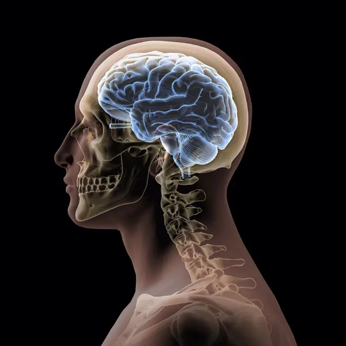
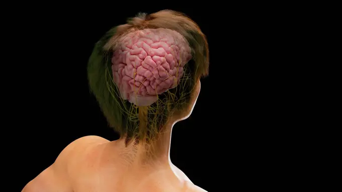
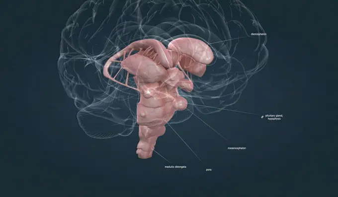
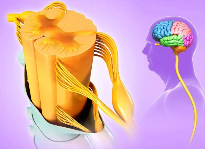
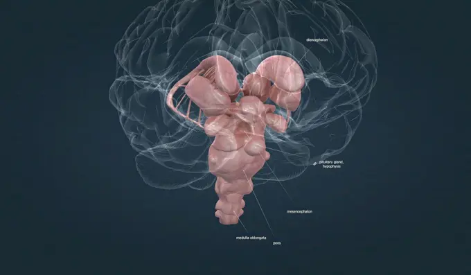
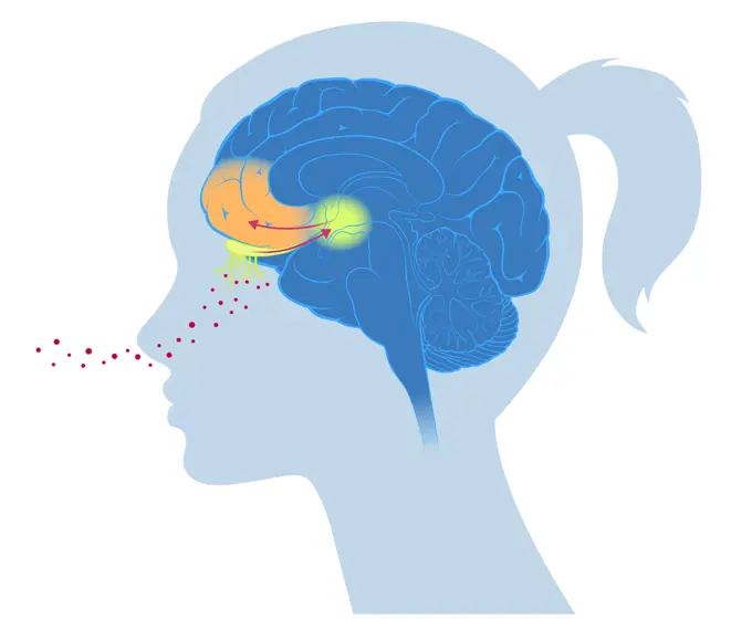
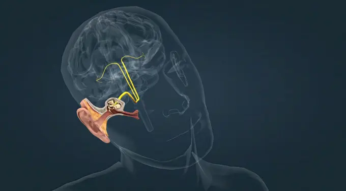
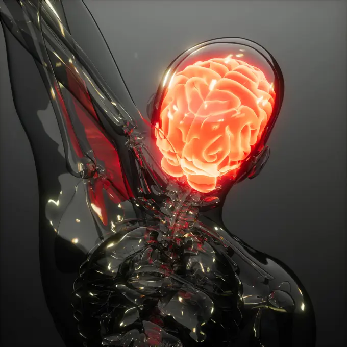
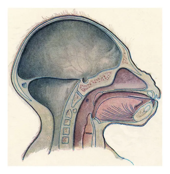
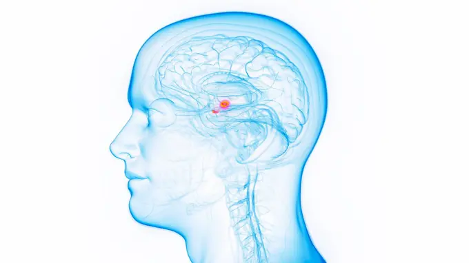
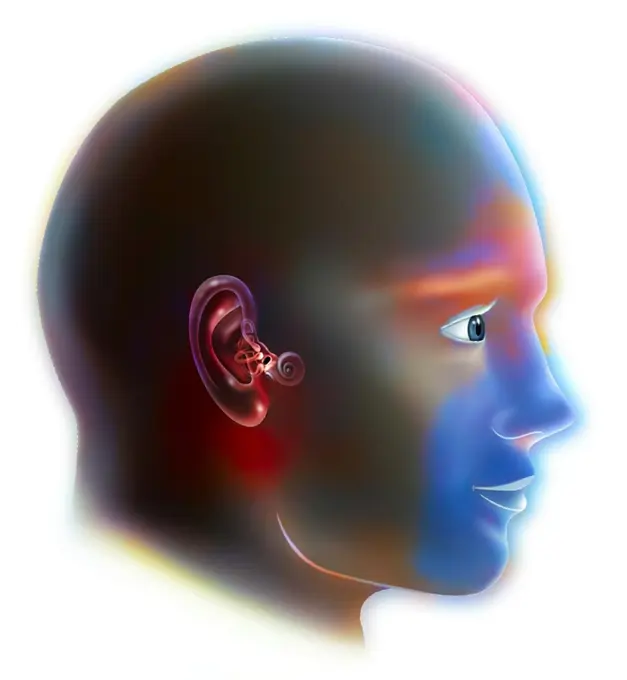
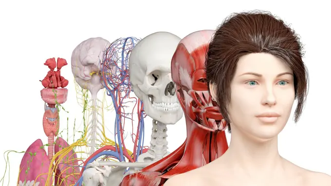
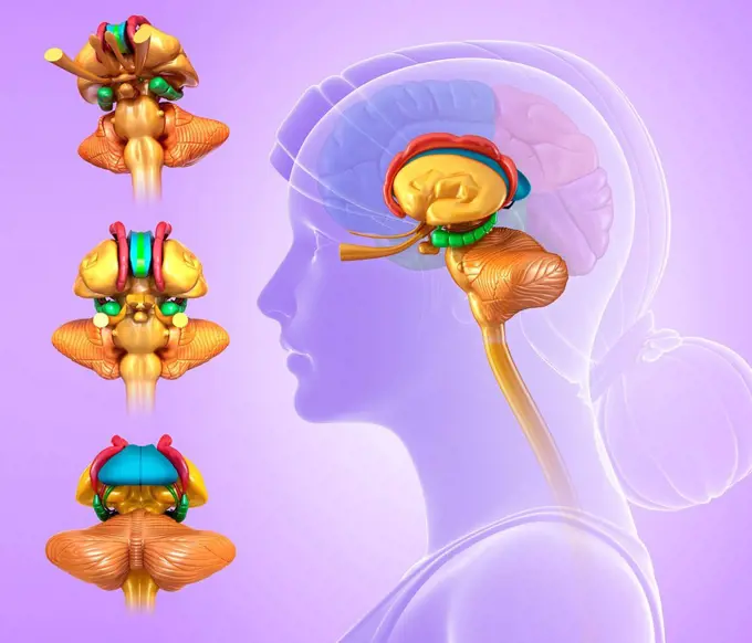
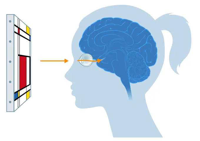
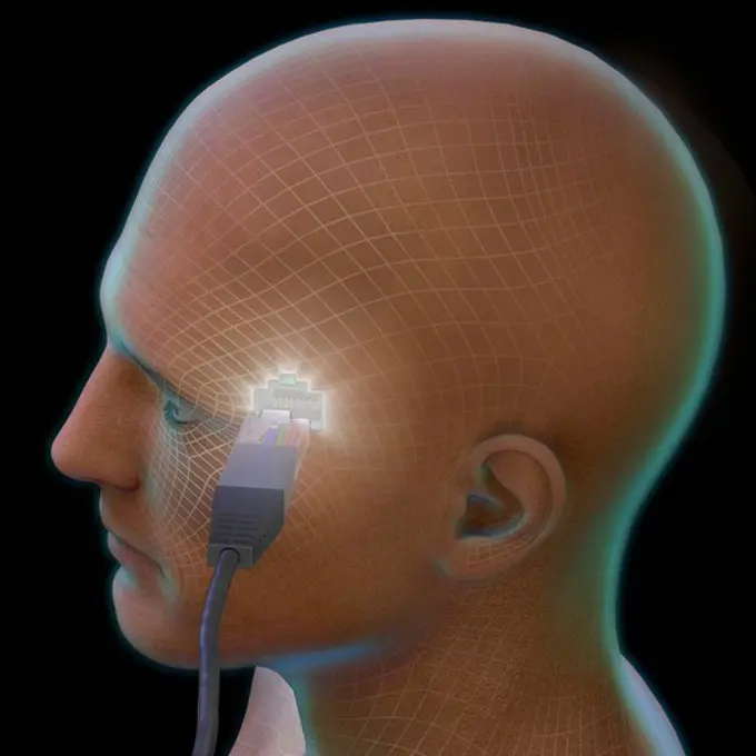
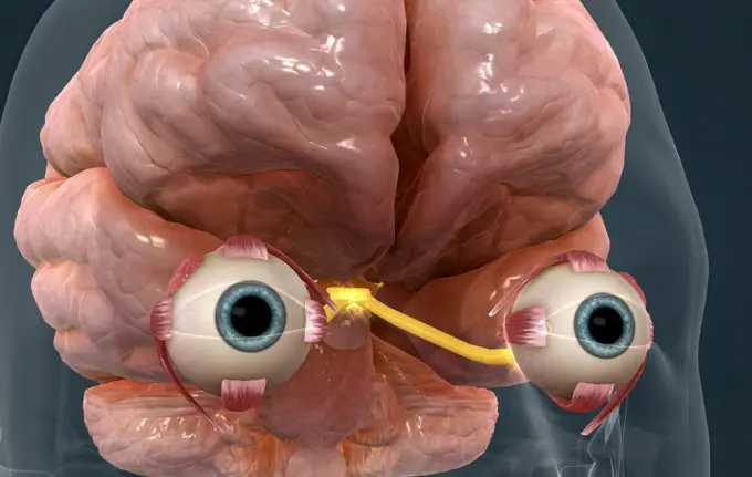
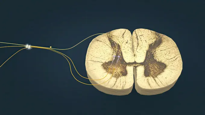
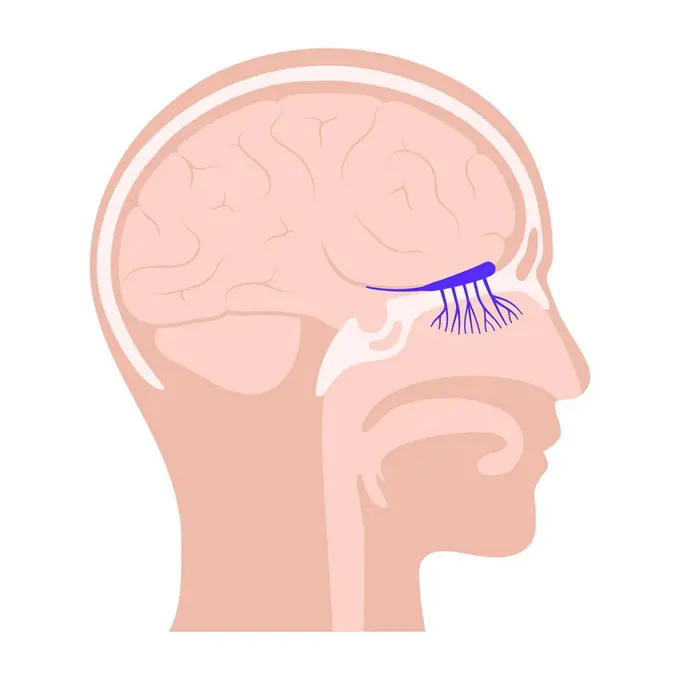
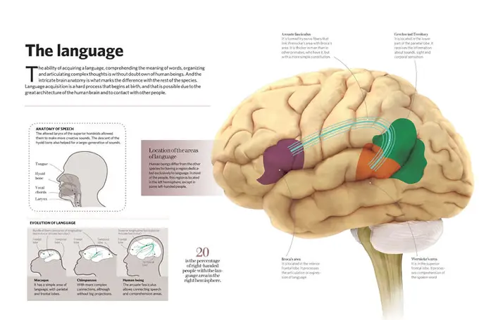
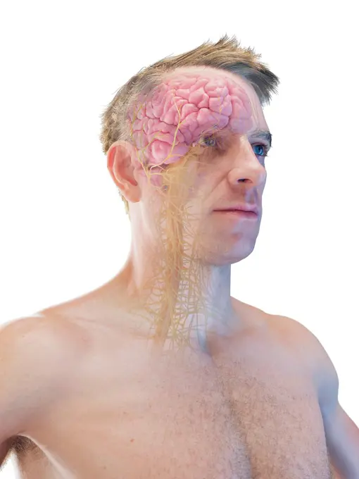
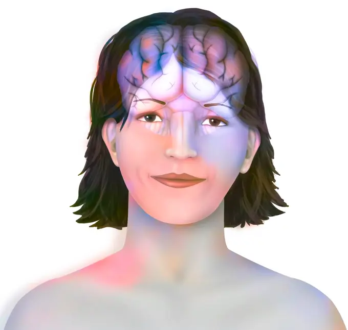
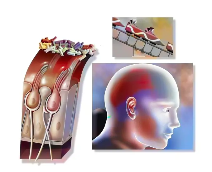
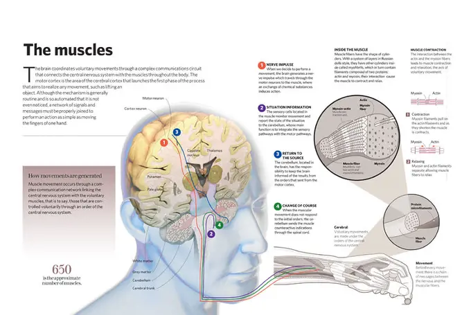
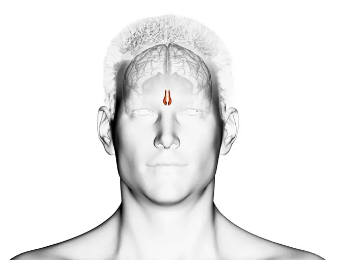
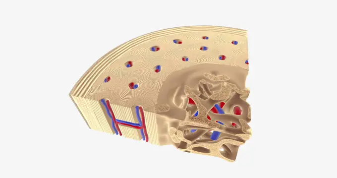
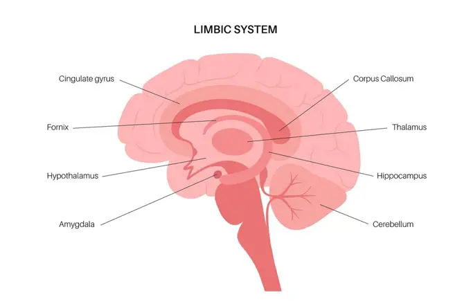
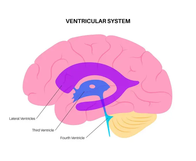
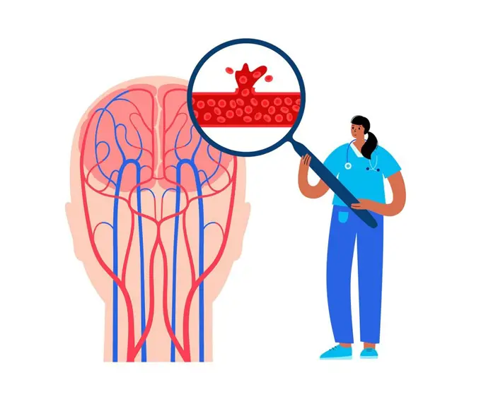
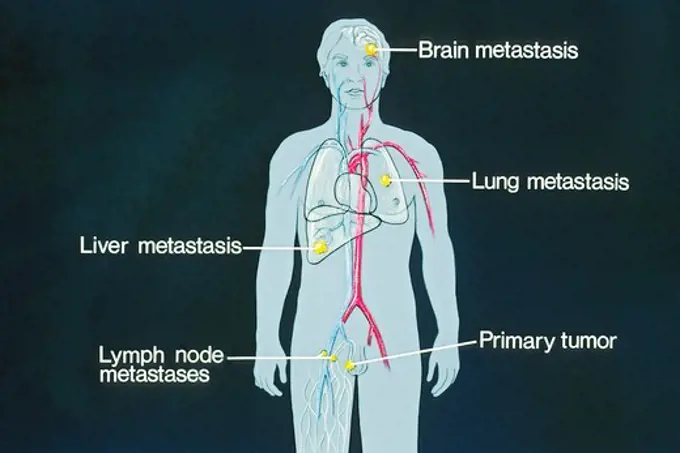
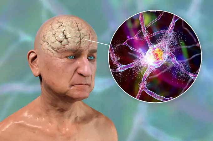
Fine ArtVintageFootageSubscriptionBoardsFree ResearchPricing
  • Fine Art
  • Vintage
  • Footage
  • Subscription
  • Boards
  • Free Research
  • Pricing

Brain Function and Anatomy

Illustrations depicting various aspects of brain anatomy and functions, focusing on processes related to memory and emotions, with a scientific style.

Profile of man's head and shoulders with semi-transparent skull and spine
156 assets in this story
4239-18641619
Illustration of a dog's brain.
4128-30416568
Central nervous system, illustration
6176-66093055
Three-dimensional anatomical color diagram of human head consisting of hinged, paper flap overlays, from "The Book of Health (1898)." by Henry M. Lyman, and Levi W. Yaggy.    This diagram appears opposite page 870 in The Book of Health
1525-56211415
The auditory cortex is the sensory system for the sense of hearing. It includes both the sensory organs (the ears) and the auditory parts of the senso 3D illustration. Sound passes through the ear and carries signals to the brain stem through the auditory nerve.
4128R-35956
Human skull, computer artwork.
4128R-13723710
Illustration of female spinal cord and brain regions.
1525-56183599
The brainstem is the posterior stalk-like part of the brain that connects the cerebrum with the spinal cord. 3D illustration. The brainstem is the posterior stalk-like part of the brain that connects the cerebrum with the spinal cord.
4128R-13586505
Illustration of spinal cord cross-section.
1899-53513365
Sensory brain sensors for smell.
1525-56183605
The brainstem is the posterior stalk-like part of the brain that connects the cerebrum with the spinal cord. 3D illustration. The brainstem is the posterior stalk-like part of the brain that connects the cerebrum with the spinal cord.
824-63224037
Prefrontal area: area active by smell and providing emotions.
1525-56183847
Sensory innervation to the external ear is supplied by both cranial and spinal nerves. 3d illustration. Sensory innervation to the external ear is supplied by both cranial and spinal nerves.
6188-58103450
Human Brain Radiology Exam science anatomy scan of human brain glowing Copyright: xZoonar.com/StanislavxRishnyakx 13445290
1525-28067567
Median section through the head of a young gorilla, vintage engraved illustration. From the Universe and Humanity, 1910.
5507-33089753
Brain organ anatomy. Human organ anatomy. Human brain. Vector, cartoon illustration
4128-30421697
Amygdala, illustration
1525-23943379
Brain and human senses organ set. Nose, ear, hand, tongue, eye. Sensory organs. See, hear, feel, smell and taste. Elements for an educational manual. Vecor illustrations isolated on white background.. Brain and human senses organ set. Nose, ear, hand, tongue, eye. Sensory organs. See, hear, feel, smell and taste. Elements for an educational manual. Vecor illustrations isolated on white background
1899-53509035
Inner ear (organ of hearing) and vestibular system.
1525-22137525
3d background texture of some brains - very realistic, and kind of nasty if this sort of thing grosses you out.
4128-30417460
Human anatomy systems, illustration
4128R-14058290
Human brain anatomy, illustration
4128R-14058208
Brain arteries and anatomy, illustration
4378-2922
Diagram showing a LAN cable entering a socket on the side of a human head.
4378-2634
Diagram showing a LAN cable entering a socket on the side of a human head.
1525-26235651
Brain damage can cause increased pressure inside your skull and headaches. The pressure also injures your brain or spinal cord. 3D illustration. Increased intracranial pressure, brain damage and spinal cord injury
824-63224050
Vision: path of light in the brain.
4128R-21848
Hypothalamus. Computer artwork of the brain showing the hypothalamus red. The hypothalamus has a number of functions, which include linking the nervou...
4102-33955
Diagram showing a LAN cable entering a socket on the side of a human head.
1525-56190179
When light hits the retina , special cells called photoreceptors turn the light into electrical signals. These electrical signals travel from the retina through the optic nerve to the brain. Then the brain turns the signals into the images you see. 3d illustration. The mechanism of vision in the human eye
6188-66528794
Depiction of an ischemic stroke, or cerebrovascular accident CVA. CEREBROVASCULAR NEUROL. DISEASE 016889_053
1525-56198364
Sensory nerve fibers innervate muscles and tendons, ligaments, joint capsule to carry proprioceptive impulses to the central nervous system 3D illustration. nerve fibers innervate muscles and tendons, ligaments, joint capsule to carry impulses to the central nervous system.
4128-19490783
Olfactory system, illustration
4409-28578093
Infographic about the unique capacity of human beings: the language. The ability to learn a language, to understand the meaning of words, to organize and articulate complex thoughts. There is also information about the evolution of language. Adobe InDesign (.indd).
1899-30609074
Eye, comparison between a normal eye and a myopic, hyperopic, astigmatic, presbyopic eye.
4128-30416647
Central nervous system, illustration
1899-53509018
Brain, cerebral hemispheres with longitudinal fissure, cerebellum and brainstem).
1899-30609344
Functioning of the macule, organ of static balance (position of the head).
824-63165377
Functioning of the macule: organ of static balance (position of the head).
4409-28578218
Infographic about the muscles in the human body, how they perform an action sent by the brain. Adobe InDesign (.indd).
824-63165378
Functioning of the macule: organ of static balance (position of the head).
6188-67434532
Frau, die wegschaut, mit visueller Demonstration der Verbindung zwischen Gehirn und Herz Woman looking away with visual demonstration of connection between brain and heart influencing person decision. Anatomical features of structure of body of woman for educational literature for doctors Copyright: xZoonar.com/EladaxVasilyevax 22957540
4128-19247043
Hypothalamus, illustration.
1525-56853780
The Healty Bone Cross Section 3D rendering. The Healty Bone Cross Section
4128-19358395
Brain limbic system, illustration.
4128-20041003
Ventricular system, illustration
4128-19247586
Haemorrhagic stroke research, illustration.
4391-169
This is a schematic drawing of sites of potential metastases, places where cancer can spread. The primary tumor and metastases to the brain, lung, liver and lymph nodes are shown.
824-63224114
All uses except for Canadian college and university publishing - Sensory pathways, motor pathways, sensory-motor pathways, reflex arc, stimulus, pain. The reflex arc is a neural circuit made up of a neuron receiving information and a neuron which induces an adapted action. This first neuron, or a chain of intermediate neurons are called afferent neurons or sensory neurons. They process the information received (interneurons). Here, the child pricks his hand on the cactus. The brain processes information. Then, the efferent neurons or motor neurons send the order to the effector organ (here the muscles of the arm and hand) after processing this information. In this case, the child will withdraw his hand from the spikes of the cactus. The zoom at the bottom right of the illustration shows a motor neuron. Motor neurons are cells that control the movement of the body. They are represented by the purple cell that runs from the spinal cord to the muscle fibers where they transmit information
4128-V58615396
4378-3153
A sectional view of the human head revealing the anatomy of the ear canal and inner ear anatomy.
4128-30419477
Female ear, illustration
4128R-15303160
Aspergilloma of the brain, illustration
4409-28579702
Infographic of the fetal development up to the 40th day of gestation, in which can observe considerable differences in the brain and facial features of the fetus. QuarkXPress (.qxp); 6259x4015.
6188-67431736
3D Isometric Flat Conceptual Illustration of Human Embryo Evolution, Medizin und pränatale Gesundheitbr3D Isometric Flat Conceptual Illustration of Human Embryo Evolution, Medicine and Prenatal Health Isometrische flache konzeptionelle Illustration des me 3D Isometric Flat Conceptual Illustration of Human Embryo Evolution, Medicine and Prenatal Health 3D Isometric Flat Conceptual Illustration of Human Embryo Evolution, Medicine and Prenatal Health Copyright: xZoonar.com/AnastasiiaxTorianykx 22948326
4128-18800804
Elderly person with dementia, illustration
4128-20040531
Pituitary gland anatomy, illustration
PREVIOUS
of 2
NEXT
2401 S. Ervay, Suite 206
Dallas, Texas 75215
United States
Browse
CategoriesGalleriesFree Research
For Creators
License Your ContentSuperStock For StudentsFrame of Mind BlogContributor PortalNew Image SpotlightLicense AI Imagery
Resources
About UsLicensingTerms & ConditionsAPI AccessPrivacyPricing
Contact
+1 866 236 0087help@superstock.com Contact form
©2026 SuperStock. All Rights Reserved. -A
This site is protected by reCAPTCHA and Google Privacy Policy and Terms of Service apply.