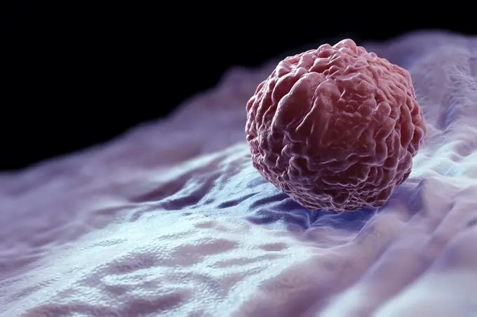
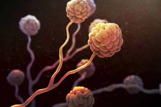
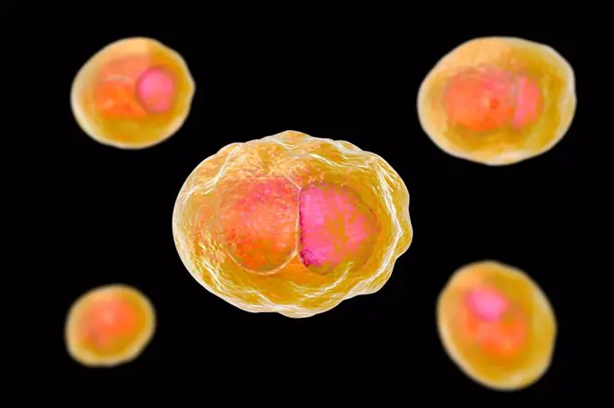
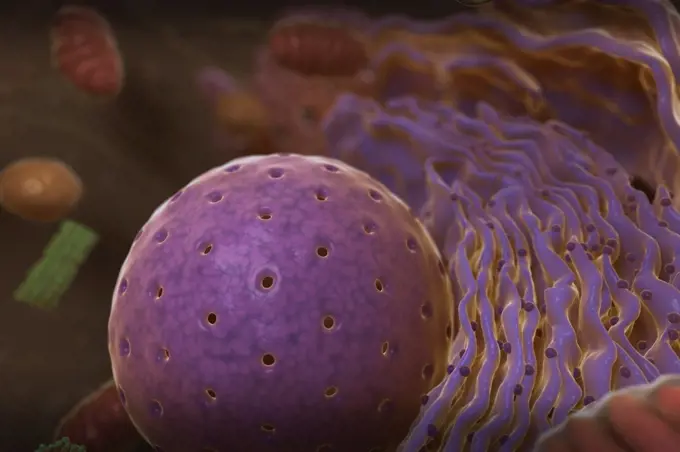
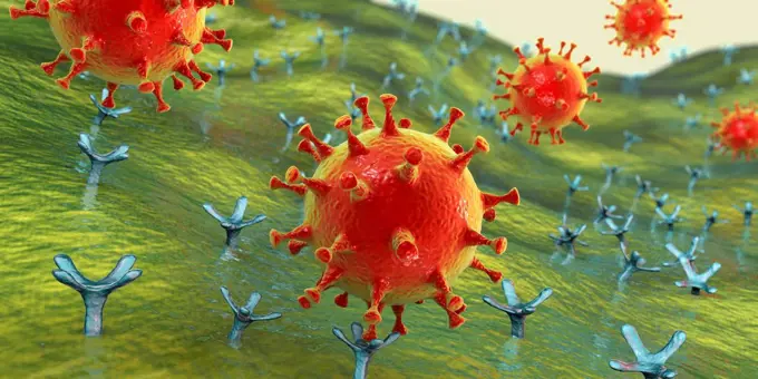
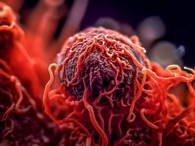
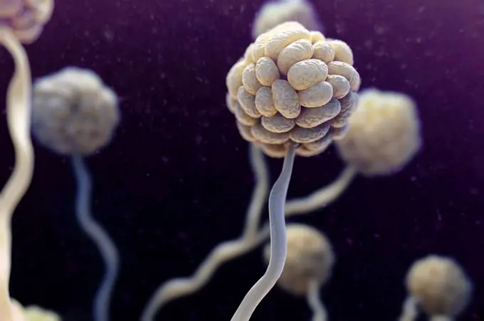
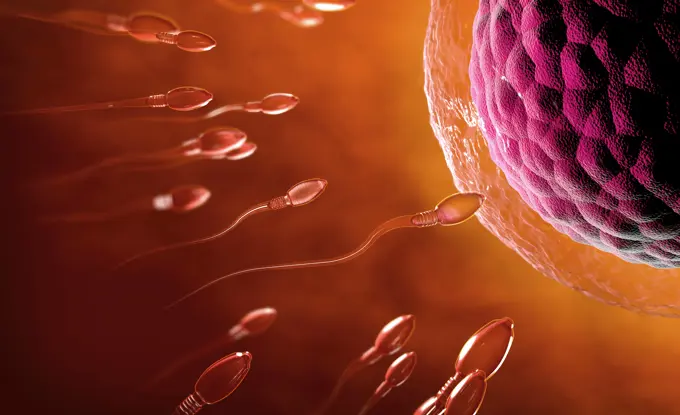
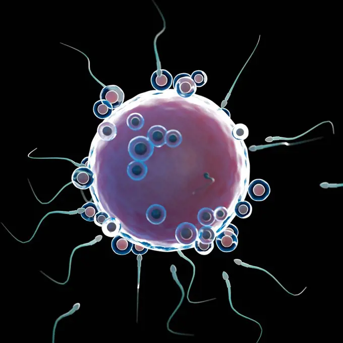
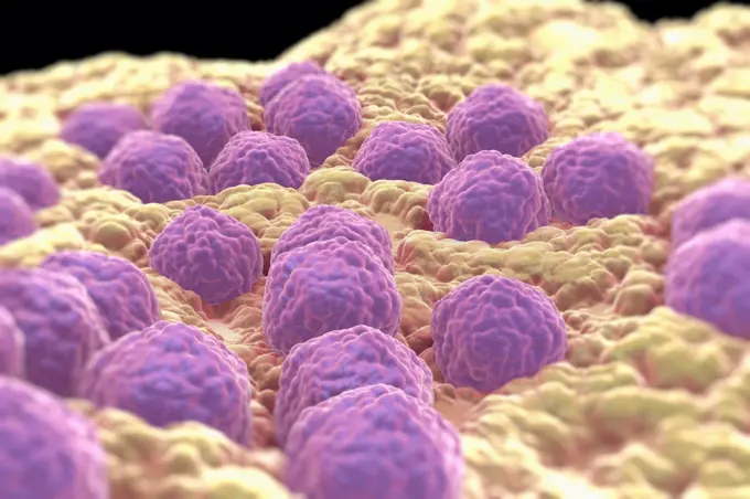
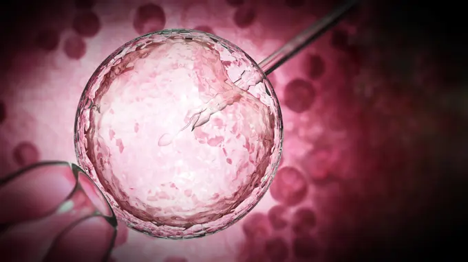
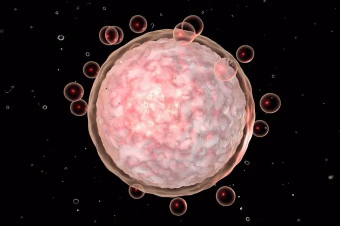
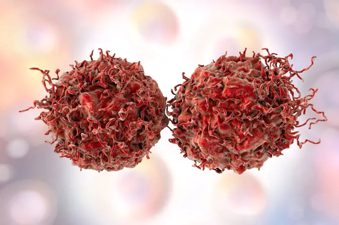
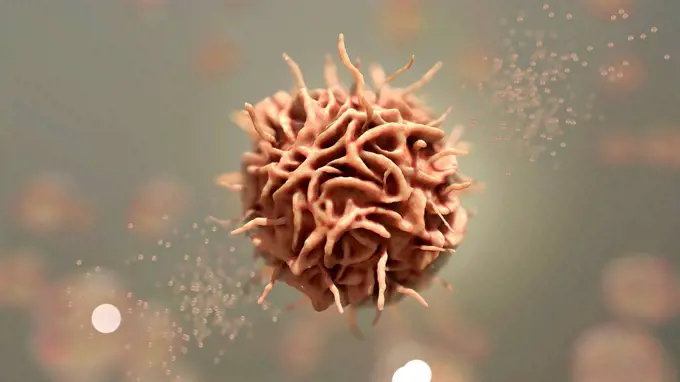
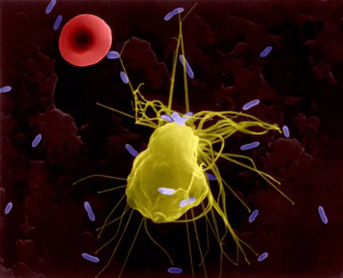
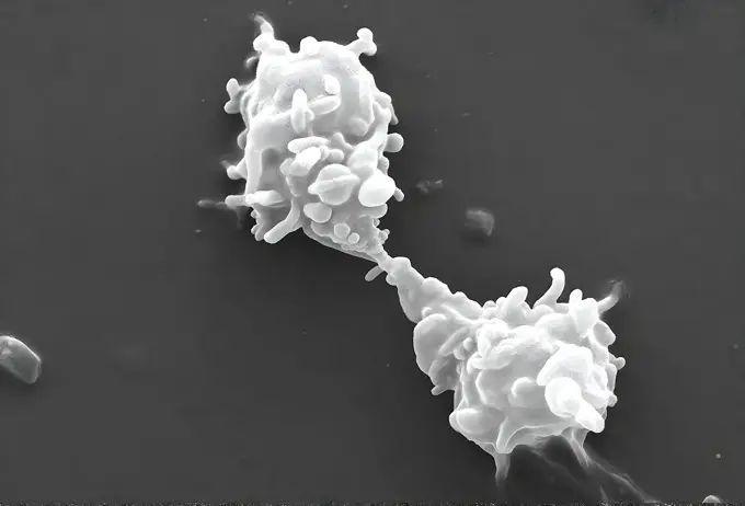
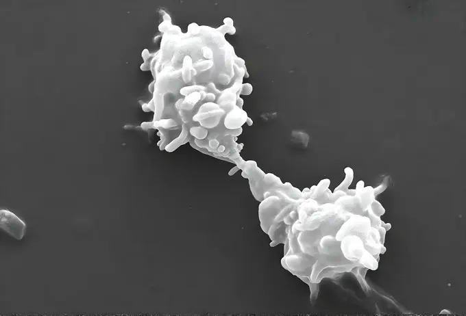
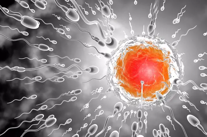
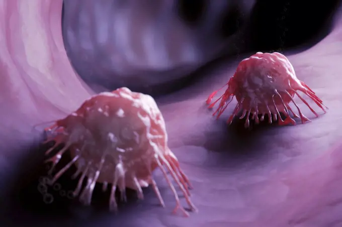
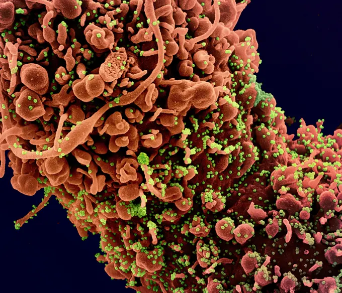
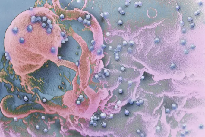
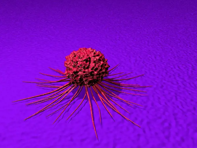
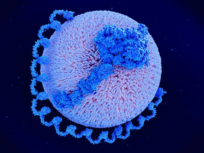
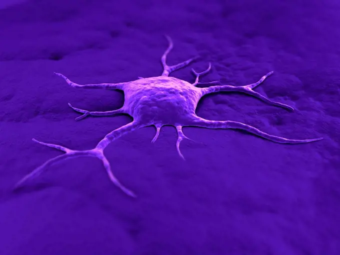
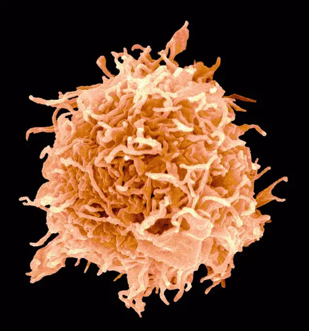
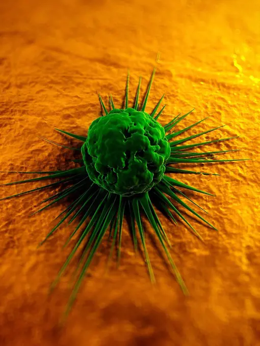
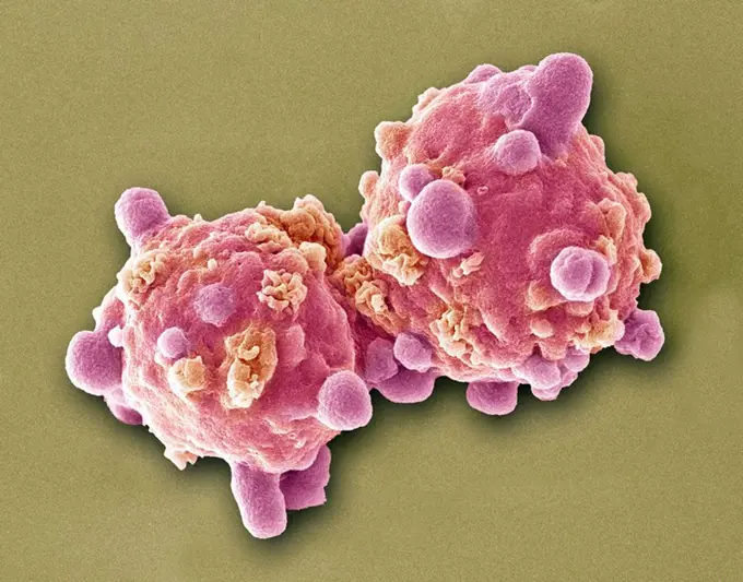
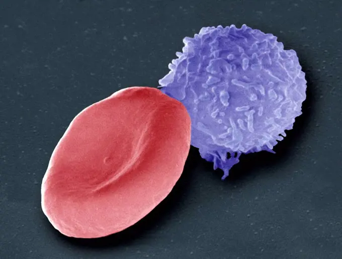
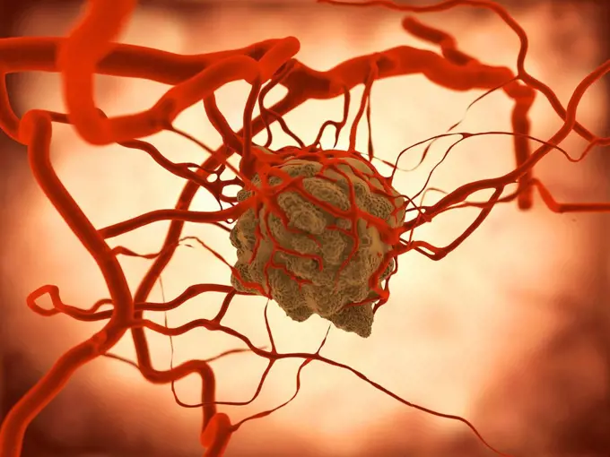
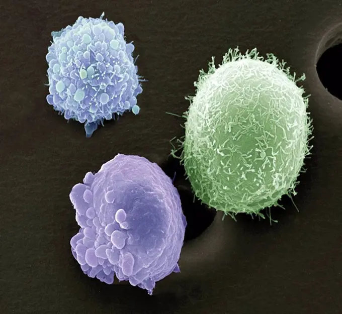
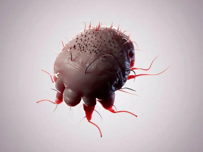
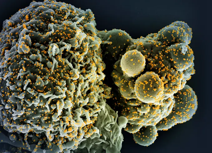
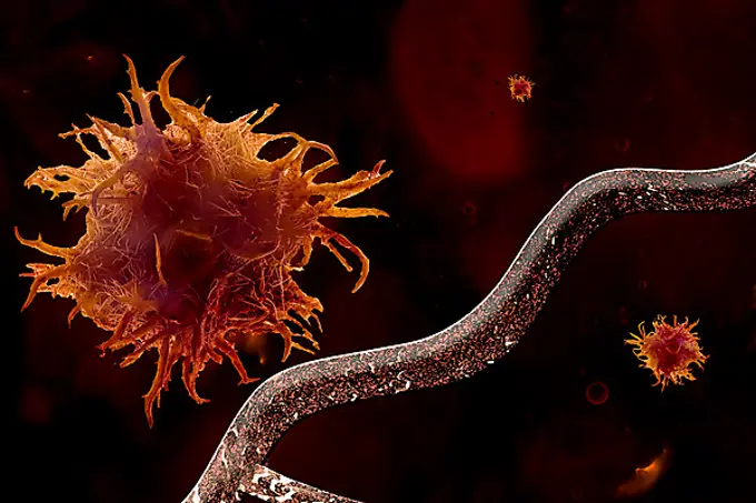
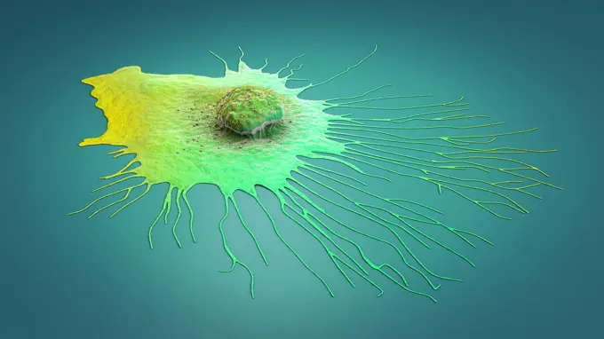
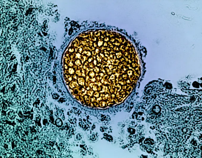
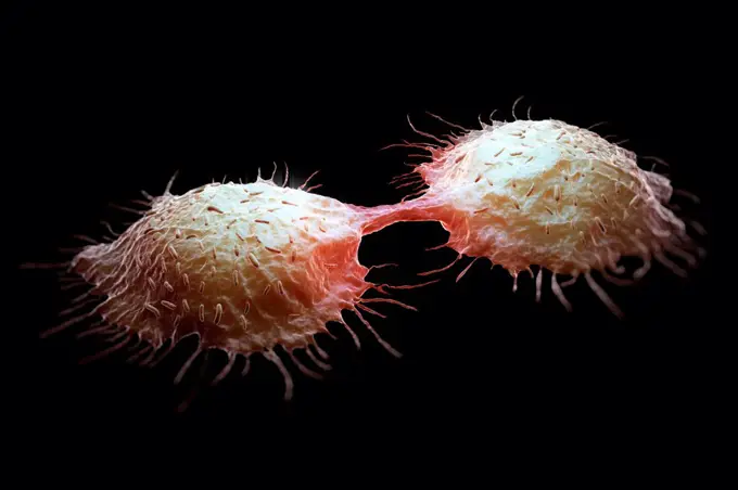
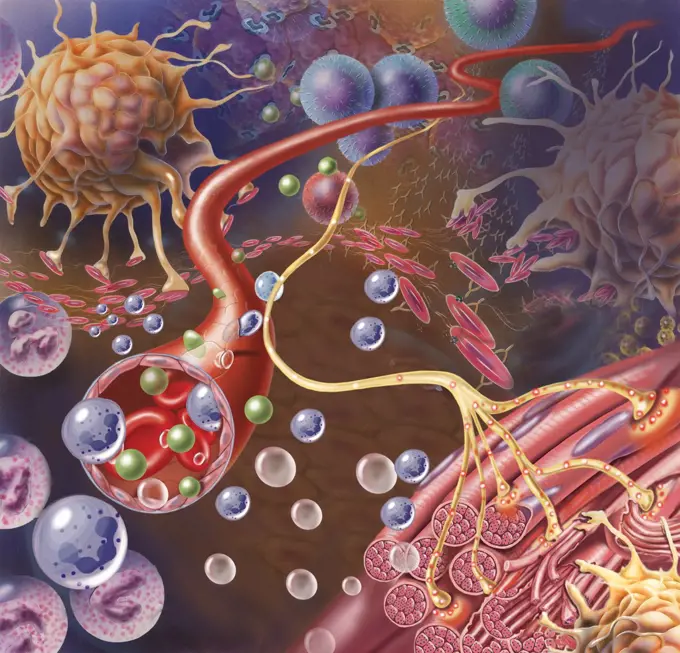
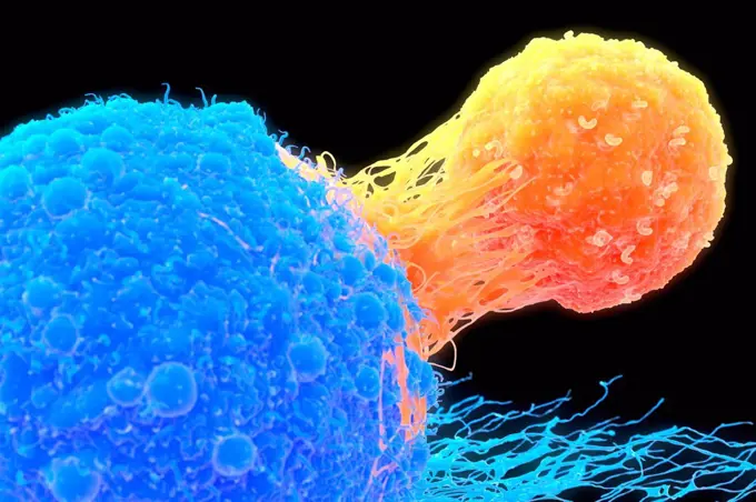
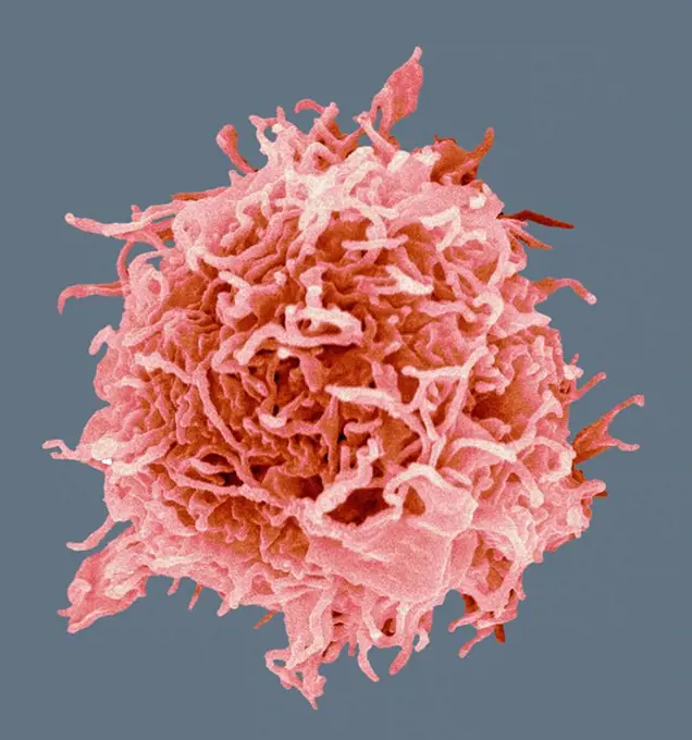
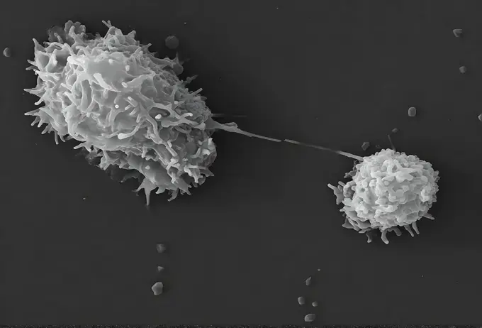
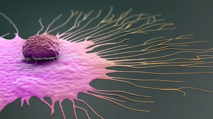
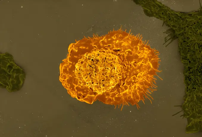
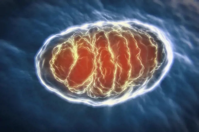
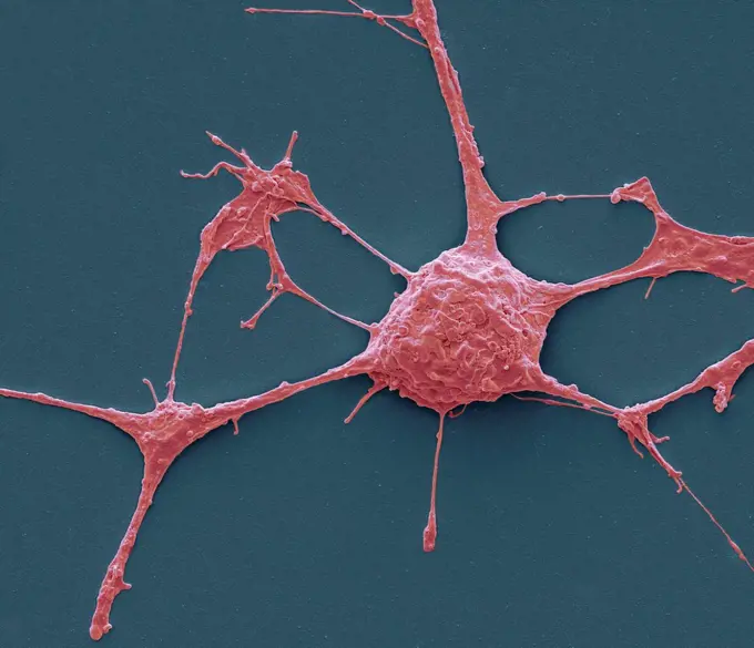
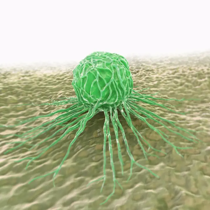
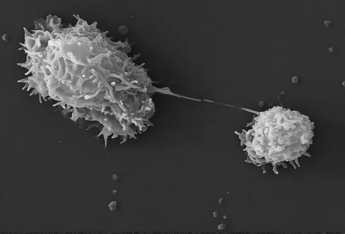
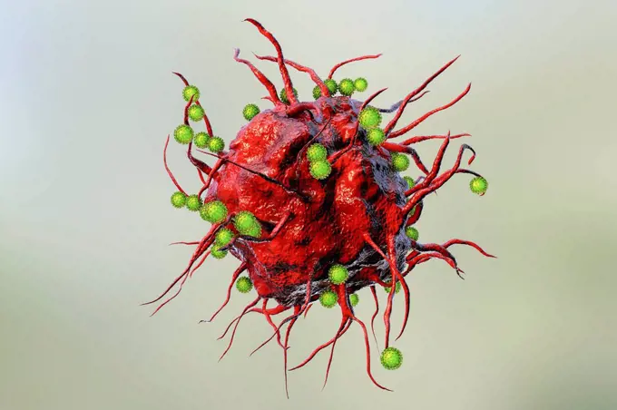
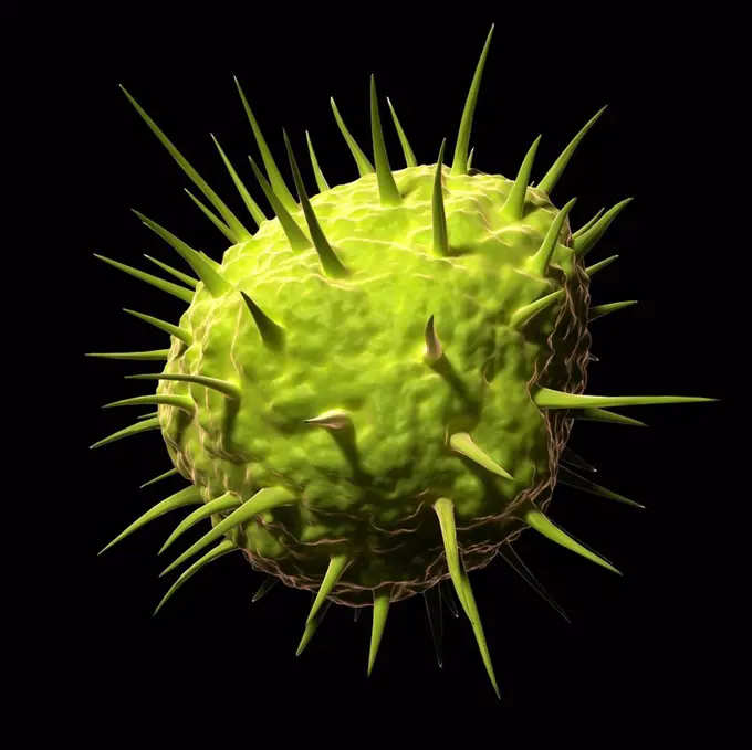
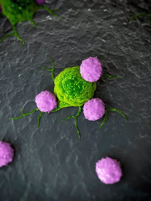
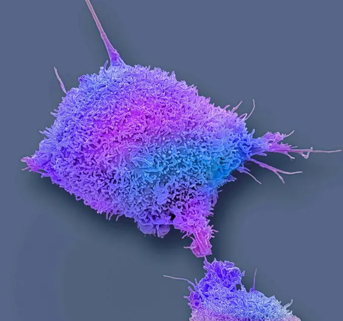
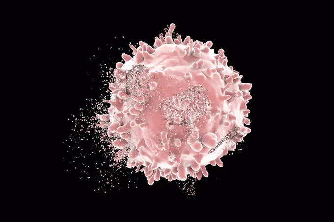
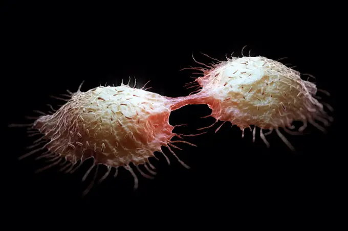
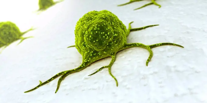
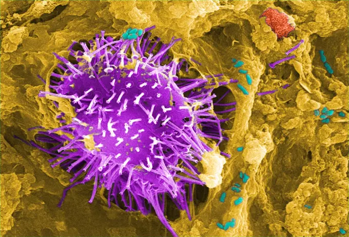
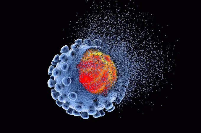
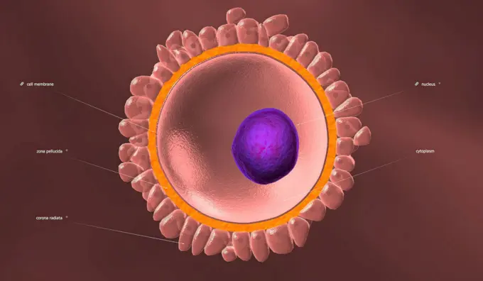
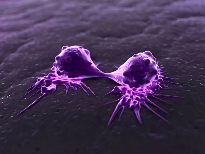
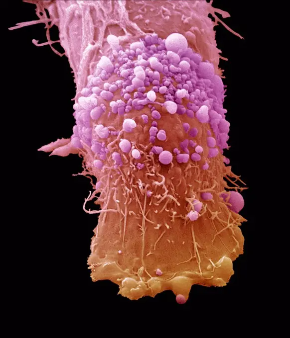
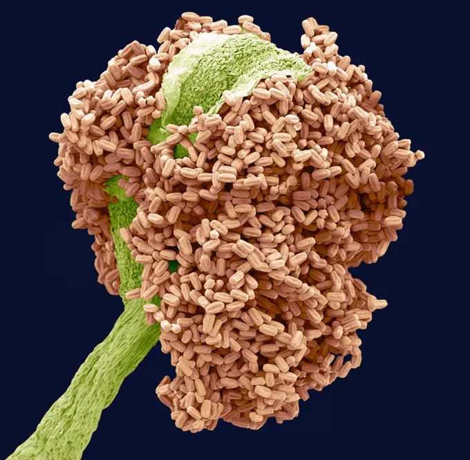
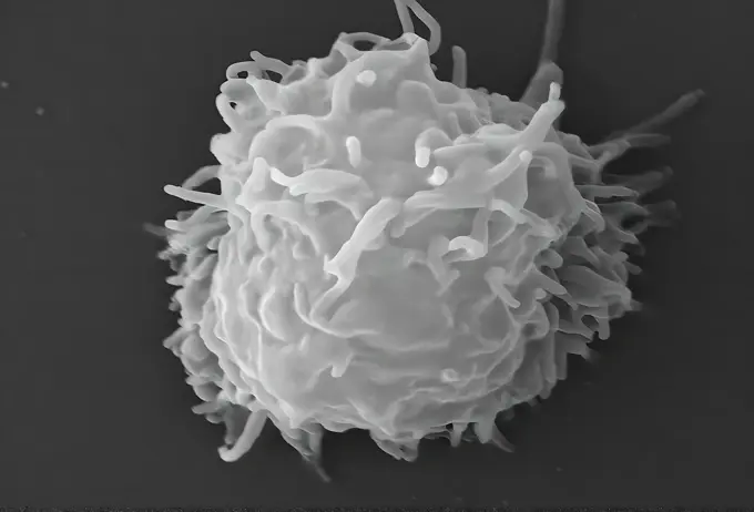
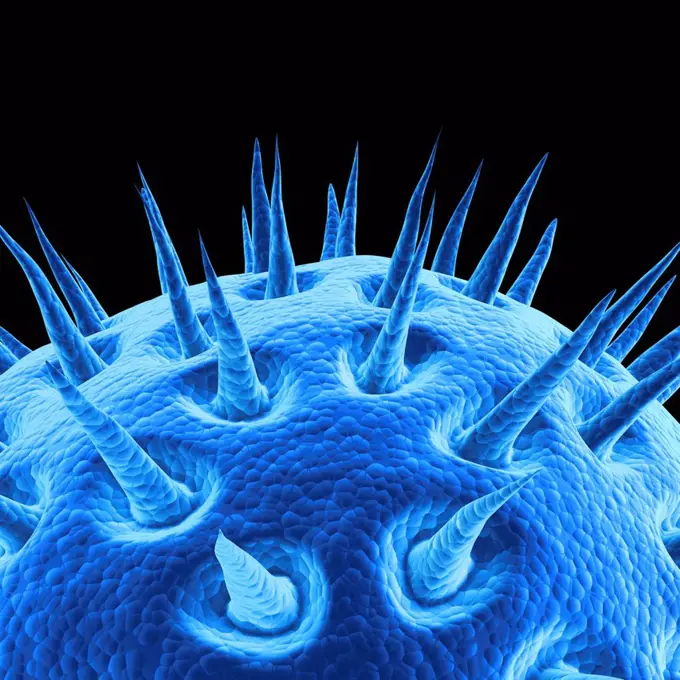
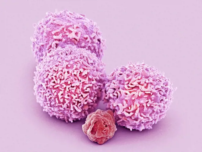
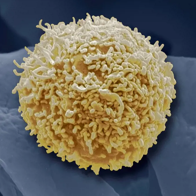
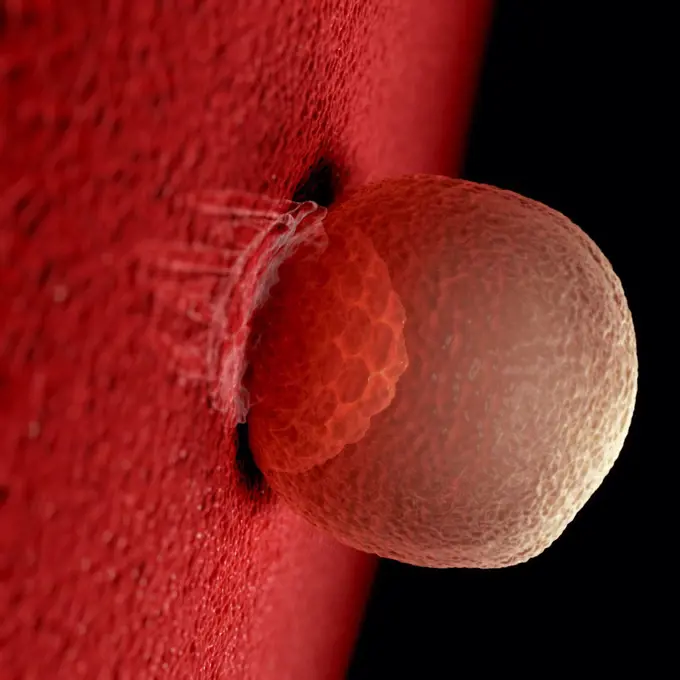
Fine ArtVintageFootageSubscriptionBoardsFree ResearchPricing
  • Fine Art
  • Vintage
  • Footage
  • Subscription
  • Boards
  • Free Research
  • Pricing

Cellular Structures and Microscopy

High-resolution images of various cells, including stem cells and cancer cells, showcasing intricate details in a scientific context.

Embryonic Stem Cell
Embryonic Stem Cell
194 assets in this story
4378-4590
Black Mold Spores
4128-V58562325
4128R-13325998
Leucocyte attacking bacteria, illustration.
4128-V58568609
4128R-15405906
Chlamydia trachomatis bacteria, illustration
4378-4030
Inner Workings of a Human Cell
4128-16073683
Covid-19 coronavirus binding to human cell, illustration
4128-48285956
Blood vessel formation in tumour, illustration
4128-V58557865
4378-4593
Black Mold Spores
6188-65537787
3D illustration of transparent sperm that swim on an egg cell 3D illustration of transparent sperm that on an egg room for swimming copyright: xzoonar.com/christophxburgstedtx 22565429
4128-19053674
Human egg cell and sperm, illustration
4128-111495102
Cancer cell spreading, illustration.
4378-3810
Rubella virus particles on a cellular surface. Rubella disease, commonly known as German measles, is the cause of congenital rubella syndrome when infection occurs during the first weeks of pregnancy.
824-63223272
Colorized scanning electron micrograph of a T lymphocyte. Credit: NIAID.
4128-111453070
Prostate cancer cells, SEM
6188-57664895
3D illustration of artificial insemination process showings sperms being injected inside the ovule. 3D illustration 3D illustration of artificial insemination process showings sperms being injected inside the ovule. 3D illustration. Copyright: xZoonar.com/CigdemxSimsekx 21275798
4128R-13575537
Human egg cell, computer illustration.
4128R-13575647
Prostate cancer cells, computer illustration.
4128-111493745
Computer generated illustration of a dendritic cell, which are part of the mammalian immune system.
4128R-13619944
Alveolar macrophage phagocytosis of E. coli, coloured scanning electron micrograph (SEM). A red blood cell is in the periphery (mammal lung pleural ca...
1899-61460856
This scanning electron microscope (SEM) image revealed a protozoan Acanthamoeba polyphaga as it was about to complete the process of cell division known as mitosis, becoming two separate organisms. Note the numerous pseudopodia protruding from the surfaces of these organisms. These pseudopodia allow amoebae to move and grasp objects in their environment. CDC/Catherine Armbruster Margaret Williams 2009
824-57659323
This scanning electron microscope (SEM) image revealed a protozoan Acanthamoeba polyphaga as it was about to complete the process of cell division known as mitosis, becoming two separate organisms. Note the numerous pseudopodia protruding from the surfaces of these organisms. These pseudopodia allow amoebae to move and grasp objects in their environment. CDC/Catherine Armbruster Margaret Williams 2009
1815-18039341
3D Rendered Illustration of sperm cells racing to fertilize egg
1525-26275258
The lungs are a pair of spongy, air-filled organs located on either side of the chest . The trachea conducts inhaled air into the lungs through its tubular branches, called bronchi. 3D illustration. Lung organs located in the thoracic cavity of the thorax
4128R-14588018
Cancer cell, illustration
4378-1906
A group of cancer cells.
824-63225820
Colorized scanning electron micrograph of a cell showing morphological signs of apoptosis, infected with SARS-COV-2 virus particles (green), isolated from a patient specimen. Image captured at the NIAID Integrated Research Facility (IRF) in Fort Detrick, Maryland.
4269-7101
Colored scanning electron micrograph of HIV viruses (blue dots) budding from a T-lymphocyte white blood cell (yellow & green).
4128R-12571800
Blood cells. Coloured scanning electron micrograph (SEM) of human blood showing a red and white cell ( lymphocyte). Red blood cells (erythrocytes) hav...
4128-15982700
Colorectal cancer cells. Coloured scanning electron micrograph (SEM) of a cancer cells from the human colon (large intestine). Cancer of the colon is ...
4128R-14588017
Cancer cell, illustration
4128-V58557946
4128-111584785
Hollyhock pollen grain. Coloured scanning electron micrograph (SEM) of a pollen grain from a hibiscus (Alcea setosa) flower. Pollen grains are the mal...
4128R-11313313
HIV particle, computer artwork.
4239-18641829
Digitally colorized scanning electron micrograph revealing ultrastructural details at the site of interaction of two spherical shaped, Middle East respiratory syndrome coronavirus (MERS-CoV) viral particles, colorized blue, that were on the surface of a camel epithelial cell, colorized red.
4128-24796227
Lipoprotein A particle, illustration
4128-V58562137
4128R-12699820
Coloured scanning electron micrograph (SEM) of breast cancer cells growing in culture. These are from the MCF-7 cell line. Magnification: x800 when pr...
4128R-14708
Cancer cell, conceptual computer artwork. The uneven surface and cytoplasmic projections are characteristic of cancer cells.
4128R-136
B_cell, SEM
4128R-15061
Cancer cell, computer artwork. The uneven surface and cytoplasmic projections are characteristic of cancer cells.
4128-V58562490
4128R-4320
Leukaemia cells, SEM
4128R-3548
Blood cells, SEM
4128R-11291843
Artwork of tumor.
4128R-5582
Skin cancer cells, SEM
4128R-20454
Scabies mite, artwork
824-65831024
Colorized scanning electron micrograph of a cell (green) infected with the Omicron strain of SARS-CoV-2 virus particles (gold), isolated from a patient sample.
4128-V58562342
1848-54783062
Tumour in an early stage, angiogenesis, illustration
6188-65540263
Wissenschaftliche Illustration einer wandernden Brustkrebszelle - 3d-Illustration Scientific illustration of a migrating breast cancer cell - 3d illustration Copyright: xZoonar.com/ChristophxBurgstedtx 22577974
824-66066016
A Coccidioides fungal spherule (yellow) found in a tissue sample. This organism causes coccidioidomycosis, also known as Valley Fever. Credit: micrograph by CDC colorization and visual effects by NIAID.
4378-5471
Colon Cancer Cells
4239-20481112
Nerve ending, seen in lower right, sends pain message from injured muscle. Blood vessel and immune cells are seen in the center and upper right of image.
4128-16073937
T-cell attaching to cancer cell, illustration
4128R-7866
B_cell, SEM
1899-61460849
This scanning electron microscope (SEM) image revealed two Acanthamoeba polyphaga protozoa, as they interacted through their pseudopodia, projecting from the surfaces of these organisms. These pseudopodia allow the amoebae to move, to grasp objects in their environment and, in this case, to communicate with each other. CDC/Catherine Armbruster Margaret Williams 2009
4128-V58562329
6188-65540222
Wissenschaftliche Illustration einer wandernden Brustkrebszelle - 3d-Illustration Scientific illustration of a migrating breast cancer cell - 3d illustration Copyright: xZoonar.com/ChristophxBurgstedtx 22577767
4128-V58562493
824-63225809
Colorized scanning electron micrograph of a macrophage.
4128-V58562141
4378-3957
A single mitochondrion. These organelles act as cell power plants as they generate adenosine triphosphate (ATP), a source of chemical energy.
6188-58133371
A micro cell division process A micro cell division process illustration Copyright: xZoonar.com/OxanaxRishnyakx 6242670
4128R-11474472
Neurone. Scanning electron micrograph (SEM) of a PC12 neurone in culture.The PC12 cell line, developed from a pheochromocytoma tumor of the rat adrena...
4378-3796
Close up view of a single breast cancer cell on a surface.
824-57659316
This scanning electron microscope (SEM) image revealed two Acanthamoeba polyphaga protozoa, as they interacted through their pseudopodia, projecting from the surfaces of these organisms. These pseudopodia allow the amoebae to move, to grasp objects in their environment and, in this case, to communicate with each other. CDC/Catherine Armbruster Margaret Williams 2009
4128-16073749
SARS-CoV-2 viruses and immune cell, illustration
4128R-11312891
Mimi virus, computer artwork.
4128R-11473183
White blood cells attacking a cancer cell, computer artwork.
4128R-15465092
Cervical cancer cell, SEM
4128R-13619880
Destruction of leukaemia blood cell, computer illustration. Conceptual image for leukaemia treatment.
4378-20013842
Colon Cancer Cells
4128R-11544472
Cancer cells, computer illustration.
1899-85635
Water Analysis SEM
4128-16073644
Eradicating covid-19 coronavirus, conceptual illustration
1525-56184232
Female egg structure with cytoplasm, nucleus and corona radiata 3D illustration. Female egg structure with cytoplasm, nucleus and corona radiata.
4128R-17152
Cancer cells dividing, computer artwork.
4128R-13047380
Skin cancer cell, coloured scanning electron micrograph (SEM). Cultured melanoma cell showing the numerous blebs and microvilli characteristic of canc...
4128R-11474743
Influenza, computer artwork.
4128R-8943
Bay tree anther, SEM
824-57659319
This scanning electron microscope (SEM) image revealed some of the ultrastructural features observed on the surface of a protozoan Acanthamoeba polyphaga. Note the numerous pseudopodia, protruding from the surface of the organisms. These allow the amoeba to move and grasp objects in its environment. CDC/Catherine Armbruster Margaret Williams 2009
4128R-21361
Virus, artwork
4128R-31106
White blood cells. Coloured scanning electron micrograph (SEM) of white blood cells (leucocytes) and a platelet (thrombocyte, red). Magnification: x6,...
4128R-15002
Lymphocyte white blood cell, coloured scanning electron micrograph SEM. Its surface is covered in characteristic microvilli. Magnification: x6000 when...
4378-1805
Blastocyst development after 6-8 days.
4128R-11544461
Cancer cell, computer illustration.
4128-V58562028
4128R-30464
Bone cancer cell. Coloured scanning electron micrograph of an osteosarcoma cancer cell. Osteosarcoma is an aggressive malignant neoplasm arising from ...
1525-19835776
Mitosis. Stage four
4128R-25481
Cancer cell, computer artwork.
4384-159
This highly-magnified, digitally-colorized scanning electron micrograph (SEM) revealed some of the ultrastructural morphology displayed on the rostral head region of a bedbug, Cimex lectularius.  Note the proximal anatomical relationships the insects skin piercing mouthparts it uses to obtain its blood meal, and how they join the head. Although bedbugs have been found naturally-infected with blood-borne pathogens, they are not effective vectors of disease.  The primary medical importance is inf
1525-28347411
Cancer of the prostate medical concept as cancerous cells in a male body attacking the reproductive system as a symbol of human malignant tumor growth diagnosis treatment and risks with 3D illustration elements.
PREVIOUS
of 2
NEXT
2401 S. Ervay, Suite 206
Dallas, Texas 75215
United States
Browse
CategoriesGalleriesFree Research
For Creators
License Your ContentSuperStock For StudentsFrame of Mind BlogContributor PortalNew Image SpotlightLicense AI Imagery
Resources
About UsLicensingTerms & ConditionsAPI AccessPrivacyPricing
Contact
+1 866 236 0087help@superstock.com Contact form
©2026 SuperStock. All Rights Reserved. -A
This site is protected by reCAPTCHA and Google Privacy Policy and Terms of Service apply.