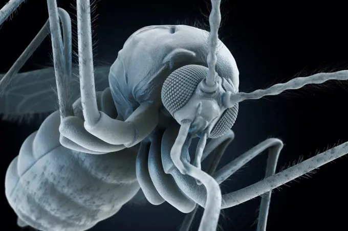
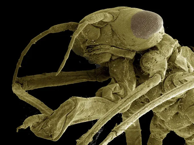
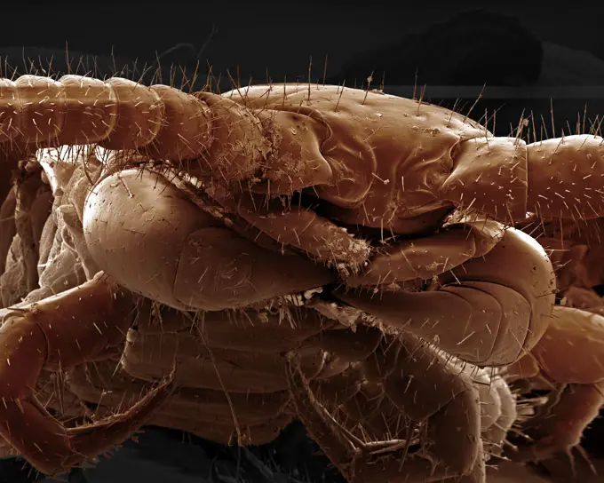
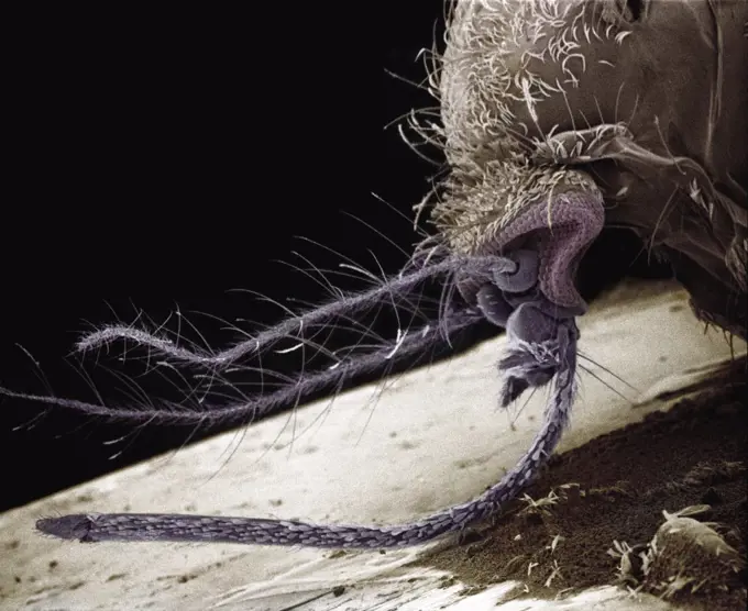
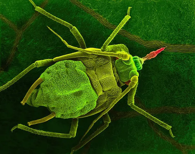
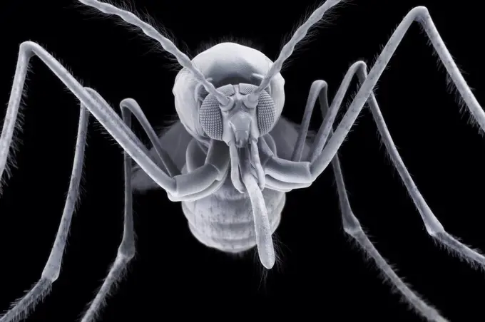
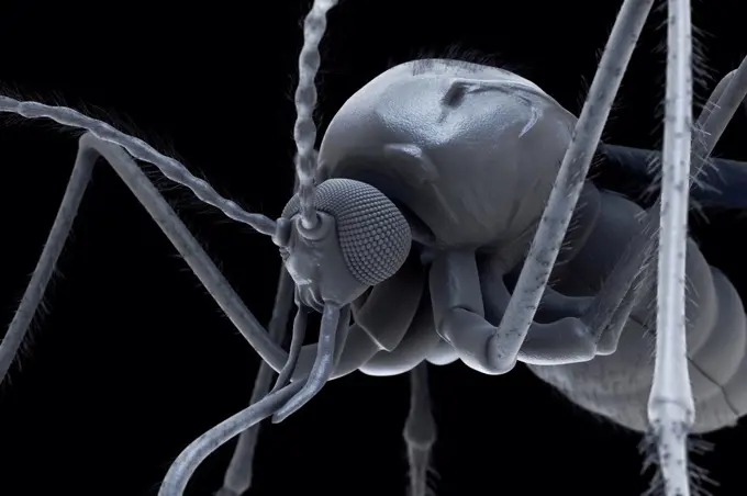
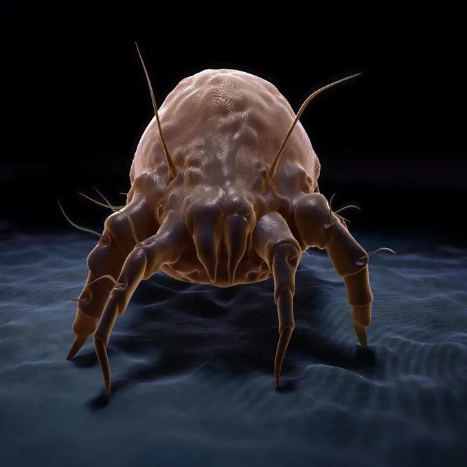
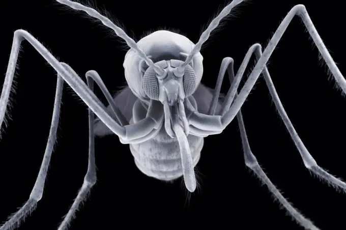
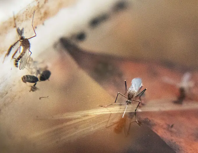
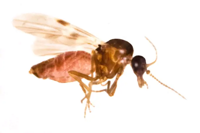
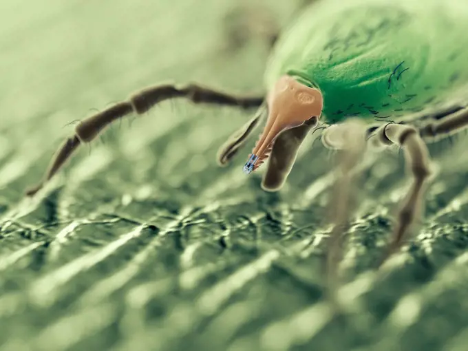
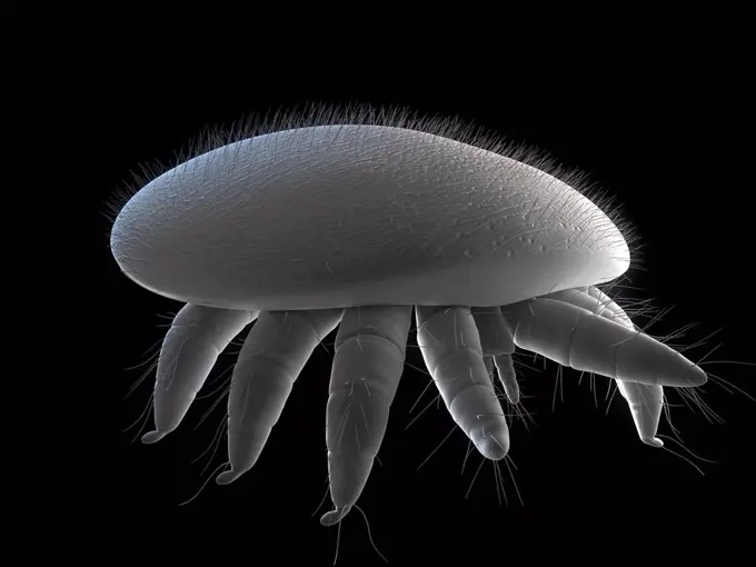
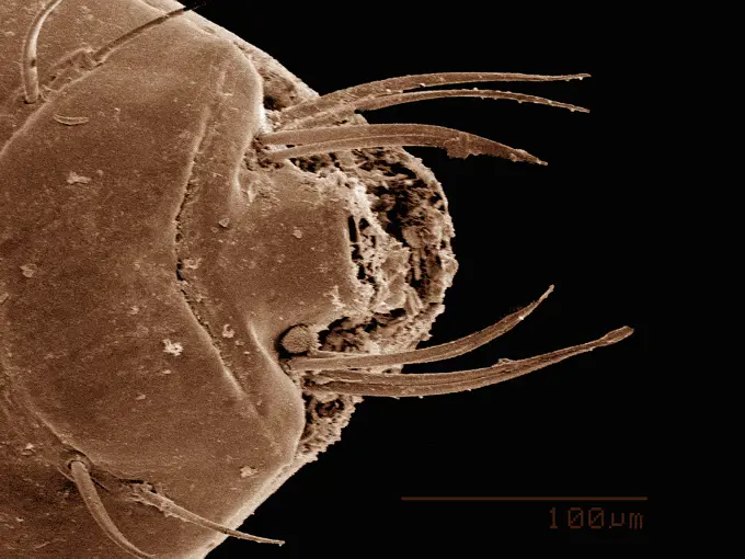
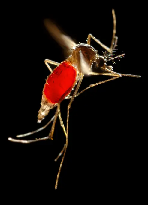
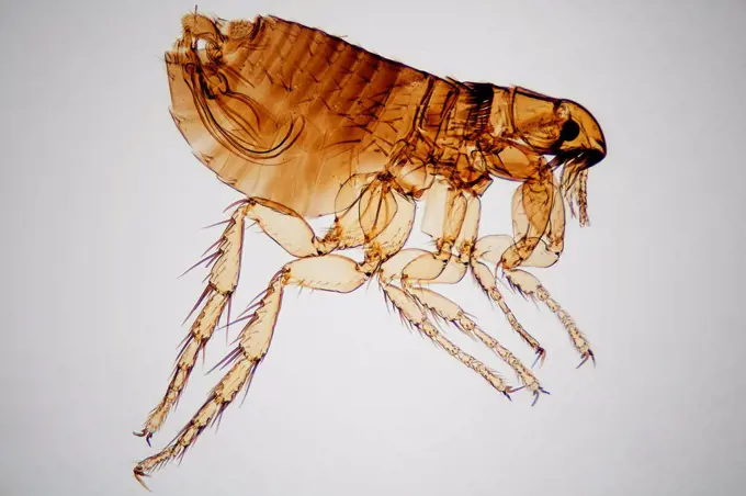
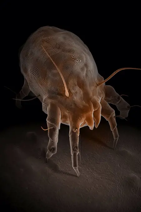
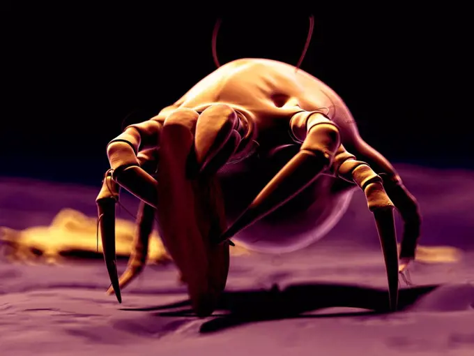
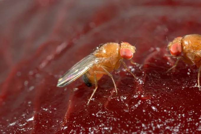
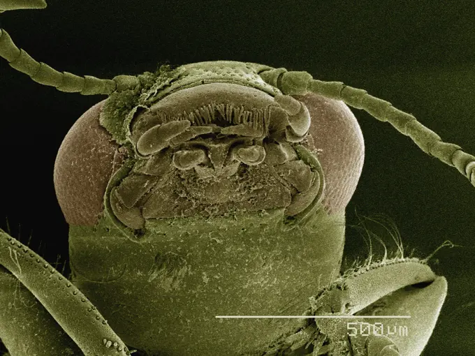
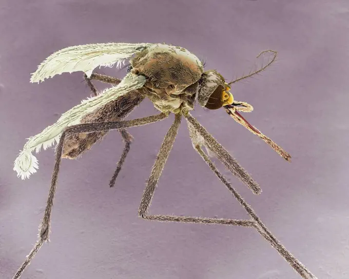
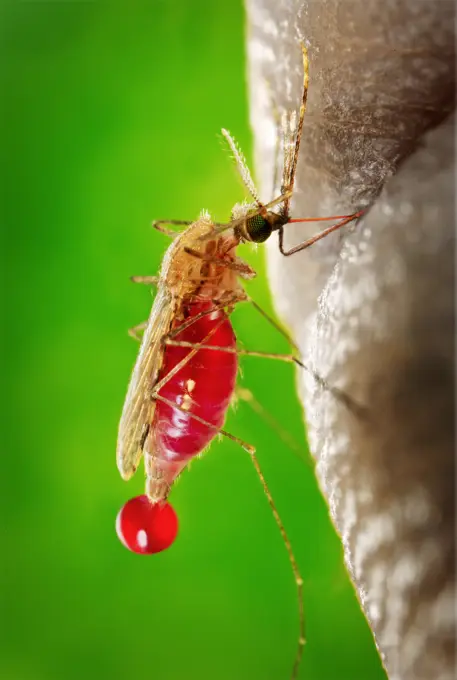
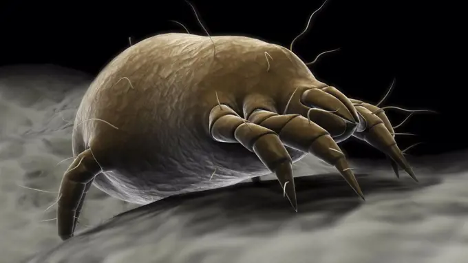
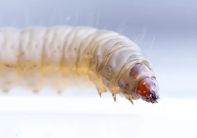
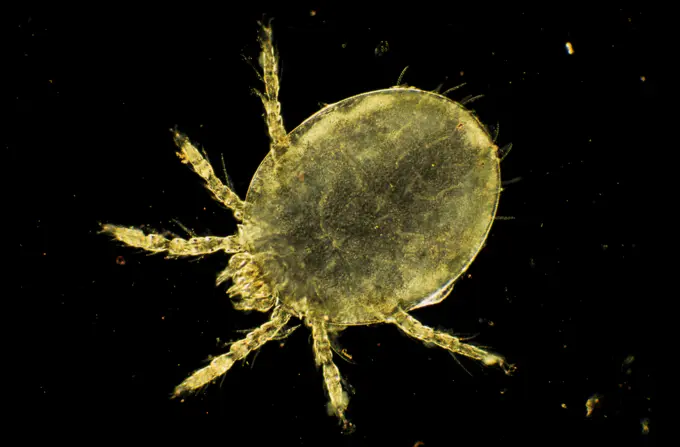
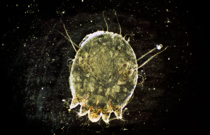
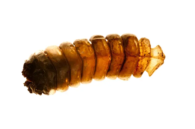
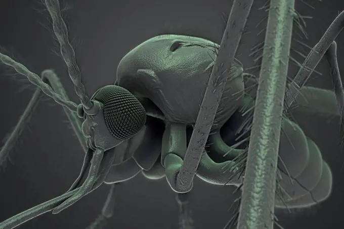
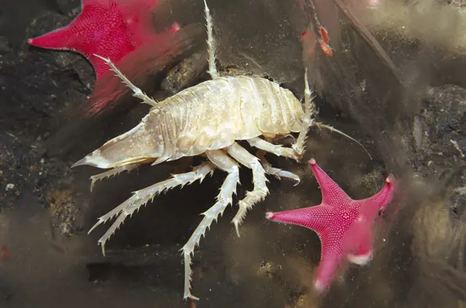
Fine ArtVintageFootageSubscriptionBoardsFree ResearchPricing
  • Fine Art
  • Vintage
  • Footage
  • Subscription
  • Boards
  • Free Research
  • Pricing

Close-Up Insect Photography

High-resolution images showcasing various insects under extreme magnification, highlighting details like textures and anatomical features.

Anopheles Mosquito
Anopheles Mosquito
210 assets in this story
4378-20014024
Anopheles Mosquito
1439-57943352
Extreme closeup of an insect under a microscope
4220-21334802
Scanning Electron Micrograph (SEM): Common Centipede.     Date:
4201-66235
Mosquito (Aedes sp) SEM close-up of the head of a female showing the modified ovipositor used to obtain blood from host species, an appendage which does not exist among males
4378-5567
Anopheles Mosquito
4128R-13620530
Coloured scanning electron micrograph (SEM) of Pea aphid (Acyrthosiphon pisum) on a bean leaf with proboscis penetrating a vein. This insect transmits...
4378-20013606
Anopheles Mosquito
4378-5611
Anopheles Mosquito
4201-66362
Human Louse (Pediculus humanus) SEM close-up view of individual between human hair at 28x magnification
4378-20013871
House Dust Mite
4220-21334705
Scanning Electron Micrograph (SEM): Crab megalops.     Date:
4378-5230
Anopheles Mosquito
4378-20014004
Anopheles Mosquito
4128R-13371571
Dust mite, illustration.
824-65830770
Culex restuans mosquitoes, collected from standing water in Annapolis, Maryland. Pupae (dark round objects)-- from which adult mosquitos will hatch-- can be seen on the left. Culex restuans is a proven vector associated with the transmission of West Nile virus (WNV) and Eastern equine encephalitis (EEE).
4413-28564
Biting Midge vector of the blue toungue disease
4128-111580223
Tick, computer illustration.
4378-2957
Parasitic head louse.
4128R-15515363
Illustration of a tick on skin
1439-57949049
Closeup of a weevil under magnification
824-63185115
Teacher. Frank Hadley Collins, Dir. Cntr. for Global Health and Infectious Diseases, Univ. of Notre Dame With a newly-obtained fiery red blood meal visible through her now transparent abdomen, the now heavy female Aedes aegypti mosquito takes flight as she leaves her host's skin surface. In this case, what would normally be an unsuspecting host was actually the CDC's biomedical photographer's own hand, which he'd offered to the hungry mosquito so that she'd alight, and be photographed while feeding. After having filled with blood, the abdomen became distended, stretching the exterior exoskeletal surface, causing it to become transparent, and allowed the collecting blood to become visible as an enlarging intra-abdominal red mass. The wings seem to be working overtime in order to keep her aloft. Dengue is a viral disease transmitted by urban Aedes mosquitoes, principally A. aegypti, a species found living in close association with humans in most tropical urban areas. Mosquito biting acti
4128R-13371549
Dust mites, illustration.
4128R-14838
Honeybee mite Varroa destructor, artwork, artwork.
1439-57952650
Extreme closeup of a tiny creature in sepia tones.
4269-7310
Aedes aegypti mosquito in flight. This mosquito is the primary vector responsible for the transmission of the Flavivirus Dengue (DF), and Dengue hemorrhagic fever (DHF), as well as Yellow fever.
4128R-15704
Bedbug Cimex sp., computer artwork.
4413-109869
Microscopic view of male cat flea on white background
4378-1112
Microscopic styled visualization of a dust mite which is associated with allergies.
4179-16995
Cat Flea (Ctenocephalides felis), Northville, MI computer enhanced
4128R-11288932
Dust mite, computer artwork.
4413-183238
Spotted-wing drosophila male on a fruit France
1439-57949031
Extreme closeup of an insect under a microscope
4378-5314
Anopheles Mosquito
6188-58045432
Libellenlarven Libellenlarve, Makro 7:1 ,model released, Symbolfoto ,property released Copyright: xZoonar.com/HelmaxSponax 324670 ,model released, Symbolfoto ,property released
4128-30418006
Tick on human skin, illustration
4128-15979449
Mosquito, computer illustration.
4220-20060442
Fossil Mantis in Amber - Dominican Republlic. 15-40 million years old. oligocene and miocene - amber is hardened tree resin which preserves organisms trapped inside - Dominican amber comes from extinct species of tropical broadleaf trees of the genus Hymenaea of the legume family       .
6019-48140381
galleria mellonella using for biology as a model
4128R-2088
Cat flea head, SEM
4128R-11543231
March fly. Scanning electron micrograph (SEM) of a female march fly (family Bibionidae). A member of a family of stout insects in the fly order, Dipte...
4128-15659924
Female Asian tiger mosquito, SEM
4141-32745
human body louse (mag. x9) pediculus humanus corporis stained, under rheinberg illumination
6188-55645660
Wooden stone with mouth tools under the microscope wooden bock with mouth tools under the microscope copyright: xzoonar.com/dr.xnorberbertxlangeex 14094889
4128-16375517
Beetle, X-ray
4141-49719
water flea, daphnia sp., amongst duckweed uk
4128R-6262
Aphid, light micrograph. Aphids, also known as plant lice and greenflies, are small plant_eating insects, and members of the superfamily Aphidoidea. A...
1848-53943741
Head of an s (Notonecta) in frontal view
4378-20014042
Anopheles Mosquito
4128R-13573077
Macrophotograph of a fossilized fungus gnat (Mycetophilidae) embedded in Baltic amber, a fossilized resin, some 30 million years old. Amber is extrude...
6188-66486115
Bakteriophage oder Phagenvirus, der ein Bakterium angreift und infiziert - 3d-Illustration Bacteriophage or phage virus attacking and infecting a bacteria - 3d illustration Copyright: xZoonar.com/ChristophxBurgstedtx 22601160
1439-57940083
Extreme closeup of a microscopic mite
4421-35830
Anomalocera patersoni, female from the marine plankton. A large copepod common at the sea's surface in the summer
1848-53943745
Head and front part of a bird flea (Ceratophyllus gallinae) exposed against a white background
824-63128573
Culex (family Culicidae) seen at the SEM. See image n0582505 on the site.
4128-19249758
Scabies mite, illustration.
4128R-14847730
Lyme disease tick, illustration
824-65830948
Mosquitos collected from standing water.
824-63194391
This image depicts a lateral view of a feeding female Anopheles gambiae mosquito. This specimen had landed a human skin surface, and was in the process of obtaining it's blood meal through its sharp, needle-like proboscis, which it had inserted into its human host. Note the red color of the proboscis, as it was filled with blood, as well as the bright red abdomen that had become enlarged due to its blood meal contents. You can also see a droplet of blood that was being expelled from the distal tip of its abdomen. A. gambiae is a known vector for the parasitic disease malaria.
4239-18641573
Microscopic visualization of a dust mite.
6019-48140380
galleria mellonella using for biology as a model
4220-21334722
Dark Field Light Micrograph: Harvest Mite Chigger.     Date:
4220-21334716
Dark Field Light Micrograph: Scabies Mite.     Date:
4410-1665
Study of tail of Prairie Rattlesnake (Crotalus viridis)
1848-53942606
Hadrobunus grandis (Hadrobunus grandis) with parasitic red mite on one of the legs
4378-5680
Anopheles Mosquito
4201-18615
Giant Antarctic Isopod (Glyptonotus antarcticus) in newly formed anchor ice, and Sea Stars (Odontaster validus), Antarctica
4220-20164072
Holly Leaf MINER (Phytomyza ilicis). x5 magnification. Grubs of Holly Leaf Miner are Holly pests.
4279-67012
DEU, 2005: European Castor Bean Tick, European Sheep Tick (Ixodes ricinus), studio picture.
4220-21334706
Light Micrograph Flattened Mayfly Nymph.     Date:
4378-5594
House Dust Mite
1848-49594345
Unidentified Fly in Colombian Amber Pleistocene
4413-109875
Microscopic view of male Squirrel flea
4384-177
Under a very low magnification of 13X, this digitally-colorized scanning electron micrograph depicted the entire ventral surface of the larval staged antlion, sometime referred to as a doodlebugs, because of the trails they leave in the soft sand as they hunt for prey.  These arthropods undergo dramatic morphologic changes when it metamorphoses into a beautiful flying antlion lacewing. Note the large mandibles to the right, which are used to apprehend prey that unwittingly fall into the conica
4128R-15537301
Illustration of a tick crawling on human skin
4128-15980087
Sidneyia marine arthropod, computer illustration.
4378-3829
A single Sarcoptes scabiei mite which is the cause of the contagious skin infection Scabies. The mite burrows under the host's skin, causing intense allergic itching.
4128R-13384884
Dust mite, illustration.
4269-27257
Anopheles gambiae. Scanning electron micrograph (SEM) of the Anopheles gambiae mosquito's anterior head. Magnification x 114. This mosquito is a vector of the parasite Plasmodium, the agent of malaria.
4128R-13447247
Human flea. Light micrograph (LM) of a female human flea (Pulex irratans). Fleas are wingless and flattened from side to side, which makes them diffic...
4128-V58573564
4378-1254
Bedbugs magnified through a microscope.
4128R-13384911
Dust mite, illustration.
4070-16279088
Money spider (Tenuiphantes sp) tiptoeing behaviour on dandelion seed head. Spiders can stand on tiptoes and produced silk to 'balloon' and travel with the airflow. In this image the silk dragline and feinter silk anchor lines are visible, UK. Controlled conditions.
6145-52203106
030819-F-5550W-001. Base: Langley Air Force Base State: Virginia (VA) Country: United States Of America (USA)
4239R-7960
Microscopic view of bacteriophage.
4141-49886
fruit fly female, drosophila melanogaster, half eye genetic variation
4384-188
The body louse, Pediculus humanus corporis, is the vector of three human pathogens: Rickettsia prowazekii, the agent of epidemic typhus; Borrelia recurrentis, the agent of relapsing fever; and Bartonella quintana, the agent of trench fever, bacillary angiomatosis, endocarditis, chronic bacteremia, and chronic lymphadenopathy.
4220-21334693
Scanning Electron Micrograph (SEM): Crab larvae.     Date:
4128-30417797
Dust mites, illustration
1848-53943742
Weevils (Oxystoma ochropus) in frontal view
4128R-13620426
Coloured scanning electron micrograph (SEM) of Dust mite (Dermatophagoides sp.). Millions of dust mites inhabit the home, feeding on dead human skin t...
4269-27758
Flat mite. The flat mite (Brevipalpus phoenicis) carries the leprosis virus, which affects citrus fruits. The disease is currently in South America but moving north. Magnified about 150x.
4128-19053648
Dust mites, illustration
4220-21334662
Scanning Electron Micrograph (SEM): False Scorpion / Pseudoscorpion.     Date:
4378-5777
House Dust Mites
1558-15241757
Microphoto of Red Mite (Dermanyssus gallinae)
4128R-11288983
Dust mite, computer artwork.
1848-51744904
Water fleas at approx. 25x magnification
4378-3382
A single Sarcoptes scabiei mite which is the cause of the contagious skin infection Scabies. The mite burrows under the host's skin, causing intense allergic itching.
1848-54789456
Hydroida sp.
PREVIOUS
of 3
NEXT
2401 S. Ervay, Suite 206
Dallas, Texas 75215
United States
Browse
CategoriesGalleriesFree Research
For Creators
License Your ContentSuperStock For StudentsFrame of Mind BlogContributor PortalNew Image SpotlightLicense AI Imagery
Resources
About UsLicensingTerms & ConditionsAPI AccessPrivacyPricing
Contact
+1 866 236 0087help@superstock.com Contact form
©2026 SuperStock. All Rights Reserved. -B
This site is protected by reCAPTCHA and Google Privacy Policy and Terms of Service apply.