Fine ArtVintageFootageSubscriptionBoardsFree ResearchPricing
  • Fine Art
  • Vintage
  • Footage
  • Subscription
  • Boards
  • Free Research
  • Pricing

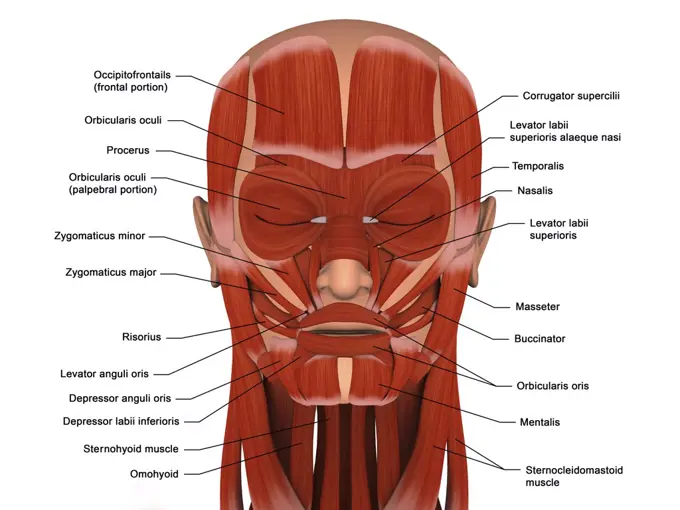
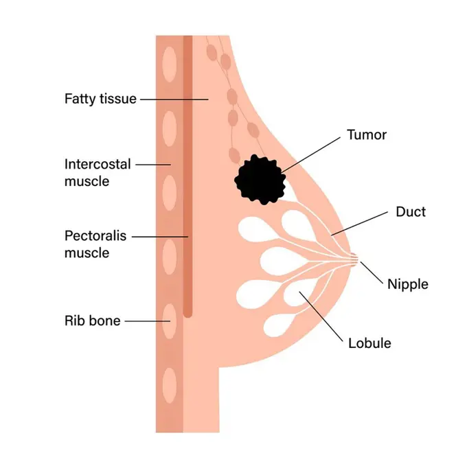
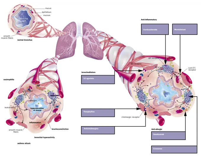
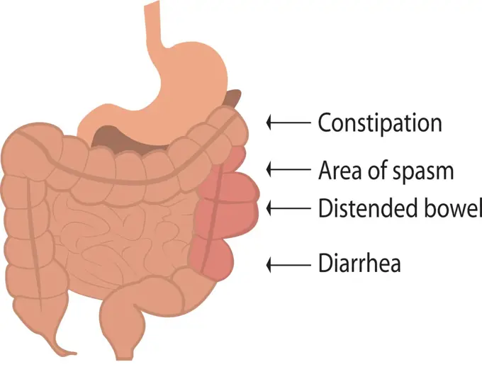
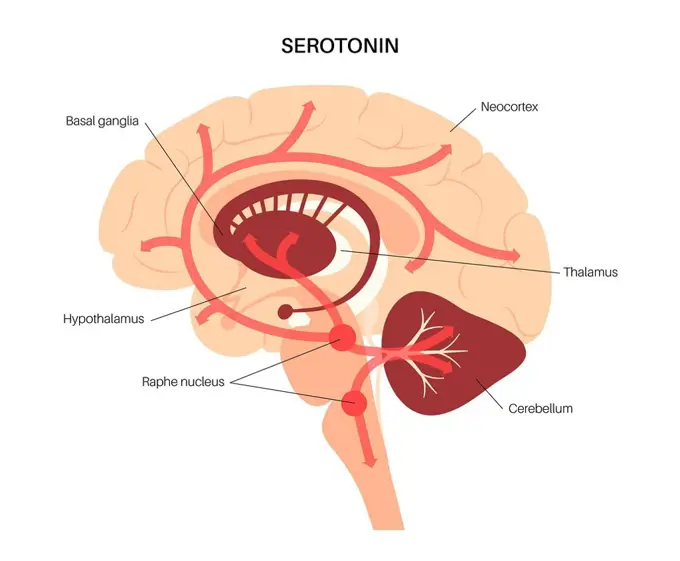
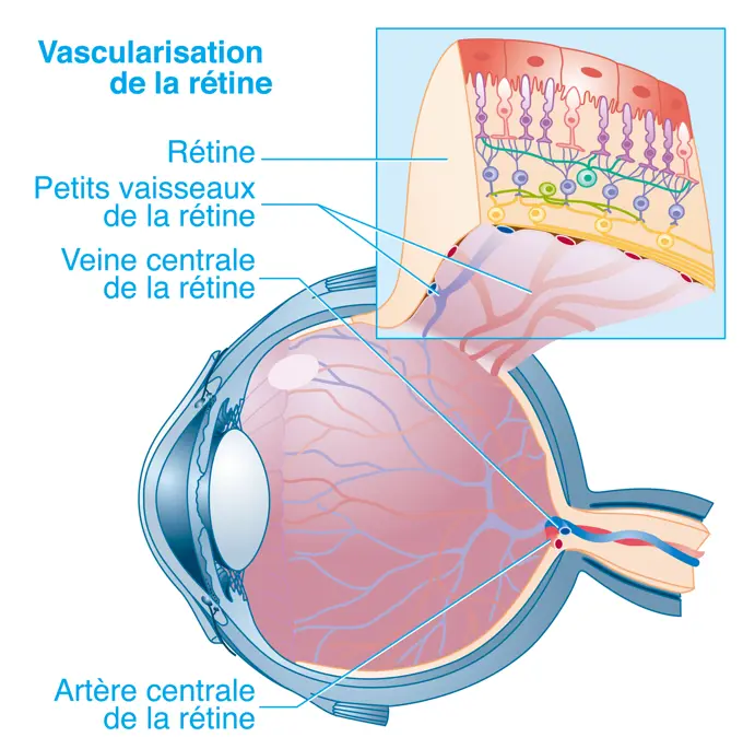
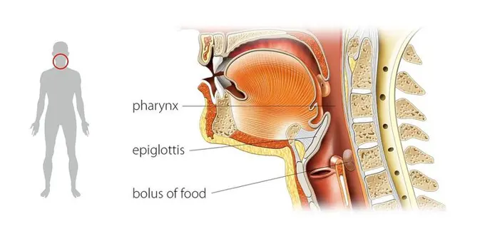
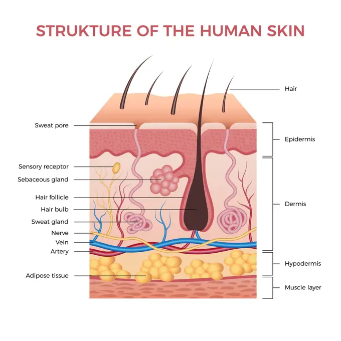
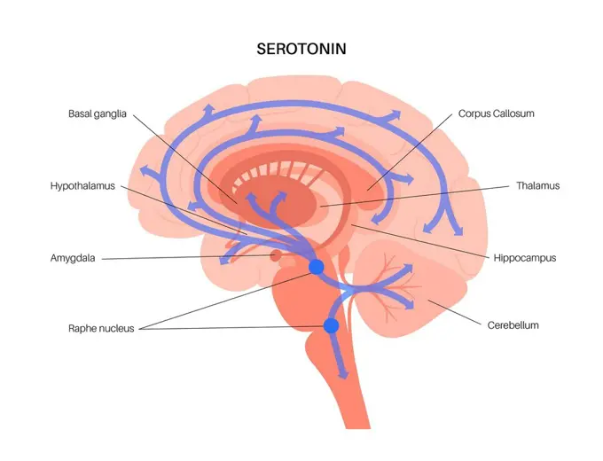
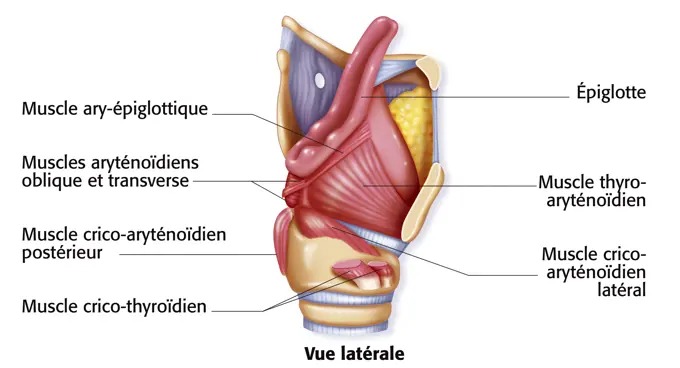
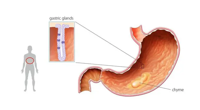
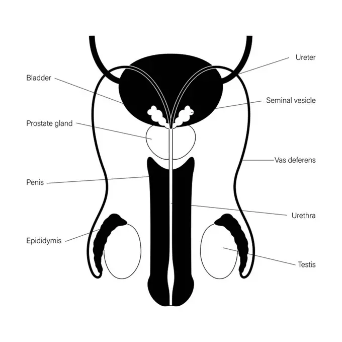
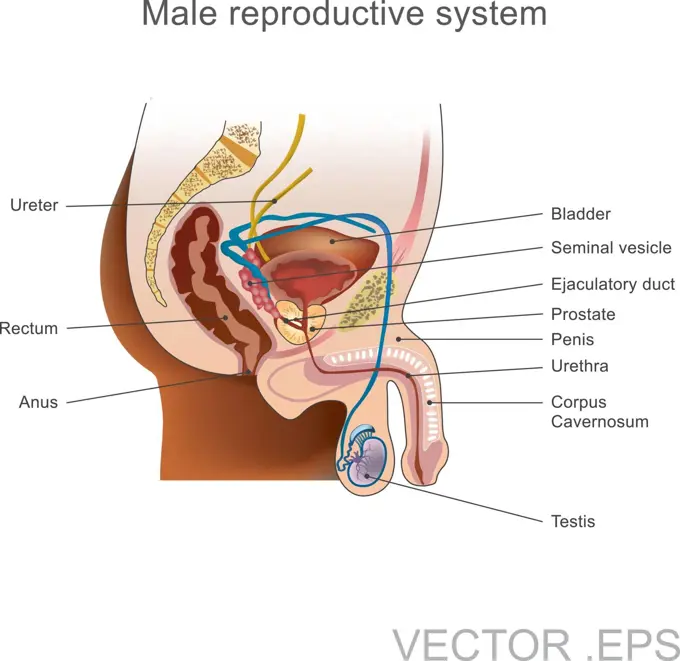
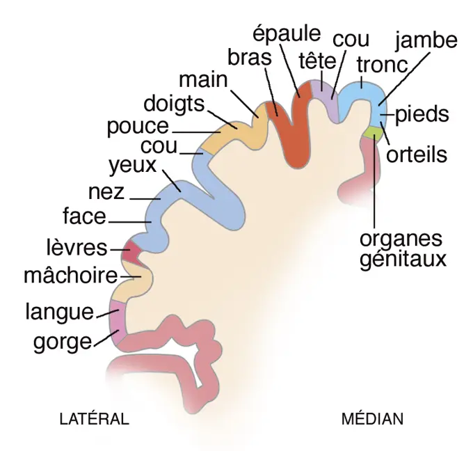
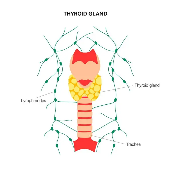
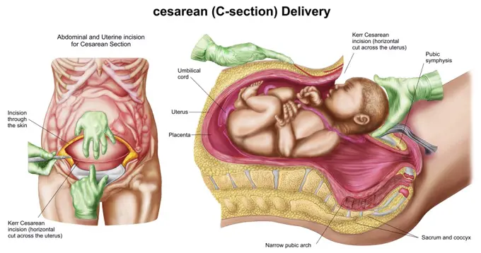
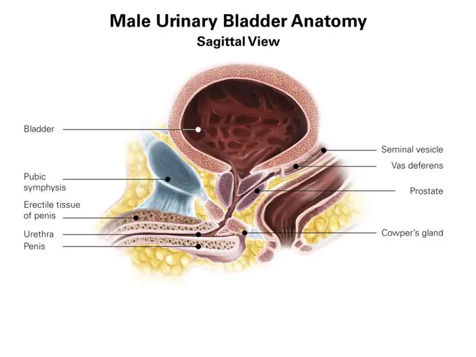
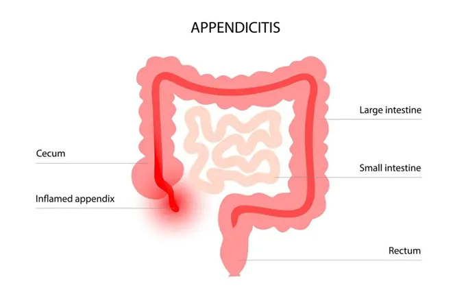
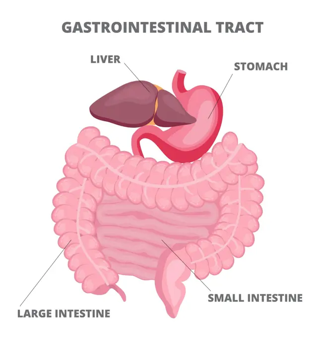
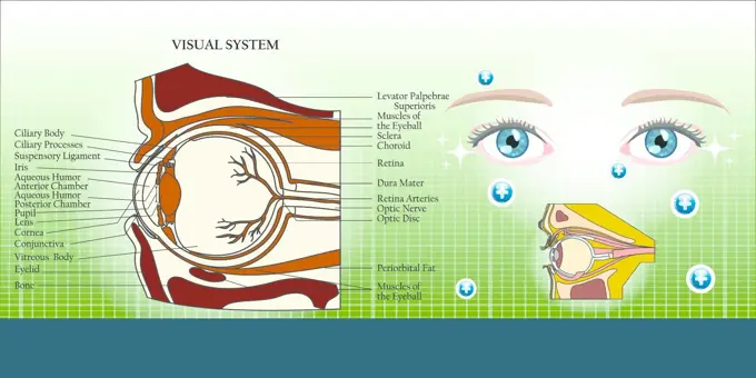
Human Anatomy Overview

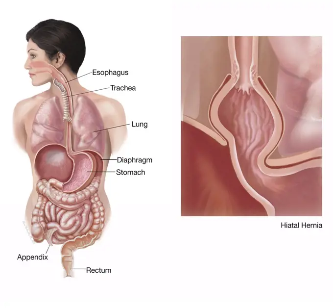
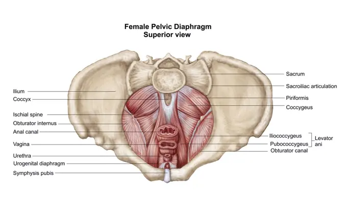
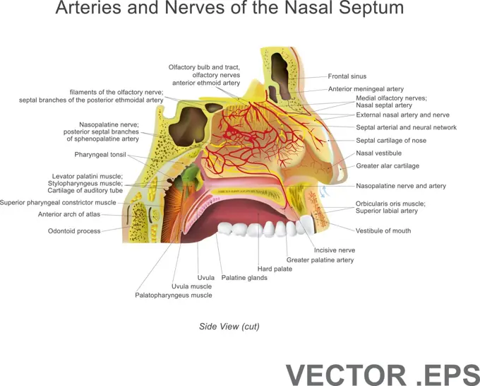
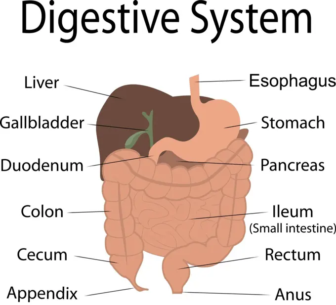
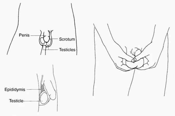
Detailed illustrations of human organ systems, including the colon and kidneys, with labeled anatomical structures and functions.

Anatomy of prostate gland.
108 assets in this story
4239-18641153
General colon anatomy, with labels.
4239R-8139
Anatomy of human kidney function.
824-63175510
Representation of the circulatory system with the direction of circulation and the exchanges with the different organs, brain, lungs, kidney, intestine, muscles: oxygen supply, carbon dioxide rejection and waste evacuation.
4239-18641139
General colon anatomy, with labels.
824-63190803
Illustration of 2 nephrons and the urine collection system in the kidney.
4128-28682162
Micturition neural control, illustration
824-57656239
Male genitals. Anatomy of the male genital organs: - in blue, corpus spongiosum and corpora cavernosa of the penis - behind the penis, in grey, testicle surmounted by the epididymis in pink, then by the vas deferens passing behind the seminal vesicle and opening into the prostate. - above the prostate, it is the bladder - in front of the bladder, the pubic symphysis, behind, the colon.
4239-18641937
Common locations of anal abscesses.
4239R-8147
Anatomy of prostate gland.
4239R-8129
Anatomy of adrenal gland, transverse section.
824-63189084
Illustration of hormone control over the menstrual cycle. - The hypothalamic cycle : gonadotropin-releasing hormone (GnRH) released in the hypothalamus causes the hypophyseal secretion of two gonadotropin hormones (LH and FSH). - The hypophyseal cycle : The FSH (freen losange) intervenes in the development and maturing of one follicle per cycle. The LH (blue triangle) allows the formation of the corpus luteum. LH and FSH trigger ovulation. - The ovarian cycle : Oestradiol (purple square) causes positive retrocontrol on the secretion of LH that is responsible for ovulation. The corpus luteum secretes progesterone (pink circle) which causes negative retrocontrol on the hypothalamo-hypothyseal complex during the luteal phase.
824-57656263
The digestive system. Representation of the digestive system from the mouth to the anus. We see the pancreas in transparency at the back of the stomach.
1899-61461914
Representation of the anatomy of the ear, from left to right: - external: pavilion, auditory canal, eardrum - middle: ossicles, Eustachian tube - internal: cochlea, auditory nerve
824-63219957
Asthma and its treatments. Representation of the lungs with the bronchial tree from which 3 zooms depart. Above left, a healthy bronchiole, below, two asthmatic bronchioles. An allergen (green star) triggers the asthma attack by activating lymphocytes producing IgE (green Y) leading to the degranulation of mast cells (purple) which release leukotrienes (black beads) and cytokines (blue beads) mediating inflammation of the mucous membrane which leads to bronchoconstriction of smooth muscle fibers, hypersecretion of mucus and obstruction of the respiratory tract.
1899-61461905
Path of sound from the ear to the brain. Anatomy of the ear, from left to right: - Outer ear and external auditory canal - Middle ear, eardrum (light purple), ossicles (beige), hammer, incus, stirrup, Eustachian tube (burgundy) - Inner ear, cochlea, vestibules and semicircular canals (purple) - Auditory nerve (yellow) With Zoom of the organ of hearing: - Cochlear canal (pink) containing the organ of Corti, including the hair cells (orange) - The cochlear nerve (yellow) transmits the electrical signal to the auditory area of the brain
824-63219040
Myocardial infarction, coronary artery with thrombus, atheromatous plaques, treatments. The atheromatous plaques (yellow) in the wall of the coronary artery cause coronary disease. In the zoom of the coronary vessel, a thrombus forms at the bottom right, in contact with the blood by platelet aggregation. Treatments act on platelets to prevent platelet aggregation, on the secretion of aldosterone by the kidneys and adrenals, but also upstream, on the liver by limiting the synthesis of cholesterol at the origin of atheromatous plaques.
4128R-28155
Head anatomy, computer artwork.
4239-18642014
Structures of the inner ear.
824-63220055
Brain and ovary, uterine endometrium, cervical mucus, targets of hormonal contraceptives. The hypothalamus regulates the secretion of FSH and LH from the pituitary gland. FSH and LH occur at the time of follicular phase and ovulation. In case of fertilization, the egg fertilized by the spermatozoa migrates to the uterus for its nesting in the endometrium. Hormonal contraception acts by blocking the hypothalamic-pituitary axis, blocking ovulation, rendering the endometrium unsuitable for nesting and / or modifying cervical mucus, making it unfit for sperm migration.
1525-56854468
The female breast anatomy includes internal milk ducts and glands and external nipples. 3D rendering. The female breast anatomy includes internal milk ducts and glands and external nipples.
824-63175511
Representation of the circulatory system with the direction of circulation and the exchanges with the different organs (supply of oxygen and evacuation of waste in the form of carbon dioxide and urine.
4239-18641968
Full gastrointestinal system on female figure with detail of hiatal hernia.
4391-217
A diagram of the human female breast, including a cutaway, and subcutaneous details.
1525-27129021
GASTROINTESTINAL TRACT Medicine Education Diagram Vector Set
4239-18641122
Colored base of human skull, inferior view, with labels.
824-63219973
Colorectal cancer and its treatments. Above right, the colon in radiological type from where a zoom of a colon seen in section with 2 cancerous tumors at 2 different stages highlighting the vascularization of the tumor. In the window at the bottom left, zoom in on one of the blood capillaries of the tumor with details of the transcription replication process thanks to VEGF (blue beads), a vascular growth factor produced by the tumor, which will bind to the VEGF receptor ( blue) present in the vessel wall. The tumor cell also produces the growth factors EGF (yellow beads) which, by binding to the EGF receptor (yellow) of the tumor cell, causes a cascade of signals responsible for cell proliferation and resistance to apoptosis.
1848-53930910
Diphtheria of the tonsils and uvula, illustration from Meyers Konversationslexikon, a German encyclopedia, 1897
4128-20042384
Arm anatomy, illustration
4239R-8122
Anatomy of adrenal gland, cross section.
4391-321
A cutaway drawing of the male urogential system, from the kidneys to the testicles.
824-63219034
Asthma, Normal bronchiole, asthmatic bronchiole with bronchoconstriction, hypersecretion, treatments. Representation of the lungs with the bronchial tree from which 3 zooms depart. Above left, a healthy bronchiole, below, two asthmatic bronchioles. An allergen (green star) triggers the asthma attack by activating lymphocytes producing IgE (green Y) causing the degranulation of mast cells (purple) which release leukotrienes (black beads) and cytokines (blue beads) mediating inflammation of the mucous membrane which leads to bronchoconstriction of smooth muscle fibers, hypersecretion of mucus and obstruction of the respiratory tract.
824-63175498
Representation of the respiratory system during inspiration (left silhouette) and expiration (right silhouette), left side, lung lodged in the rib cage, right side lung in section with bronchioles and some alveoli. Zoom in on the level of the pulmonary alveoli.
4409-17349067
Salivary glands.
824-63181807
Illustration of a network of presynaptic and postsynaptic neurons attached to each other by the intersynaptic space which allows nervous impulses to flow, from the upper neuron (top) to the lower neuron (bottom).
4128-28575188
Lymphatic system, illustration
4239R-20483612
Anatomy of the cochlear duct in the human ear.
824-63220059
Brain and ovary, uterine endometrium, cervical mucus, targets of hormonal contraceptives. The hypothalamus regulates the secretion of FSH and LH from the pituitary gland. FSH and LH occur at the time of follicular phase and ovulation. In case of fertilization, the egg fertilized by the spermatozoa migrates to the uterus for its nesting in the endometrium. Hormonal contraception acts by blocking the hypothalamic-pituitary axis, blocking ovulation, rendering the endometrium unsuitable for nesting and / or modifying cervical mucus, making it unfit for sperm migration.
4128-19358575
Female radical mastectomy, illustration.
1538R-57922
824-63219979
Breast cancer and its treatments. A silhouette of a woman with a brain section with the pituitary gland. The pituitary gland stimulates the secretion of estrogens by the ovary (uterus in frontal section), which will bind to the estrogen receptors of cancer cells (bottom center). On the silhouette a mammary gland with the axillary ganglia which relate to it. A first zoom starts from this gland to show the anatomical structure of the mammary gland with a main galactophore channel receiving the lobules, themselves made up of acinus. An enlarged acinus is seen in section. A cancerous tumor is located on the main milk duct. In the foreground, zoom of a blood vessel surrounding cancer cells seen in section the tumor with its numerous vessels in the background. The HER2 receptor (green), EGFR (pink) and the estrogen receptor (orange) are present on the cancer cell whose nucleus is cut to show the mitotic spindle. A strand of DNA in the replication phase is extracted from a chromosome.
1525-26034201
Hand drawn illustration of the torso muscles isolated on white, artistic anatomy graphic study
4430-10695
zoology/animals monkeys chimpanzees left: musculature of head & neck right: median cut through head drawing after Professor G. Ruge to Hans Kraemer ""Weltall und Menschheit"" Berlin 1900,
4128-19490367
Human hormones, illustration
4239-18641605
Facial muscles of the human head (with labels).
4128-19358519
Female breast cancer, illustration.
824-63219960
Asthma, Normal bronchiole, asthmatic bronchiole with bronchoconstriction, hypersecretion, treatments. Representation of the lungs with the bronchial tree from which 3 zooms depart. Above left, a healthy bronchiole, below, two asthmatic bronchioles. An allergen (green star) triggers the asthma attack by activating lymphocytes producing IgE (green Y) causing the degranulation of mast cells (purple) which release leukotrienes (black beads) and cytokines (blue beads) mediating inflammation of the mucous membrane which leads to bronchoconstriction of smooth muscle fibers, hypersecretion of mucus and obstruction of the respiratory tract.
5507-43151542
Irritable bowel syndrome (IBS) in a large intestine
6188-67437915
3D Isometrische flache konzeptionelle Illustration des Oropharynx, Bildungs-Schemabr3D Isometrische flache konzeptionelle Illustration des Oropharynx, Bildungsschema 3D Isometric Flat Conceptual Illustration of Oropharynx, Educational Schema 3D Isometric Flat Conceptual Illustration of Oropharynx, Educational Schema Copyright: xZoonar.com/AnastasiiaxTorianykx 22973335
4128-28575155
Lymphatic system, illustration
1525-27160149
Hand drawn vector illustration of Wuhan corona virus, covid-19. The entry of the virus into the lungs through breathing. Protect with medical mask. White background and black outlines.
4128-20040724
Prostatitis, illustration
4128-20040739
Prostatitis, illustration
4128-19490877
Nasal epithelium, illustration
4297-1678
Anatomical illustration of the underside of the human brain
824-63190784
Illustration of the various sensitivities of the lemniscus system. They are made of axons that leave special skin receptors (fine touch) and receptors located in the joint capsules and ligaments (conscious proprioreceptive sensitivity). They ascend the spinal cord, penetrating the medulla oblongata and relay to the nucleus gracilis and cuneiform. After crossing on the median line (sensitive decussation), these pathways form the medial lemniscus (Reils ribbon) which goes all the way up the brainstem behind the pyramidal tracts to reach the thalamus, the last relay before the parietal lobe in the cortex.
4128-19358436
Serotonin pathway in the human brain, illustration.
824-57656258
Vascularization of the retina. Highlighting the retina, the cells of the retina and its vascularization.
4409-17349019
Swallow-1.
4409-28578827
Infographic of the peripheral nerves which carry information to and from the brain and the spinal cord, and the types of nerve responses determined by external stimuli. QuarkXPress (.qxp); 6259x4015.
824-63175538
Representation of the organization of the visual cortex. In the vision mechanism, the hypothalamus (small purple circle) regulates circadian rhythms (cycles of active and non-active periods), the pretectum (small orange circle) controls the pupil and lens reflex, the colliculus upper (green oval) directs eye and head movements.
1525-26529698
Skin anatomy. Human normal skin dermis epidermis adipose layers recent vector biological infographic. Illustration anatomy skin human epidermis structure. Skin anatomy. Human normal skin dermis epidermis adipose layers recent vector biological infographic
4128-20040539
Pituitary gland anatomy, illustration
4128-19358416
Serotonin pathway in the human brain, illustration.
824-63190810
Lateral illustration of the larynx, from top to bottom: - hyoid bone - thyrohyoid membrane - epiglottis - the aryepiglottic muscle - the oblique and transverse arythenoid muscles - the thyroarytenoid muscle - thyroid cartilage - the lateral cricoarytenoid muscle - the posterior cricoarytenoid muscle - cricoid cartilage - the cricothyroid muscles
4409-17349022
Digestive process-1.
4128-20040817
Male reproductive system, illustration
5507-39240891
The male reproductive system.
5507-45577502
Anatomy of the ear.
5507-33664153
Human head hair growth cycle. Biological catagen, telogen phases. Hair infographics.
4128-20042511
Pelvis anatomy, illustration
4128-19358556
Invasive ductal carcinoma, illustration.
4128-20042326
Arm anatomy, illustration
4128-19358546
Types of breast cancer, illustration.
1899-53508986
Representation of the contamination of a hen by the H5N1 virus.
824-63163648
Representation of the contamination of a hen by the H5N1 virus.
4391-323
A line drawing showing frontal view of mouth and lateral view of face.
4128-19490795
Olfactory system, illustration
4128-19358562
Invasive lobular and carcinoma in situ, illustration.
4128-28575150
Lymphatic system, illustration
4239-18641159
Digital illustration of the abdominal digestive organs.
4239R-20483524
Pelvic diaphram of human female, superior view.
5507-39240885
Arteries and Nerves of the Nasal Septum.
5507-43004629
Digestive system in human body
4391-138
Three illustrations from 'What You Need to Know About Testicular Cancer:' 1)a diagram of the abdominal-pelvic area, listing the penis, scrotum, testicles; 2) a diagram of the self examination of the testicles; 3) a side view of the penis, testicles and epididymis.
824-63175526
Representation of the territories of the somatosensory cortex associated with the different parts of the body.
4128-20040959
Thyroid gland, illustration
4239R-20483643
Illustration showing Caesarean delivery of fetus.
4239-18641142
Sagittal view of male urinary bladder.
4128-19358353
Inflamed appendix, conceptual illustration.
4408-13460
Clerambourg Grains Laxatifs, Medical - Potions, Medications, & Cures
4128-19056866
Human tooth anatomy, illustration
1525-27373399
Gastrointestinal tract vector is shown on white background. Large and small intestines, liver, stomach are isolated. Medicine, healthcare concept in cartoon style.. Gastrointestinal tract vector is shown on white background.
4128-19249234
Mesenteric ischaemic, illustration.
6188-68372980
3D isometrische flache Vektor-Illustration der Mesenterialarterie 3D Isometric Flat Illustration of Mesenteric Artery, Abdominal Blood Flow System LicenseRF Copyright: xZoonar.com/AnastasiiaxTorianykx 23277042
4128-28682565
Ventricular system of brain, illustration
5507-48281611
Illustration of a nail with the names of the parts of the finger
1525-24549018
Pancreatic function in a human body vector illustration infographics
5507-37452607
Visual system and eye anatomy illustration background
4128R-15290420
Comparison of microfilariae morphology, illustration
5507-43624709
Type of obesity illustration (Japanese) . Abdominal sectional View (subcutaneous fat).
PREVIOUS
of 2
NEXT
2401 S. Ervay, Suite 206
Dallas, Texas 75215
United States
Browse
CategoriesGalleriesFree Research
For Creators
License Your ContentSuperStock For StudentsFrame of Mind BlogContributor PortalNew Image SpotlightLicense AI Imagery
Resources
About UsLicensingTerms & ConditionsAPI AccessPrivacyPricing
Contact
+1 866 236 0087help@superstock.com Contact form
©2026 SuperStock. All Rights Reserved. -A
This site is protected by reCAPTCHA and Google Privacy Policy and Terms of Service apply.