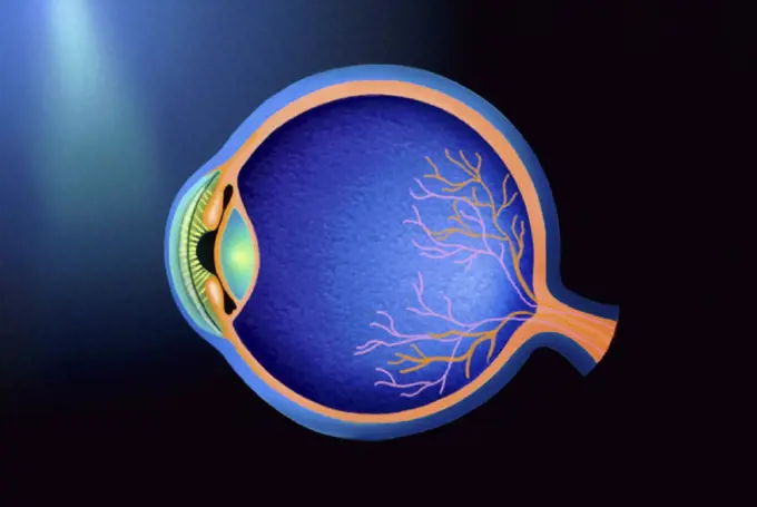
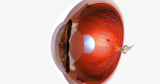
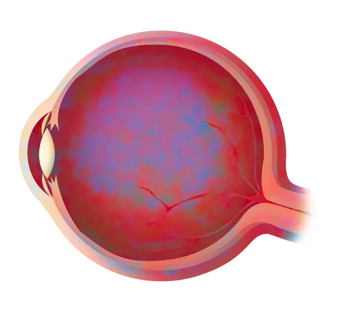
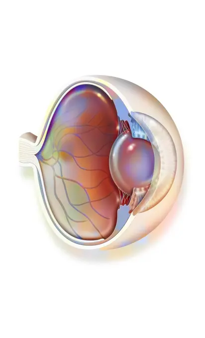
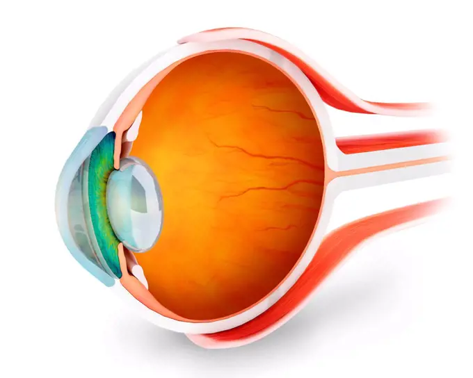
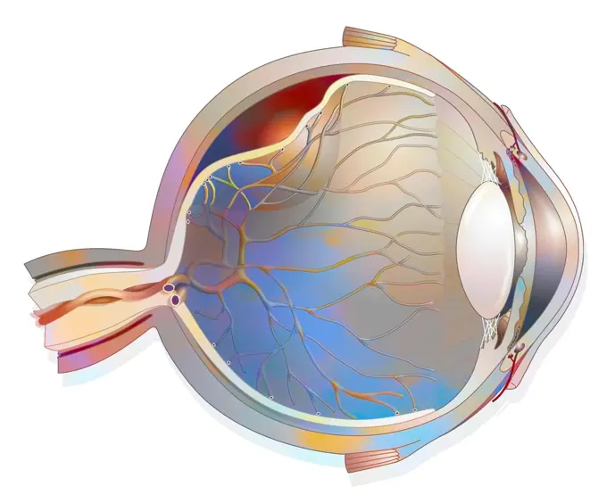
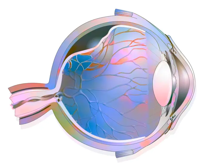
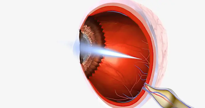
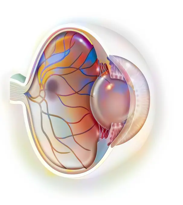
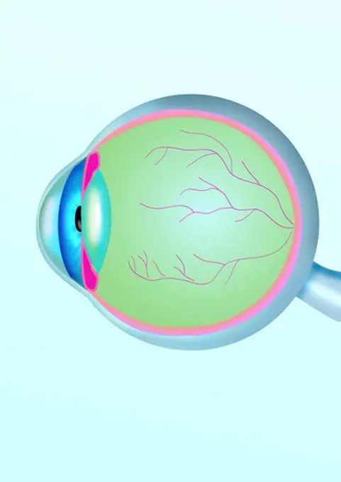
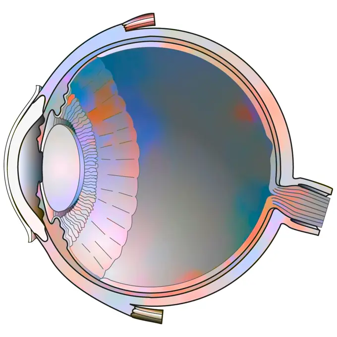
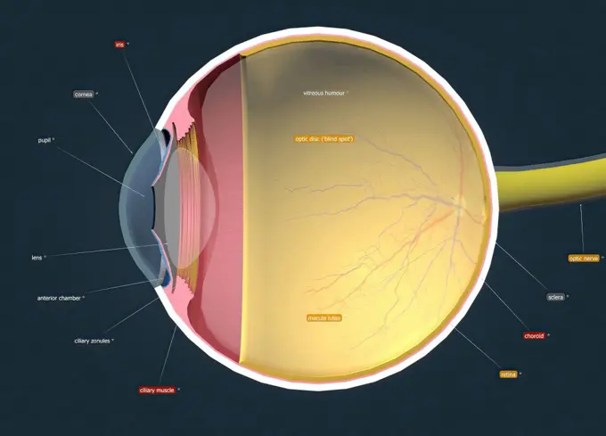
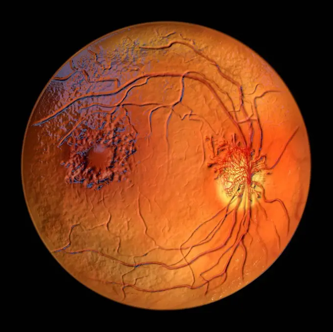
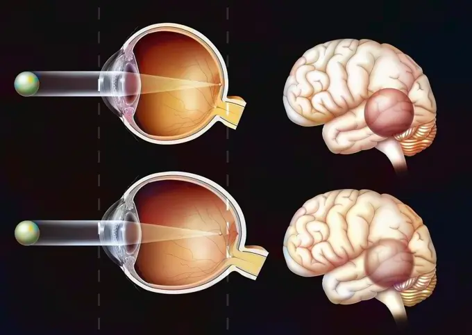
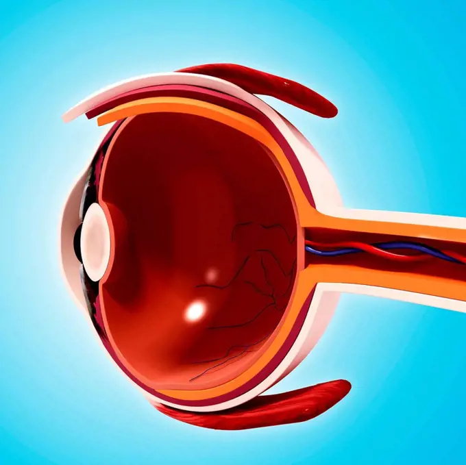
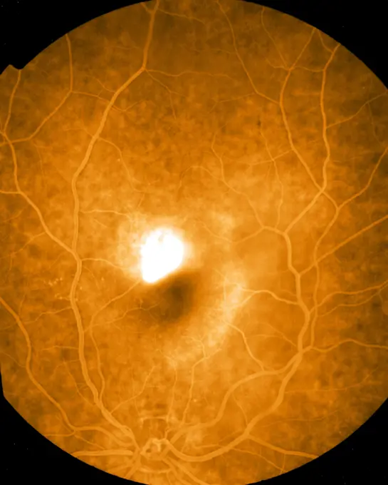
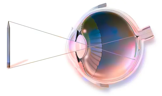
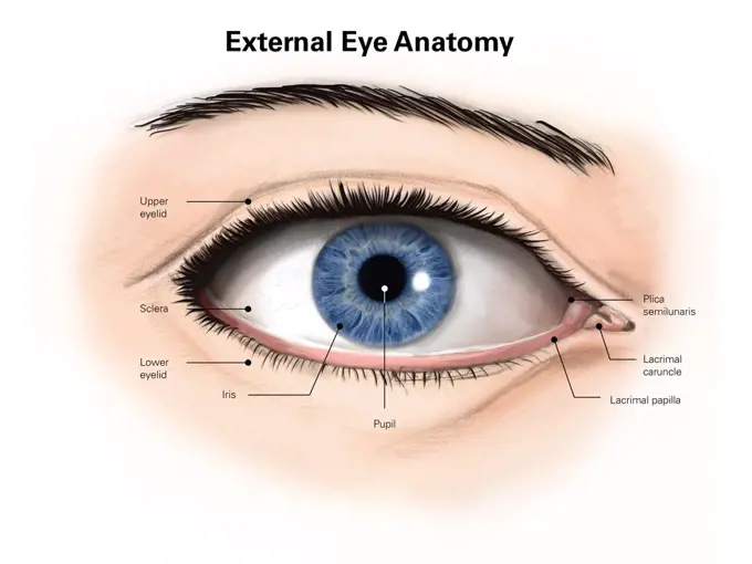
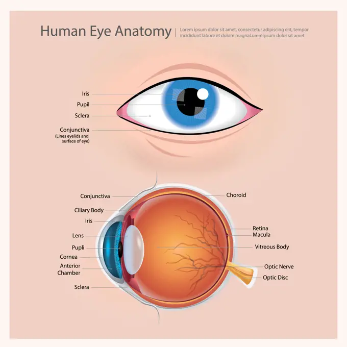
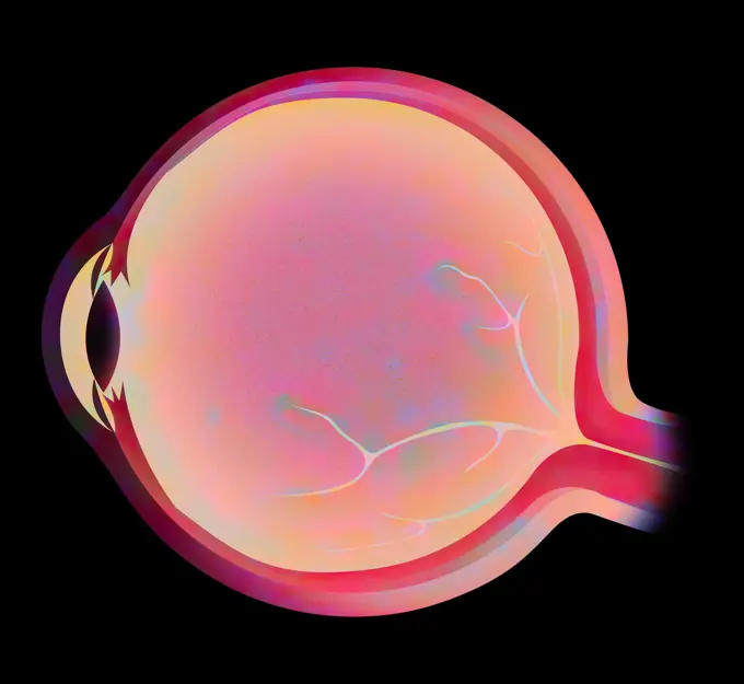
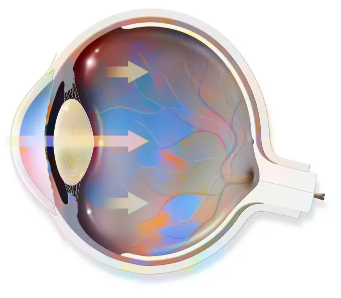
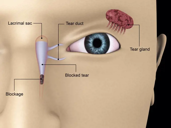
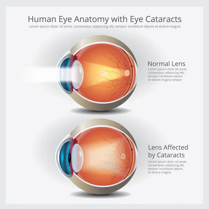
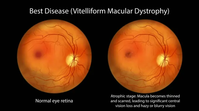
Fine ArtVintageFootageSubscriptionBoardsFree ResearchPricing
  • Fine Art
  • Vintage
  • Footage
  • Subscription
  • Boards
  • Free Research
  • Pricing

Human Eye Anatomy

Detailed 3D renderings and illustrations showing the cross-sectional view of the human eye, highlighting structures like the lens, retina, and optic nerve.

Human Eye
Human Eye
94 assets in this story
1525-56853527
Myopia, is a common visual condition in which the eye s ability to focus on distant objects is impaired. 3D rendering. Myopia, is a common visual condition in which the eye s ability to focus on distant objects is impaired.
6188-66528953
Anatomy, eye Anatomy, eye 016889_199
824-63164088
Anatomy of the eye with lens, retinal veins and arteries.
824-479
Cross section of a human eye
824-63180612
Cross-section of the left eye. From left to right, the optic nerve (yellow), followed by the retina, the choroid all the way to the ciliary muscle, attached to the lens by suspensory ligaments (red pink), the sclera (light beige), in front of the lens, the iris (green) with the pupil in the centre and in front of the iris, the cornea which surrounds the anterior chamber filled with aqueous humour.
824-63164581
Sagittal view of the eye anatomy showing lens, retina, cornea, iris, choroid.
824-219
Human Eye
824-63195466
Illustration of a cataract with its cloudy lens that does not allow light to reach the retina correctly.
824-63193771
Illustration where the back of the eye is affected by diabetic retinopathy, retinal damage caused by complications of diabetes. Hemorrhaging and exudates (irregular red and yellow areas) are visible, caused by the permeability of retinal capilliaries, as well as neovascularisation (formation of abnormal, malfunctioning capilliaries).
4239-18641906
Retina of a normal eye.
6188-66486432
3D-Darstellung eines Querschnitts des menschlichen Auges in einer Seitenansicht und einer Frontalansicht 3D illustration of a cross-section of the human eye in a side view and a frontal view Copyright: xZoonar.com/ChristophxBurgstedtx 22603166
1428-353A
Close-up of the human eyeball
4239R-8466
Anatomy of human eye, perspective.
4128-30420969
Autosomal recessive bestrophinopathy, illustration
824-63164104
Eye: detachment of the retina, which detaches from the underlying choroid.
824-63164105
Eye: detachment of the retina, which detaches from the underlying choroid.
1525-56722694
Myopia, is a common visual condition in which the eye s ability to focus on distant objects is impaired. 3D rendering. Myopia, is a common visual condition in which the eye s ability to focus on distant objects is impaired.
4128-20041465
Retina damage from diabetes, illustration
824-57656250
The eyelids. Representation in median sagittal section of the eye and the eyelid with highlighting of their structures. On the upper eyelid, from left to right: the orbicularis muscle, the Meibomian gland (in yellow), the levator muscle of the eyelid, the conjunctiva. On the lower eyelid: the Meibomian gland, the conjunctiva.
824-63195457
Illustration of a snakes pit organ. The organ is located between the eyes and nostrils which allows it to sense the heat of its prey. Infrared rays produced by body heat are sensed by the blue membrane, which transforms the infrared rays into an electrical signal that is sent to the brain.
4128R-11291095
Human eye anatomy, computer artwork.
4239-69152028
Transverse section of eye anatomy with labels.
824-63164084
Eye: treatment of macular degeneration by RNA interference.
1525-56190158
Cutaway View of a Human Eye 3d illustration. Cutaway View of a Human Eye
4128R-11283324
Eye anatomy, computer artwork.
4239-18641737
Comparison of a normal eye and an eye with hyperopia.
824-63165340
Anatomy of the eye and eyelid (viewed from 3/4) with iris, pupil.
4128-20041577
Retina damage from diabetes, illustration
4269-25470
ARMD. Eye angiography of an Age-Related Macular Degeneration (ARMD).
1899-53509310
Age-related macular degeneration affecting the macula, the central area of the retina.
824-63164582
Age-related macular degeneration affecting the macula, the central area of the retina.
4252-4103
Human eye, side view
6243-73130473
Fundus photography of the back of the eye - Normal human retina
824-63165318
Anatomy of the eye and eyelid (viewed from 3/4) with iris, pupil.
4128-20041170
Healthy retina, illustration
1899-30609119
Anatomy of the eye and eyelid (viewed from 3/4) with iris, pupil.
4269-25469
ARMD. Eye angiography of an Age-Related Macular Degeneration (ARMD).
1525-26235816
Anatomy and Structure of the Eye 3D illustration. Anatomy and Structure of the Eye
1525-56184192
Structure of the Human Eye and anatomy of vision 3d illustration. Structure of the Human Eye and anatomy of vision
824-63164906
Horizontal section showing all the main structures of the eye: lens, retina.
1525-56184181
Structure of the Human Eye and anatomy of vision 3d illustration. Structure of the Human Eye and anatomy of vision
4128-20041495
Retina damage from diabetes, illustration
1899-30609111
Comparison between normal vision and that of a myopic eye.
4128R-11291097
Human eye anatomy, computer artwork.
1525-56184320
Cell Structure and Functions, Cell Organelles 3D illustration. Cell Structure and Functions, Cell Organelles
4269-27663
ARMD. Eye angiography of an Age-Related Macular Degeneration (ARMD).
824-63164746
Anatomy of an eye and vision, image reflection (pencil) on the retina.
4239R-20482703
External anatomy of the human eye (with labels).
1525-24561103
Human Eye Anatomy Vector Illustration
6188-66528954
Anatomy, eye Anatomy, eye 016889_200
824-63164638
Anatomy of the eye whose arrows represent light and revealing the lens, retina, cornea, iris, choroid.
1428R-354
Close-up of the human eyeball with retinal damage caused by glaucoma
824-63165158
Different possible corrections of a hyperopic eye: spectacle lenses - external lenses.
1889R-30848
Illustration of an onion
824-63195455
Illustration of a felines eye. The pupil is very large, which allows a maximum amount of light to enter the eye and reach the retina. The retina has few cones (colour perception) and a large quantity of rods (light sensitivity) and has a layer of mirror cells (yellow zone) allowing the light beam which hasnt touched any rods when entering the eye to touch them when reflecting the beam back towards the cells.
824-63180727
Illustration of treatment for AMD using gene therapy. A gene that produces an anti-AMD medicine is injected into a deactivated virus which cannot spread in the cells of the retina. The virus is injected into the eye with a ratio of one virus per cell treated. The medicine gene mixes with the retina cells and produces anti-VEGF agents that stop the blood vessels multiplying that are responsible for AMD.
1899-53509481
Comparison between normal vision and that of an eye with cataract.
1899-53509057
Eye, position and dimension of the rigid lenses.
4239R-20483362
Conceptual image of human eye anatomy.
824-63220050
Infectious conjunctivitis and its treatments. View of an eye with inflammation of the conjunctiva. Zoom on the conjunctiva with from top to bottom 1 bacterial infection (green) and 1 viral infection (red-orange). In yellow, purulent discharge due to the presence of bacteria (green), the cells of the conjunctiva are destroyed. Goblet cells secreting mucus and edema of the conjunctiva in the area infected with the virus. Right from top to bottom, a bacteria seen in section, a window with an infected connective cell (Herpes Simplex virus) detailing the process of viral replication transcription of viral DNA, DNA polymerase, viral replication
824-57656261
The eyelid. Representation in median sagittal section of the eye and the eyelid with highlighting of their structures.
4239-69152034
Medical illustration depicting the differences between simple and compound eyes.
1525-27277592
Human Eye Anatomy Vector Illustration
1899-29981229
Eye, organ of vision. The human eye. Illustration from Brockhaus and Efron Encyclopedic Dictionary ca. between 1890 and 1907
1899-61461932
The eyelid. Representation in median sagittal section of the eye and the eyelid with highlighting of their structures
4285-3642
MY RETINA PHOTOS
1899-53509503
Eye, the visualized image is refracted by the lens and the cornea on the retina.
1525-27535824
Human Eye Anatomy Vector Illustration
1525-56184186
Structure of the Human Eye and anatomy of vision 3d illustration. Structure of the Human Eye and anatomy of vision
4128-20041482
Retina damage from diabetes, illustration
4128-V58577170
4128-20041796
Fluorescein angiogram of a retina, illustration
6188-68373056
3D Isometrische flache Vektor Illustration von Demodex Folliculorum 3D Isometric Flat Illustration of Demodex Folliculorum, Demodicosis Affecting the Eyelid and Causing Red Eyes LicenseRF Copyright: xZoonar.com/AnastasiiaxTorianykx 23277207
4239-18641718
Diagram illustrating blocked tear ducts in the human eye.
1525-26870431
Human Eye Anatomy Vector Illustration
824-63203292
Optical Coherence Topography (OCT) image, enabling the detailed analysis of the retina and macula structures. The left eye is visible on this image.
824-63214036
Illustration of the electronic eye implant patented by Google to replace glasses. The lens capsule is opened and the natural lens is shattered then removed with a gel injected to stabilize the implant. This implant will analyze the curve of the lens and adapt eyesight to both long and short sight depending on the pathology. It is connected to a computer which handles calibration of the lens, battery charge, updates, maintenance and data transmission.
4128-30420952
Best disease, illustration
824-63164109
Eye: detachment of the retina, which detaches from the underlying choroid.
1899-61461928
The cornea. Anatomy of the eye highlighting the cornea
4378-20393357
Testicle
4128-30420941
Best disease, illustration
824-57656257
The cornea. Anatomy of the eye highlighting the cornea.
1838-76240479
OCT scan (Optical Coherence Tomography) of human retina, both eyes
824-63151120
Anatomy, eye
4128-30420940
Autosomal recessive bestrophinopathy, illustration
4409-17287449
LA LUZ QUE SE APAGA - 2002 - OLEO/CERA/MADERA - 185x157. Author: JOSE MARIA SICILIA. Location: GALERIA SOLEDAD LORENZO. MADRID. SPAIN.
1525-66390431
Cataract eye close up view. Eyeball with cloudy lens. Anatomically accurate human organ of vision with aging problems isolated on white background. Vector realistic illustration.. Cataract eye close up view. Eyeball with cloudy lens. Anatomically accurate human organ of vision with aging problems
4128-30420938
Autosomal recessive bestrophinopathy, illustration
4128-30420953
Retinitis pigmentosa, illustration
5507-31781235
Vector flat illustration of the anatomy of the lacrimal gland.
5507-45485811
Illustration of Demodicosis
4128-30420916
Normal eye and an eye with Best disease, illustration
6188-55467012
cataract cataract Copyright: xZoonar.com/VladimirxNenovx 4568165
PREVIOUS
of 1
NEXT
2401 S. Ervay, Suite 206
Dallas, Texas 75215
United States
Browse
CategoriesGalleriesFree Research
For Creators
License Your ContentSuperStock For StudentsFrame of Mind BlogContributor PortalNew Image SpotlightLicense AI Imagery
Resources
About UsLicensingTerms & ConditionsAPI AccessPrivacyPricing
Contact
+1 866 236 0087help@superstock.com Contact form
©2026 SuperStock. All Rights Reserved. -B
This site is protected by reCAPTCHA and Google Privacy Policy and Terms of Service apply.