Fine ArtVintageFootageSubscriptionBoardsFree ResearchPricing
  • Fine Art
  • Vintage
  • Footage
  • Subscription
  • Boards
  • Free Research
  • Pricing

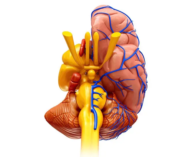
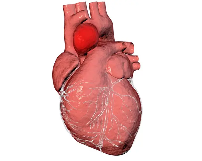
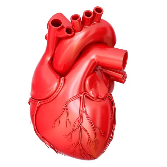
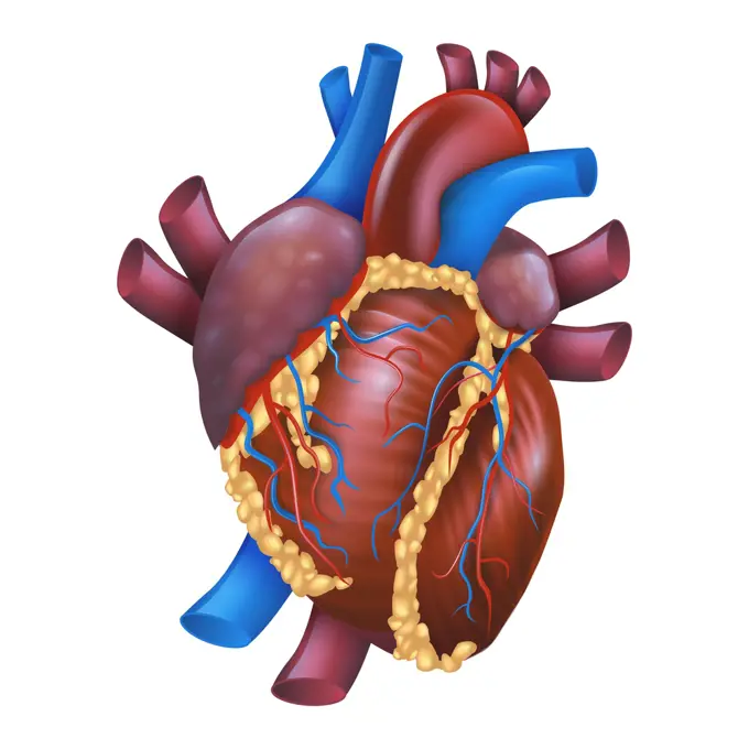
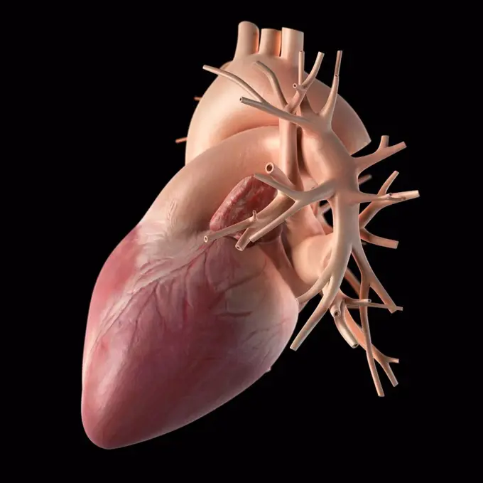
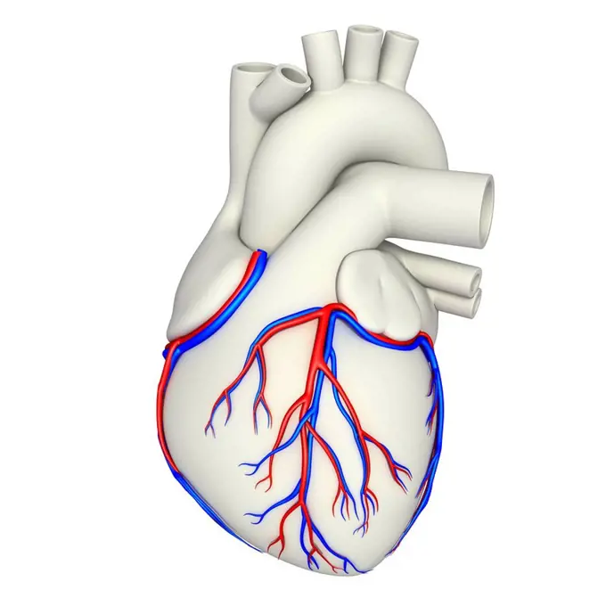
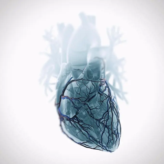
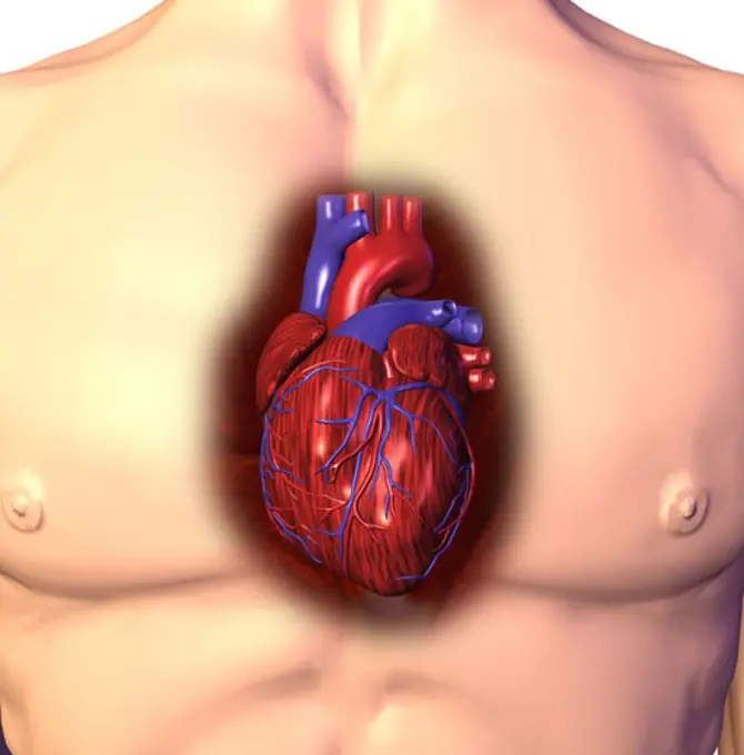
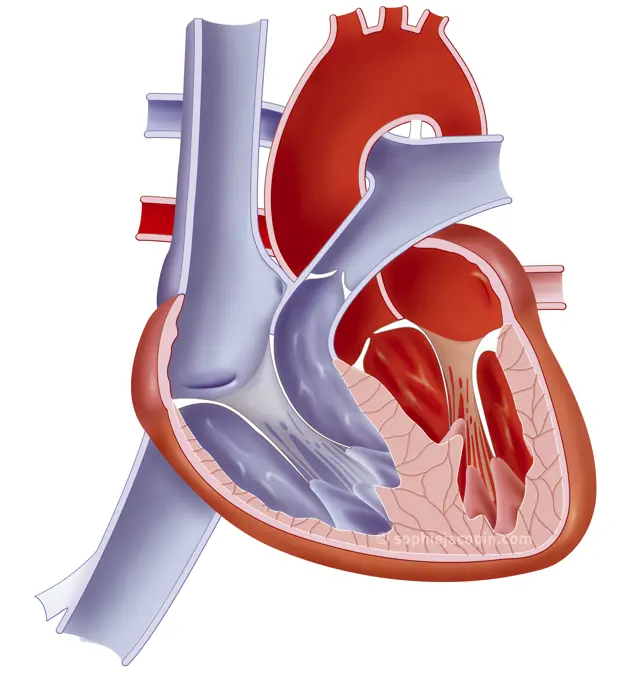
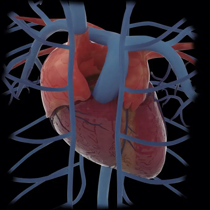
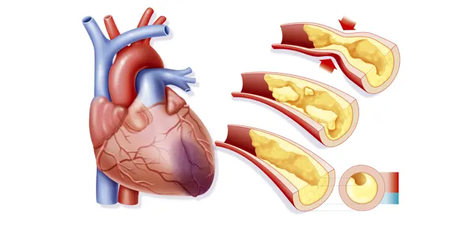
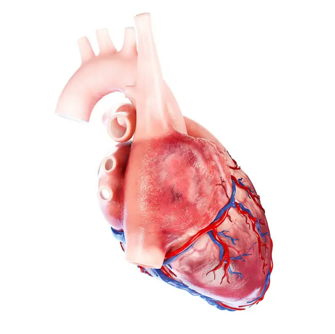
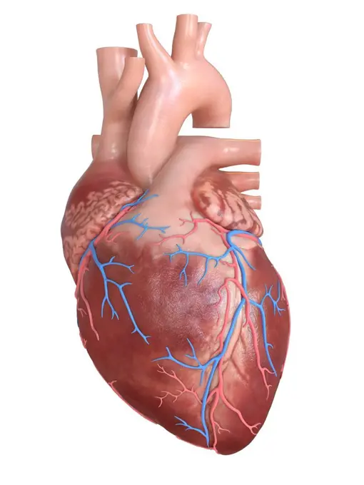
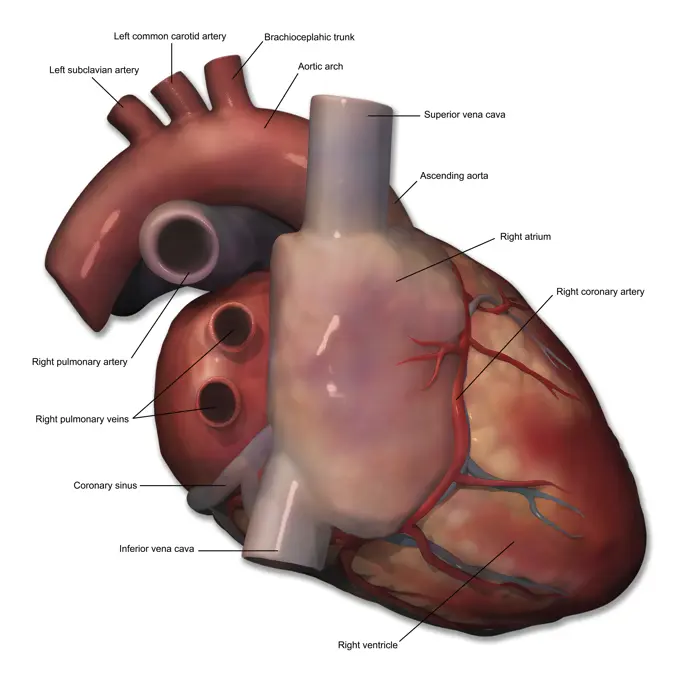
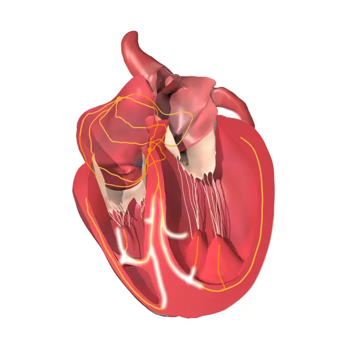
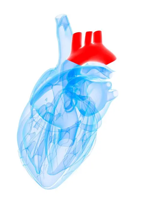
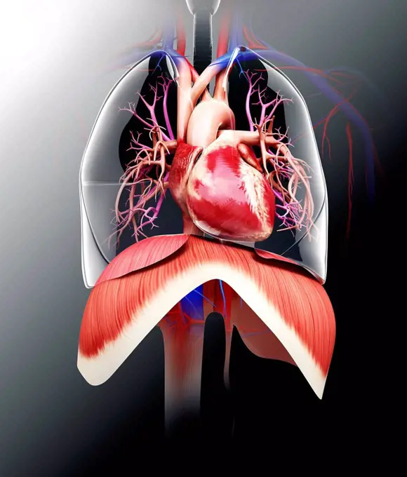
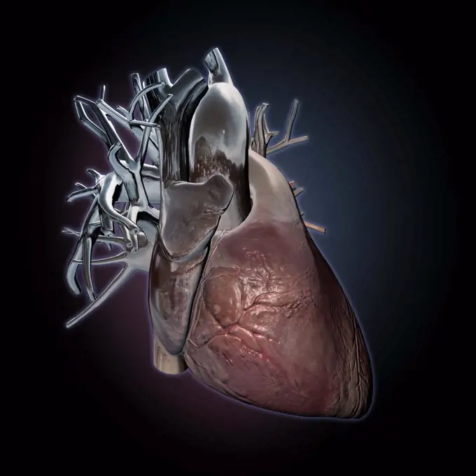
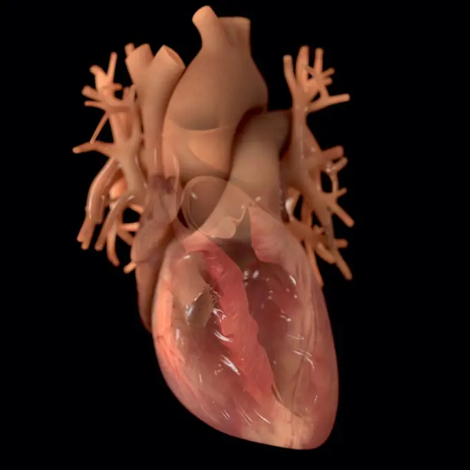
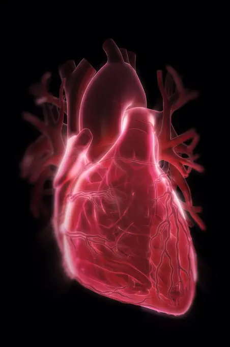
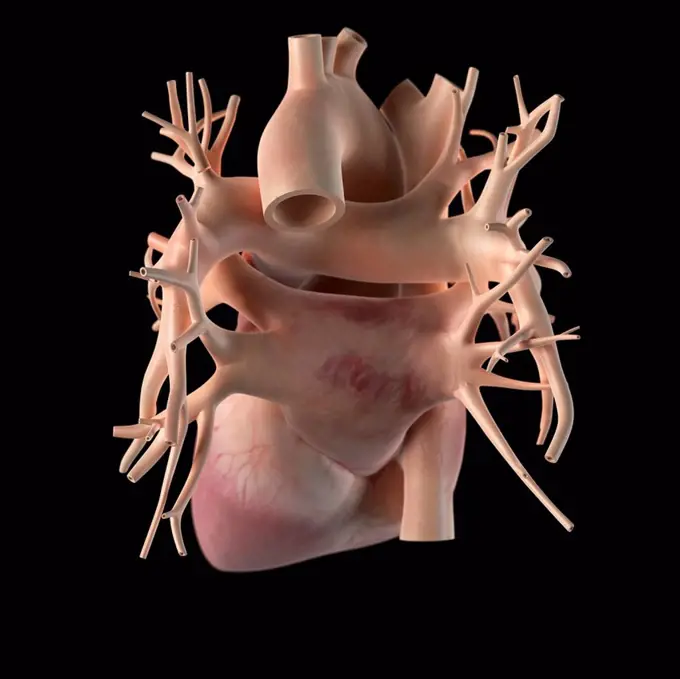
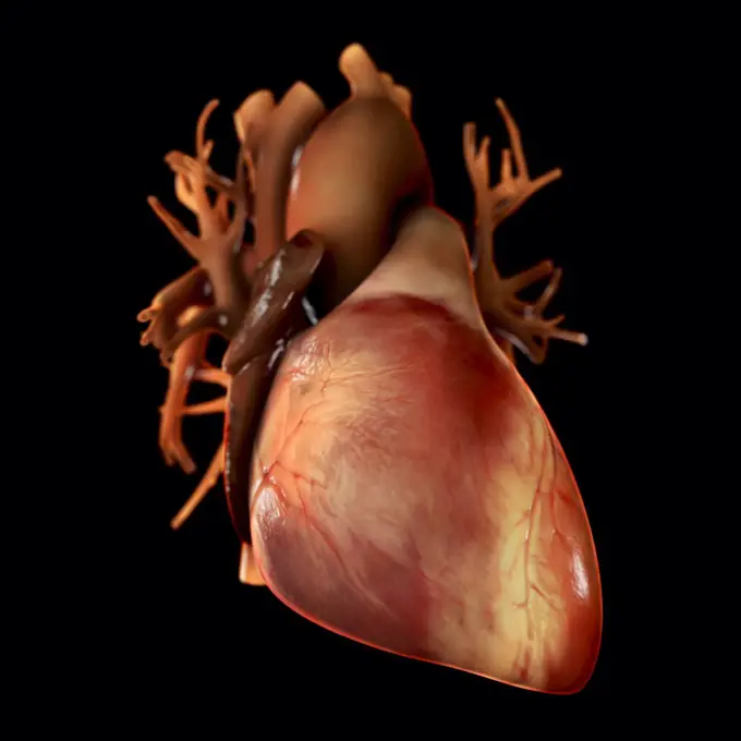
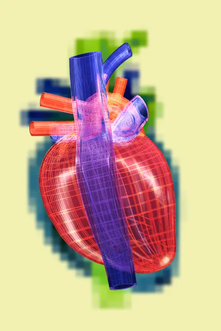
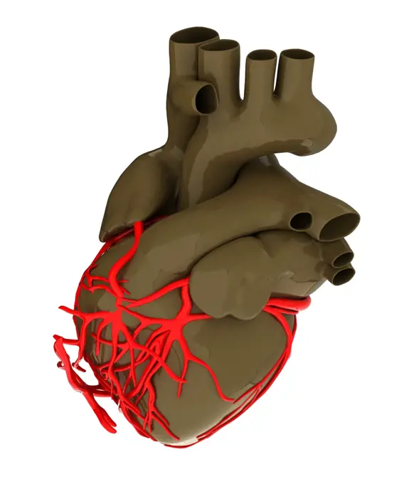
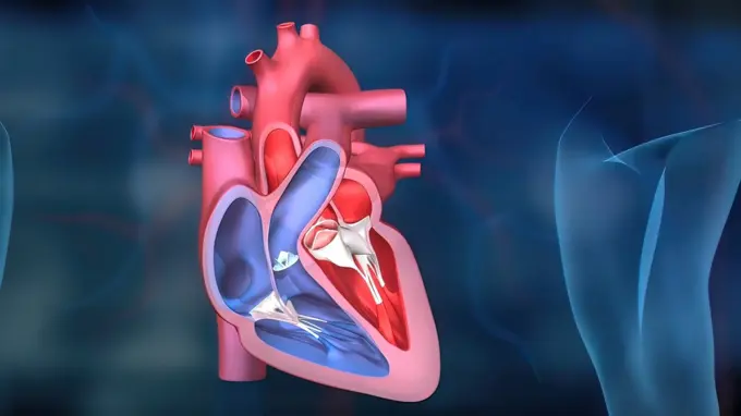
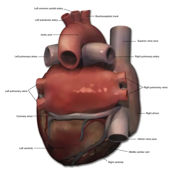
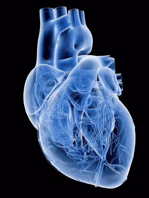
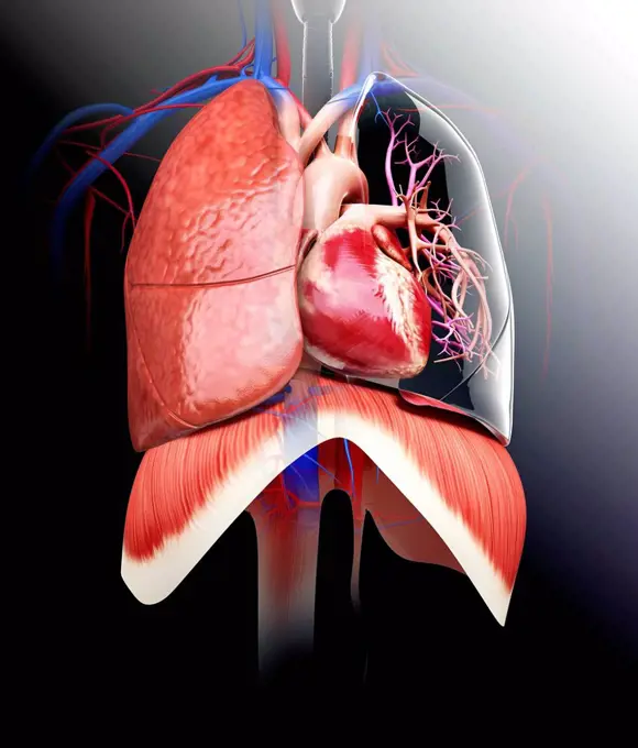
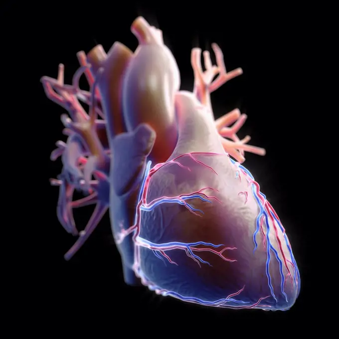
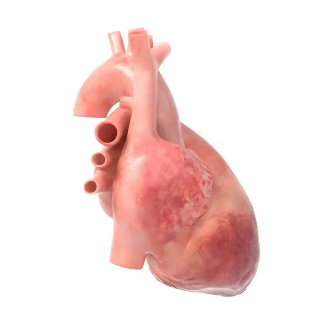
Human Heart Illustrations

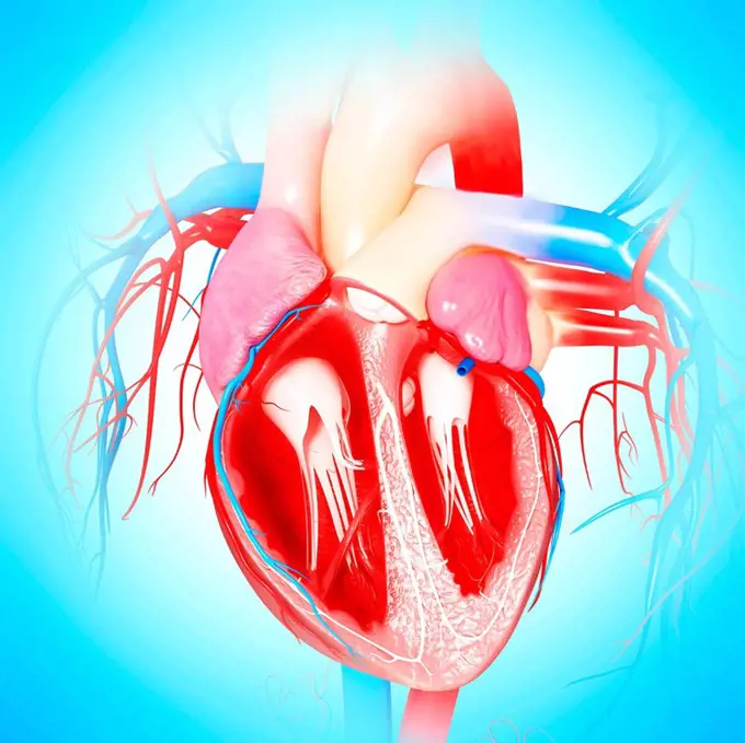
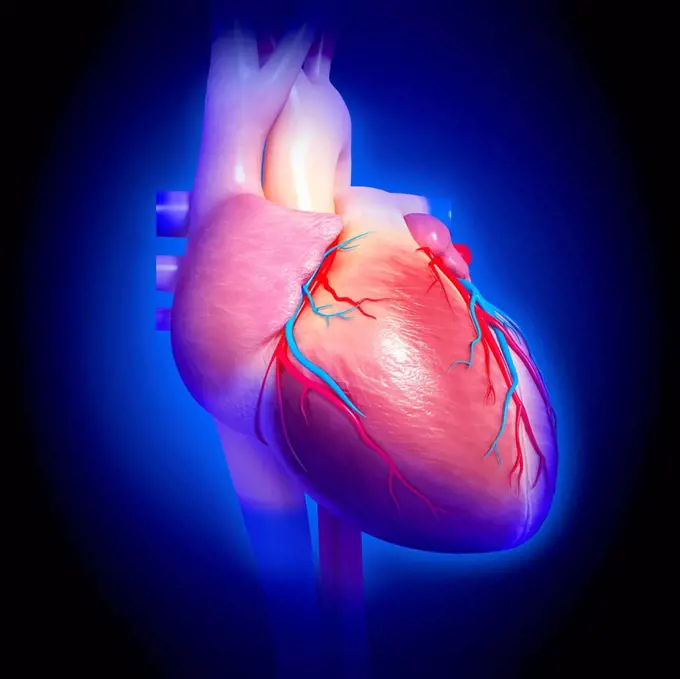
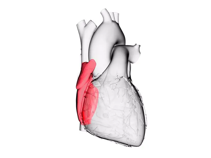
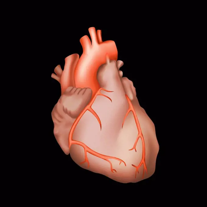
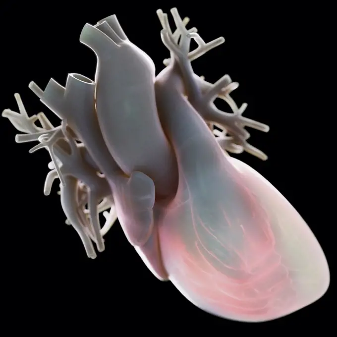
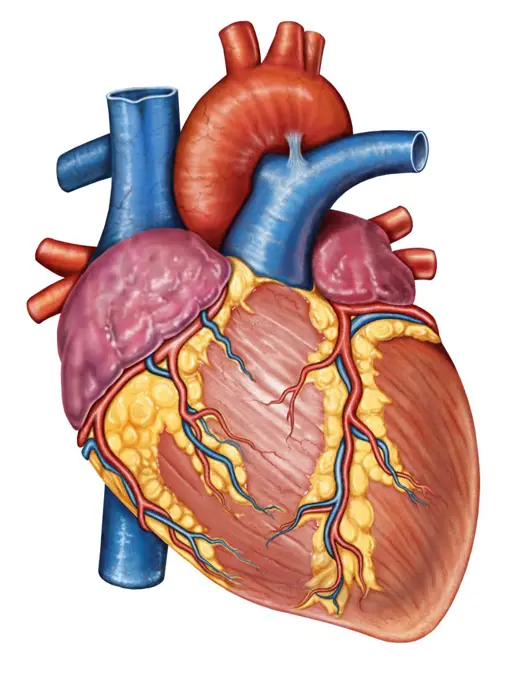
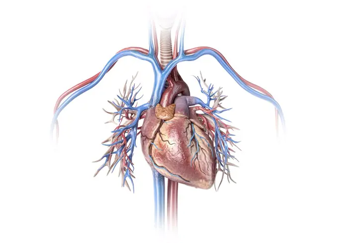
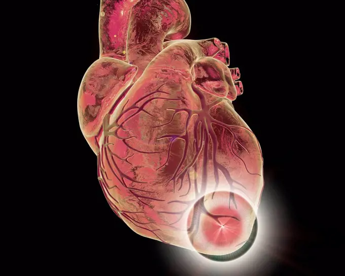
Highly detailed computer-generated images of the human heart, including illustrations depicting its structure and various conditions.

Aortic aneurysm, illustration
209 assets in this story
4128R-14060507
Aortic aneurysm, illustration
4128R-11287959
Human heart, computer artwork.
4128R-13724087
Illustration of the human heart.
1525-20518187
Human heart
1525-27190123
Realistic Vector Illustration Human Healthy Heart. Image Projection Anatomy Heart. Poster for Detailed Study Structure Cardiovascular System Organism. Isolated on White Background
4128-30417193
Human heart, illustration
4128-30417194
Human heart, illustration
4128-19053738
Human heart, illustration
5507-39365193
Internal organ of the person heart
4128R-11323160
Human heart, computer artwork.
1525-25693128
Artificial model of human heart isolated on white background
824-63188673
Illustration of the anatomy of the heart showing the intra-cardiac blood flow direction.
4128R-15514999
Illustration of a human heart
4128-19053732
Human heart, illustration
4128R-11323145
Human heart, computer artwork.
4128R-24842
Human heart, computer artwork.
4128R-14637676
Human heart coronary veins and arteries, illustration
4128R-24193
Human heart, computer artwork.
4128-15666305
Right atrium, illustration
4128R-15290282
Human heart, illustration
4128-19055880
Human heart, illustration
4128R-13723567
Illustration of the heart's coronary blood vessels.
4128-15666301
Right ventricle, illustration
4378-5745
Human Heart
4378-1696
Diagram of a heart.
4378-3417
Front view of the heart with coronary blood vessels visible set against an atmospheric white backdrop.
1525R-242931
3d rendered medical illustration of a human heart
824-63185610
A frontal illustration of a whole heart.
4128-15981661
Coronary blood vessels, computer illustration.
4239-V53647222
4128R-29448
Chest anatomy, computer artwork.
4128-111453002
Human heart, illustration
1574R-018884
Human Heart
1525-56722423
Normal blood flow in the heart 3D rendering. Normal blood flow in the heart
4378-3887
Side view of the heart anatomy and its coronary blood vessels.
824-63165992
Heart and right coronary artery, 3D CT scan.
4128R-11287968
Coronary arteries, computer artwork.
1428-1338
Human heart with wire frame, appearing like frozen mechanical heart on black background
4378-1654
Diagram of a heart.
4239R-8097
Gross anatomy of the human heart.
1525-20518376
Human heart
4239-18642052
Human heart with vessels and bronchial tree close-up. Isolated on white background.
824-63219037
The heart, anterior view, anatomy. The heart, and its large vessels: From left to right: The right atrium or right atrium which receives the delivery of the upper and lower cava veins the arch of the aorta and the departure of the brachiocephalic trunk, of the primitive carotid artery left and artery under left keyboard the pulmonary trunk above the right ventricle the left auricle surrounding the base of the pulmonary trunk above the left ventricle. The left and right coronary arteries supply the ventricles.
4128R-13928491
Heart attack, conceptual computer illustration.
824-63223301
Anatomy of an infant's heart, heart organ at birth At birth, the cardiovascular system changes from placental circulation to autonomic circulation. The foramen ovale between the right atrium and the left atrium closes around the age of 4 months. While the arterial duct between the aortic arch and the pulmonary trunk closes around the third week. It is then called an arterial ligament. It is shown in white. Then the venous ligament of Arantius to the left of the inferior vena cava closes and becomes the venous ligament of the liver. Blue and red colors indicate oxygenation of the blood. Blue represents deoxygenated blood and red represents oxygenated blood.
4239-18641531
3D rendering of human heart and thoracic veins.
4128R-15515113
Illustration of a diseased heart valve
824-63206049
Illustration of the causes of a myocardial infarction, necrosis of part of the secondary heart muscle through an oxygenation flaw (ischemia). The infarction occurs when one of more coronary arteries become blocked, the heart muscle cells that are irrigated by these arteries are no longer oxygenated, which can lead to their death. The infarct area will no longer contract properly. The obliteration of the coronary artery is linked to an atheromatous plaque, a deposit which can cause a reduction of the arterial lumen (stenosis). It can be caused by a clot, a piece of the existing atheromatous plaque that detaches itself. It can be caused by a coronary spasm, a dramatic reduction of the arterial lumen linked to the vasomotricity of the arteries.
1525-76043919
Main internal human organ heart. Anatomical representation of muscle that pumps blood. Hand drawn simplified image of heart man, vector. Main internal human organ heart
4128-30416240
Human heart, illustration
4128R-14637675
Human heart coronary veins, illustration
4128-15666318
Mitral valve, illustration
4128R-29460
Human heart, computer artwork.
1899-12943
Conducting system of the heart VII
4239-69151977
Right lateral view of human heart anatomy with annotations.
4128R-12573819
Doctor examining the human heart, computer illustration.
1899-12940
Conducting system of the heart IV
4128R-25853
Aortic arch, computer artwork.
4128R-28681
Human heart, computer artwork.
4128R-11313045
Human cardiovascular system, computer artwork.
1428-1323
Human heart and major vessels on black background
4128R-12576717
Heart and coronary arteries. Artwork of the external anatomy of a human heart, seen from the front. The surface blood vessels are the coronary arterie...
4462-21421711
Close up of human heart
4239-69151973
Anterior view of human heart anatomy with annotations.
4378-3528
Front view of the heart with coronary blood vessels visible set against a black background.
4378-1672
Diagram of a heart, showing an anterior cut.
1525-20518377
Human heart
1525-56211418
The heart and the blood vessels 3D illustration. The heart and the blood vessels
1525-20518171
Human heart
1525-25338380
3d rendered medical illustration of a human heart
4378-20014152
Human Heart
4128R-11323157
Human heart, computer artwork.
4378-1652
Diagram of a heart.
824-63126599
Heart, illustration
1525-56928727
Heart. Human internal organ. Medicine and cardiology. Pumping blood through body. Element for textbook and medical education. Arteries and red veins. Cartoon flat illustration. Human Heart. Medicine and cardiology.
4128R-11296625
Human heart anatomy, computer artwork.
824-63195024
Representation of a cardiac pacemaker or defibrillator consisting of a box placed under the skin and connected to the heart by a probe introduced by the left brachiocephalic vein, to the right ventricle.
4128-111453001
Human heart, illustration
1525-20518180
Human heart
5507-45949845
Artery system in human body
4378-1677
Diagram of a heart.
4239-18641280
Human Heart
4128-18632086
Internal structure of the heart, illustration
1525-26244056
As the heart beats, it pumps blood through a system of blood vessels, called the circulatory system. The vessels are elastic, muscular tubes that carry blood to every part of the body. 3D illustration. Heartwork system. Pumps Blood Through The Human Body
4239-69151976
Posterior view of human heart anatomy with annotations.
4128R-20582
Heart with coronary vessels, artwork
4128R-11313604
Human heart and lungs, computer artwork.
4128R-12580564
Human heart, computer illustration.
4378-3883
Three-quarter view of the heart and its coronary blood vessels.
4128-30416229
Human heart, illustration
1525-24374622
Cross-section of the human heart showing the blood-pumping function of the veins and arteries, with arrow symbols showing the direction of venous flow.
1525-28173770
Heart of Dugong, vintage engraved illustration. Zoology Elements from Paul Gervais.
4128R-28897
Atherosclerosis. Computer artwork of a narrowed artery, due to a cholesterol plaque.
1525-56854582
In a normal heartbeat, the electrical signals follow a path from the atria into the ventricles. 3D rendering. In a normal heartbeat, the electrical signals follow a path from the atria into the ventricles.
1525-25616142
Human heart. Medicine, internal organs. 3d vector icon
4128R-14637695
Human heart pulmonary valve, illustration
1525-23339120
DNA and heart. 3d illustration
4128-19055895
Human heart, illustration
4128R-28734
Human heart, computer artwork.
4128R-14058253
Brain veins and anatomy, illustration
PREVIOUS
of 3
NEXT
2401 S. Ervay, Suite 206
Dallas, Texas 75215
United States
Browse
CategoriesGalleriesFree Research
For Creators
License Your ContentSuperStock For StudentsFrame of Mind BlogContributor PortalNew Image SpotlightLicense AI Imagery
Resources
About UsLicensingTerms & ConditionsAPI AccessPrivacyPricing
Contact
+1 866 236 0087help@superstock.com Contact form
©2026 SuperStock. All Rights Reserved. -A
This site is protected by reCAPTCHA and Google Privacy Policy and Terms of Service apply.