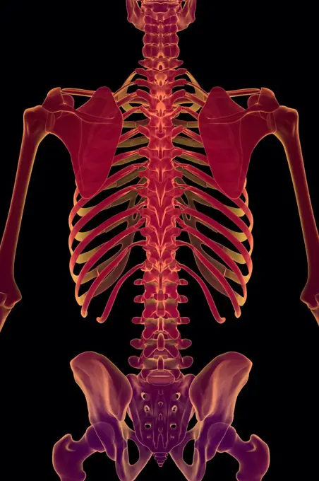
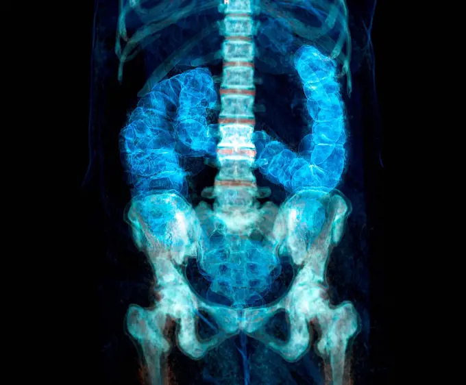
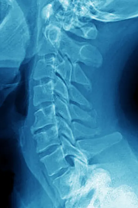
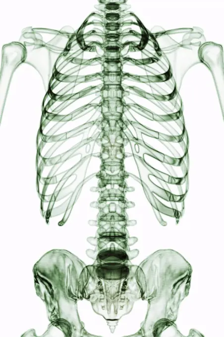
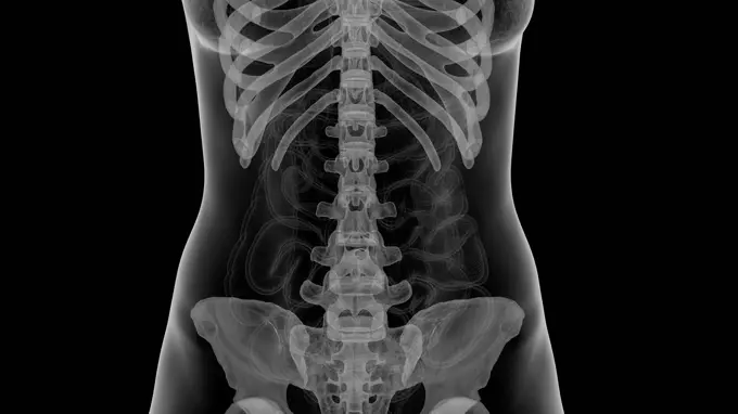
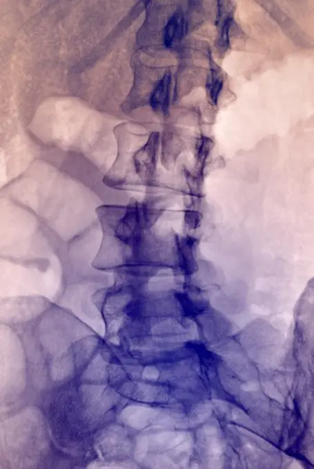
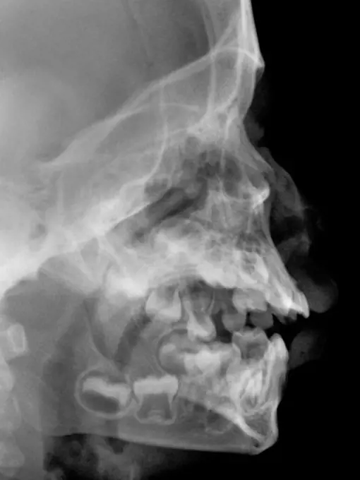
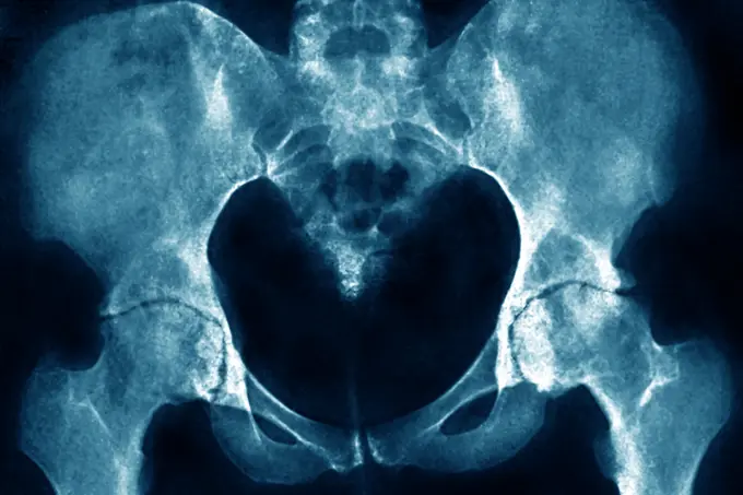
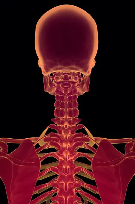
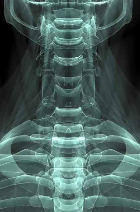
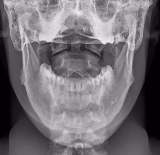
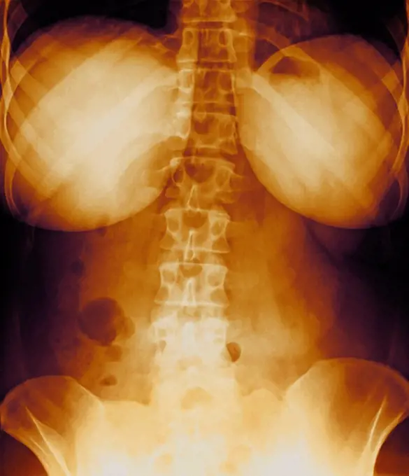
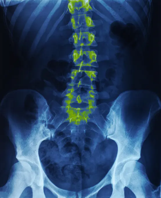
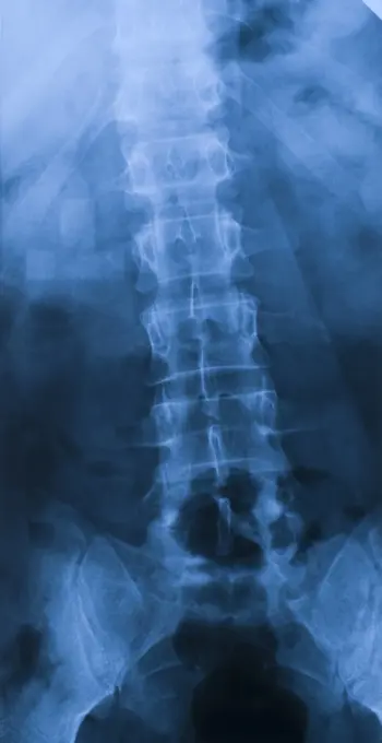
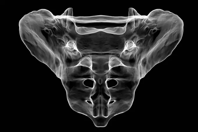
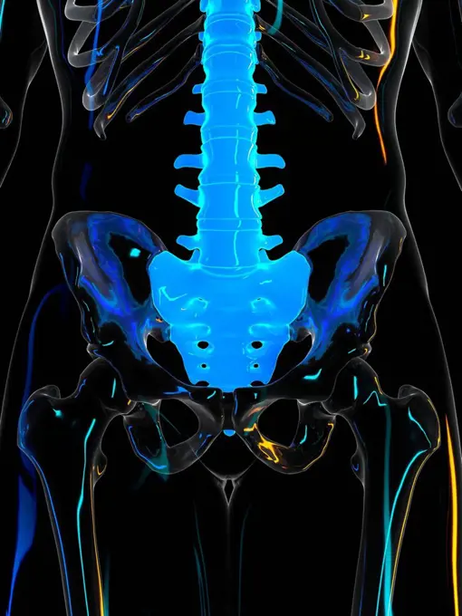
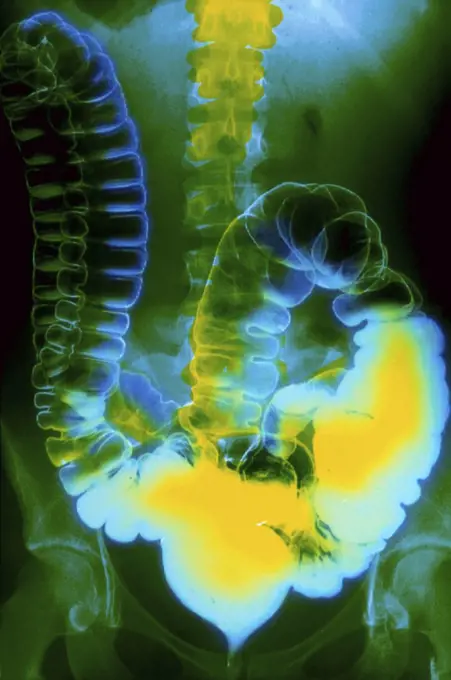
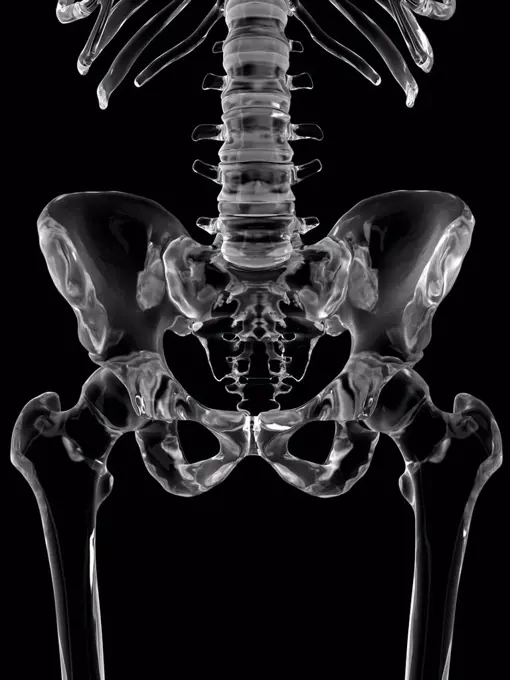
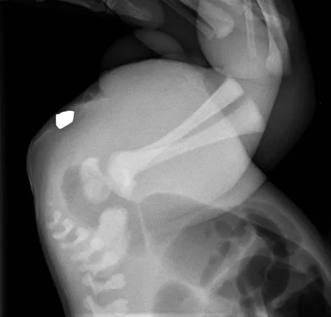
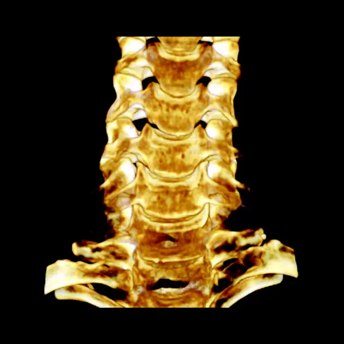
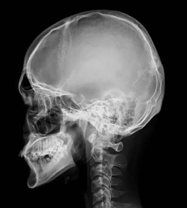
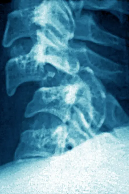
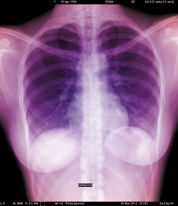
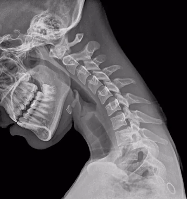
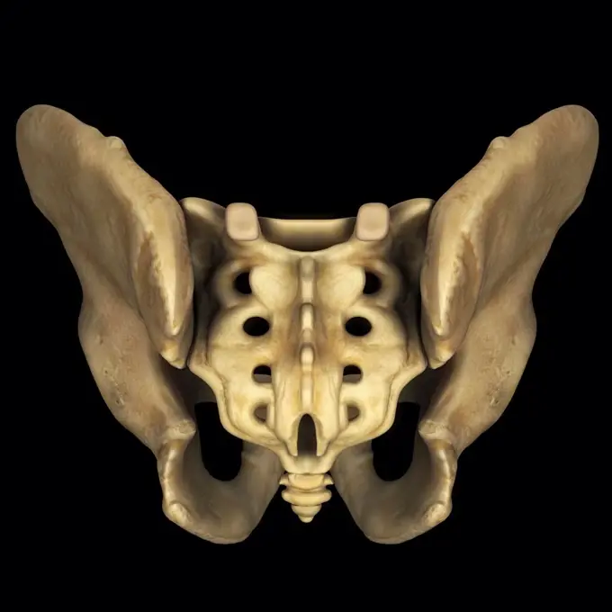
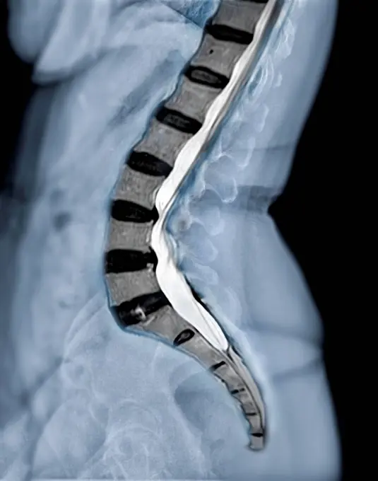
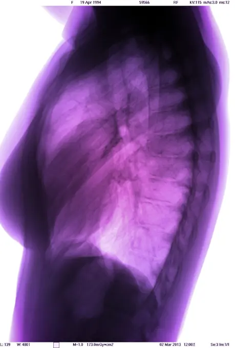
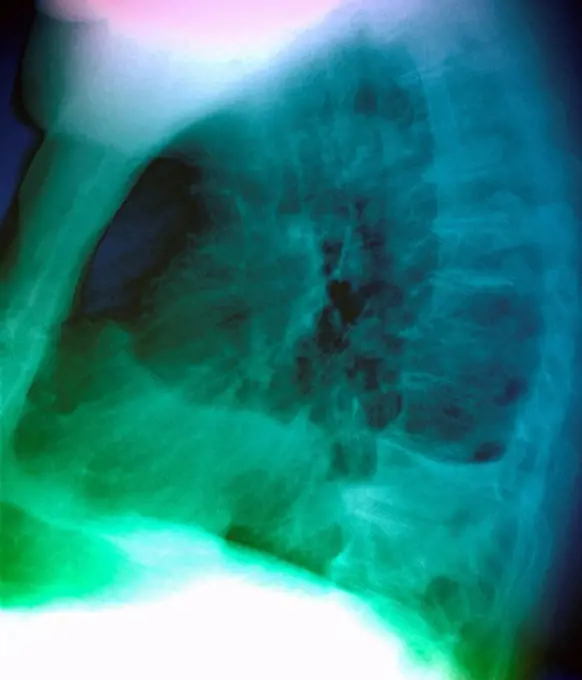
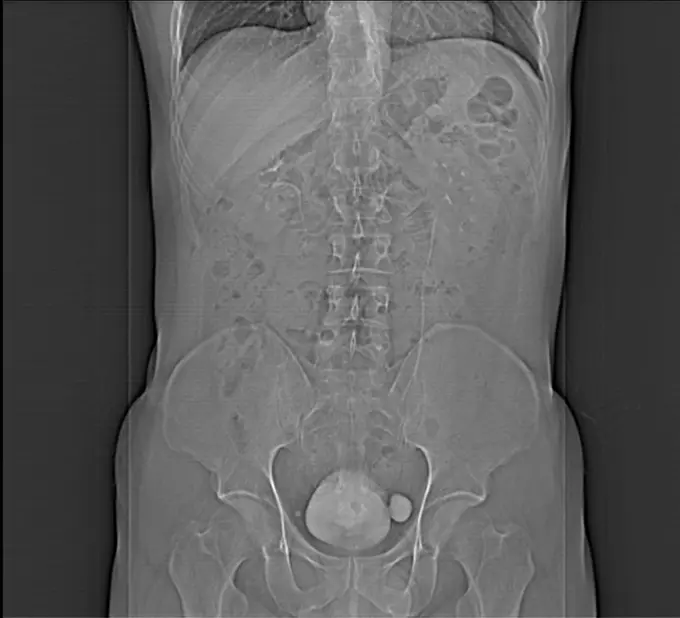
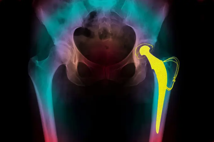
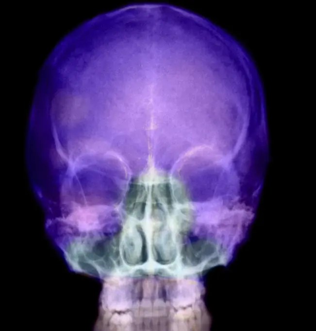
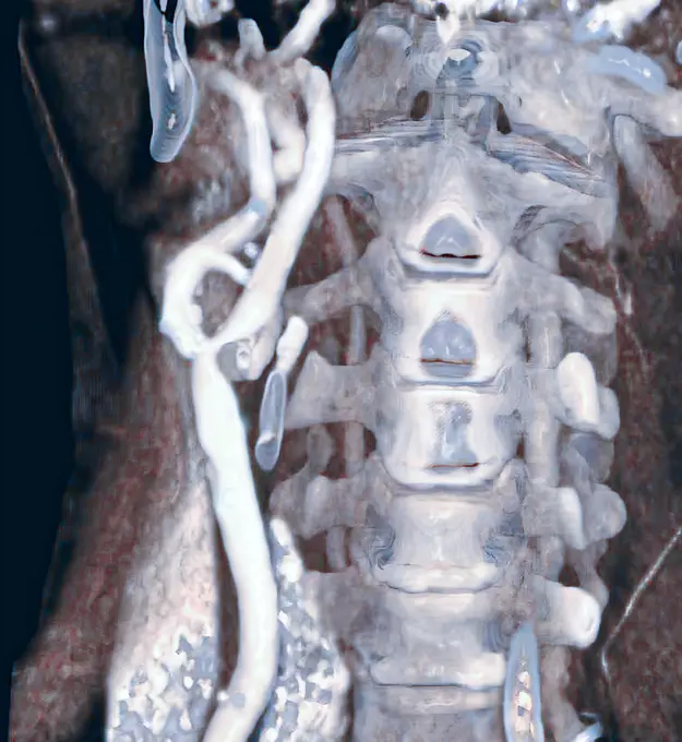
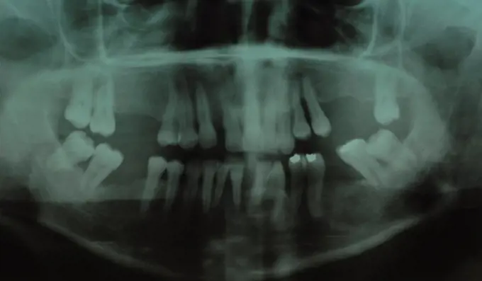
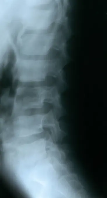
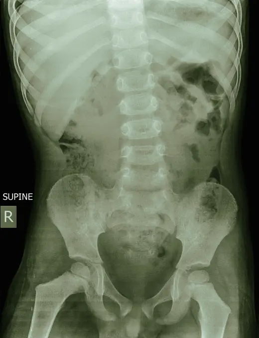
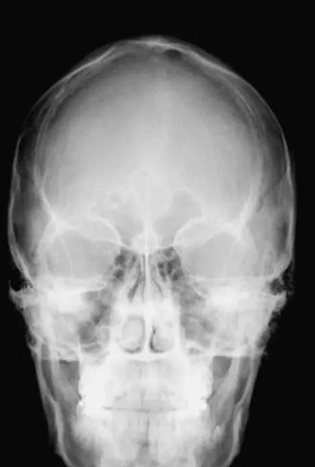
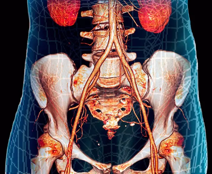
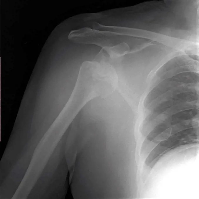
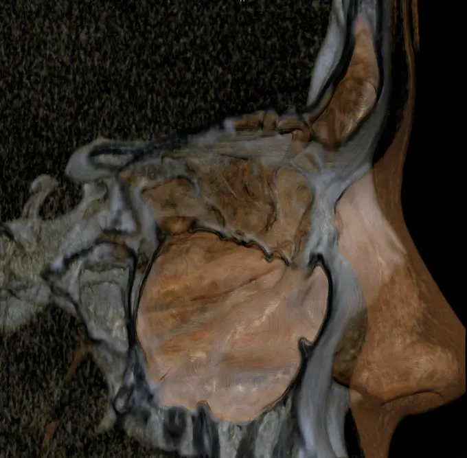
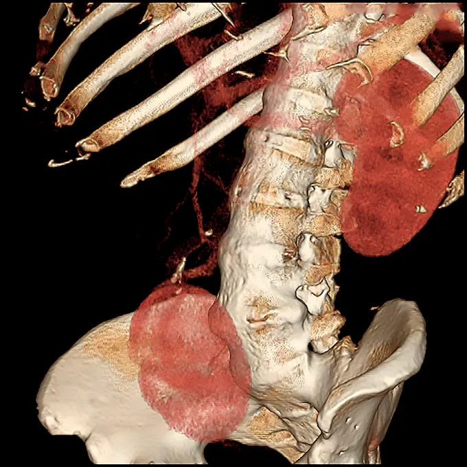
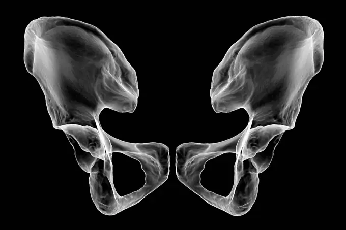
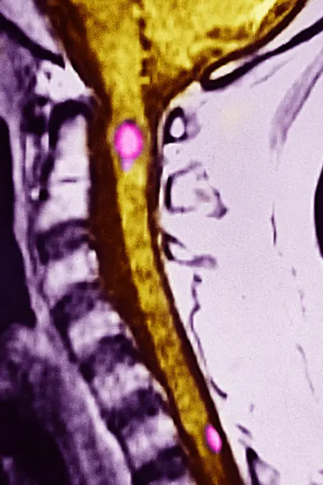
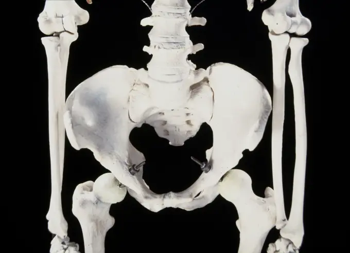
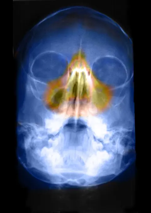
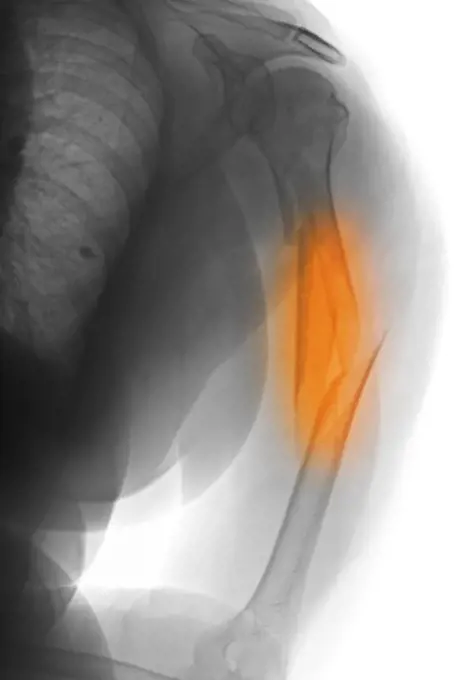
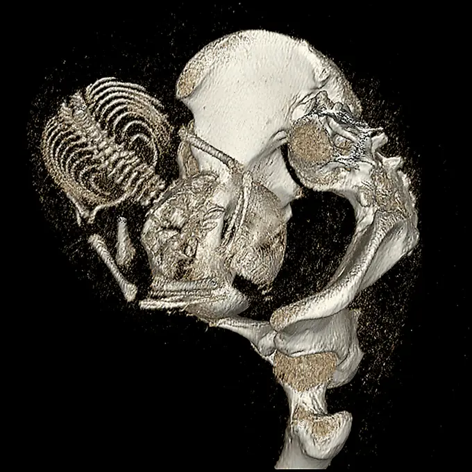
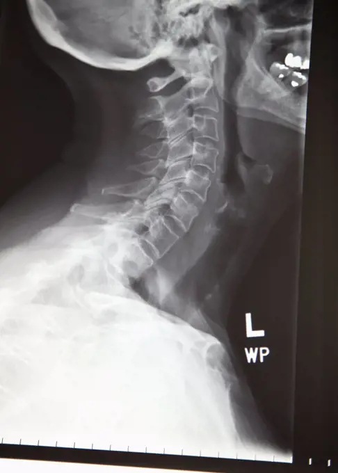
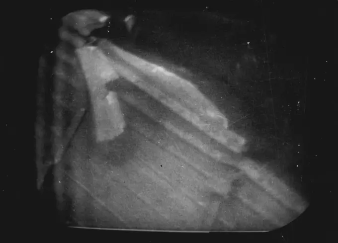
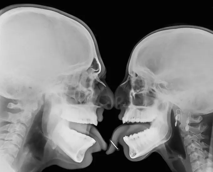
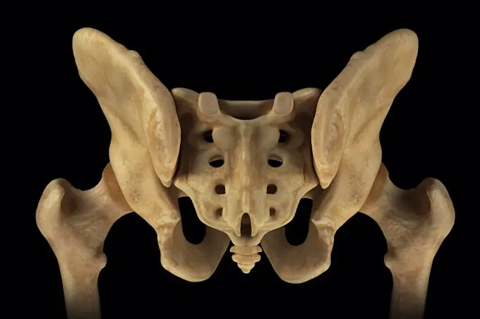
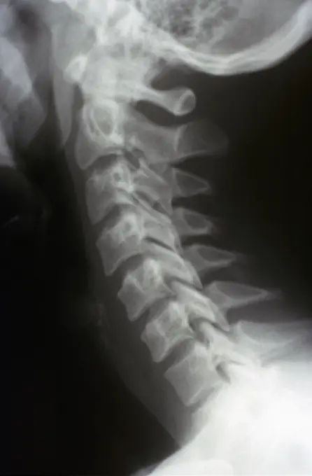
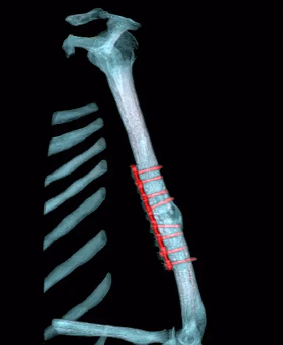
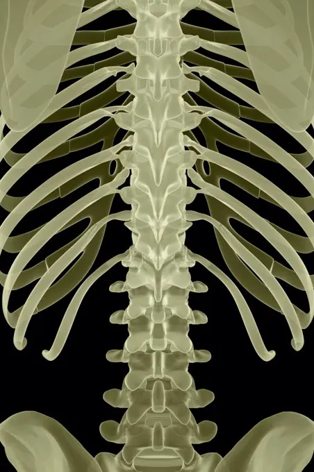
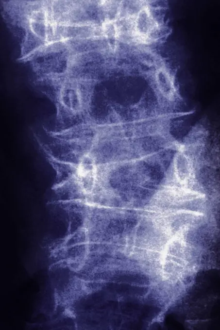
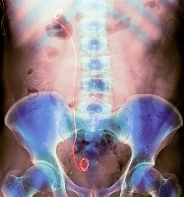
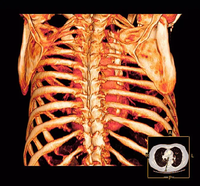
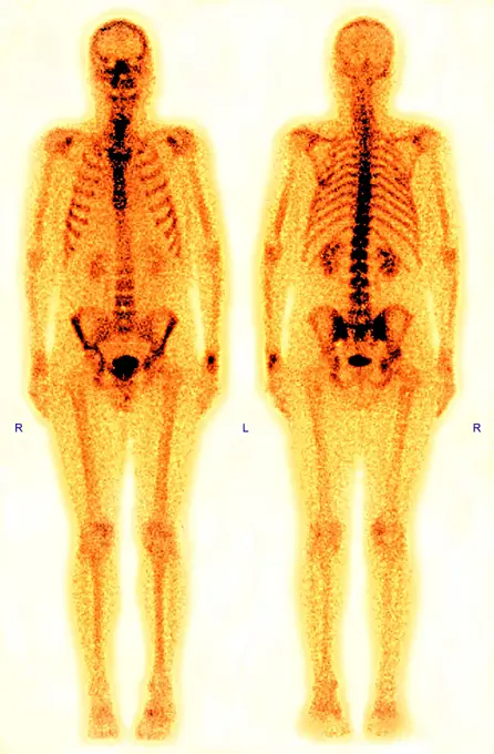
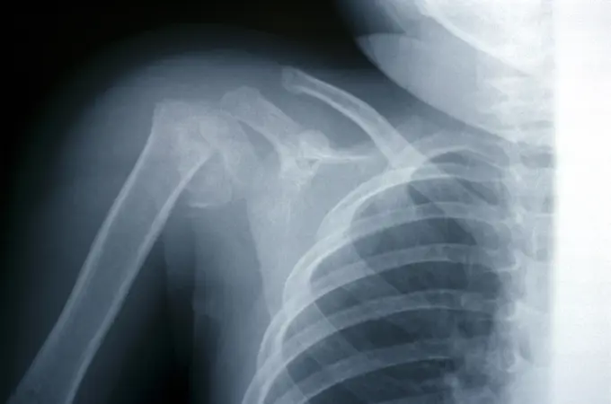
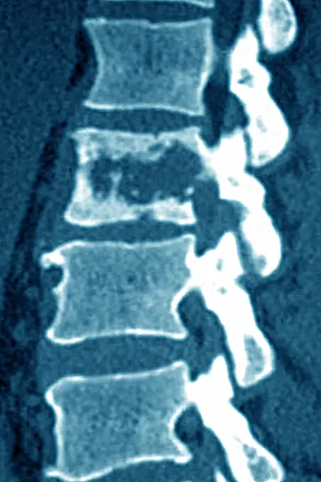
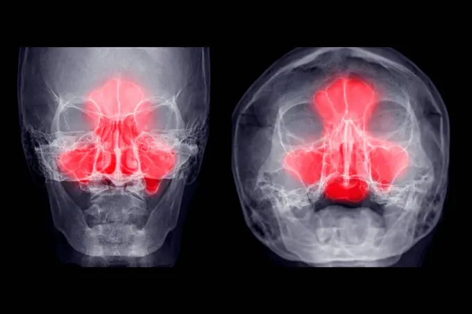
Fine ArtVintageFootageSubscriptionBoardsFree ResearchPricing
  • Fine Art
  • Vintage
  • Footage
  • Subscription
  • Boards
  • Free Research
  • Pricing

Human Skeletal Imaging

A collection of various medical imaging techniques showcasing the human skeletal system, including x-rays and CT scans highlighting bones and anatomical features.

Rear view of the bones of the upper body of the male skeleton.
120 assets in this story
4128-28514546
Large intestine, CT scan
1899-53511733
Cervical osteoarthritis visualized by a sagittal x-ray of the neck. (Photo by: CAVALLINI JAMES/BSIP/UIG)
4378-109
Front view of stylized bones of the trunk.
1525-21552808
X-Ray Image Of Human Chest for a medical diagnosis
4128-30422729
Skeletal system, illustration
1558-14105870
X-ray picture of the spine
4128-19056646
Femur, illustration
4297-1340
Abdominal x-ray of a 38 year old woman showing the lumbar spine, a portion of the pelvis and ribs
4378-415
Rear view of the bones of the upper body of the male skeleton.
4297-1105
Normal lateral skull x-ray of a 2 year old boy
824-63225974
Osteoarthritis of the hips (coxarthrosis) visualized by a pelvic x-ray of the front.
4378-123
Rear view image of the bones of the head and neck.
4128-19248780
Human spine, illustration.
4378-1951
X-ray image showing the spine.
4128R-12667
Normal neck. Frontal X_ray of the joint between the skull and the neck.
4128R-6790
Female torso, X_ray
1899-53511591
Osteoarthritis of the cervical vertebrae C5-C6. Spinal x-ray in sagittal view.
4269-5169
X-ray of the lower backbone of a child showing light scoliosis.
4128R-12580426
Human pelvis bones, computer illustration.
6188-65147137
x ray of Columna vertebralis with Os sacrum frontales Rontgenbild der Beckenwirbelsäule ,model released, Symbolfoto Copyright: xZoonar.com/AndyxNowackx 4561829 ,model released, Symbolfoto ,property released
4128-28968825
Sacrum bone, illustration
4128-19056651
Lower spine, illustration
824-687
X-Ray Showing Large Intestine Using Barium Enema
4128R-12580449
Human pelvis bones, computer illustration.
4128-28970825
Invertogram X-ray
4269-1067
Three-dimensional computed tomographic (CT) scan reconstruction of cervical rachis, composed of 7 cervical vertebras.
4128-30420017
Head and neck bones, X-ray
824-63226736
Dislocation of the cervical vertebrae, with spinal trauma, between the sixth and seventh cervical vertebrae, visualized by sagittal section radiography.
4128-28968803
Anatomy of the pelvis bones, illustration
824-63194005
A healthy lung x-ray on an 18-year old patient.
4128R-12680
Normal flexed neck. X_ray of the flexed cervical spine of a 20 year old female.
4297-1381
Normal x-ray of the pelvis of a 23 month old boy
4128-28766995
Femoral artery, CT scan
4378-403
Front view of stylized bones of the cervical spine and sternoclavicular joint.
4128-18680908
Intussusception of the intestines, X-ray
4297-1138
Abdomen x-ray of a 76 year old man with a history of bladder cancer that was treated surgically. There are multiple surgical clips in the pelvis highlighted in orange. A has been placed in the kidney
4378-3431
The bones of the male pelvis viewed from a rear perspective.
4128-28769203
Compressed spinal cord, X-ray
1525-28468522
X ray film for human skull
824-63193993
A healthy lung x-ray on an 18-year old patient.
4128R-7718
Pleural effusion, X_ray
6188-55645094
Abdomen man, man abdomen man, man, ct recording copyright: xzoonar.com/dr.xnorbertxlangeex 13790168
4269-25312
Prosthetic hip. X-ray of prosthetic hip showing the shaft of steel inserted into the femur (yellow), with the artificial ball articulating with the bone socket of the pelvis.
824-63178590
X-ray of the sinuses of a 35-year-old woman. Hypertrophy of the right and left middle and inferior turbinates
824-63215603
X-ray of the sinuses.
4128R-12777
Normal spine. Coloured X_ray of the spine of a 24 year old man.
1899-53508459
3D CT scan of stenosed carotid artery, responsable for stroke.
1899-64961
Black and white x-ray of teeth of young adult
6188-56014261
X-Ray of a human spine X-Ray of a human spine, a vertical picture Copyright: xZoonar.com/OlgaxPopovax 3862162
1439-57940358
An X-ray image showing a front view of a human torso, highlighting the ribcage, spine, and pelvis.
1525R-77007
X-ray image of human skull
4128R-30919
Abdominal aorta. Coloured 3D computed tomography (CT) angiogram of the pelvis of a 39 year old patient. At top is the abdominal aorta. The aorta is th...
4201-21246162
Great Egret (Ardea alba) chick x-ray after being hit by car, International Bird Rescue, Fairfield, California
824-29057216
Retinal detachment of the left eye, visualized by radial section MRI.
4128-19358084
X-ray showing a dislocated shoulder, where the head of the humerus is detached from the shoulder joint.
824-63175251
CT scan of internal ear.
6188-64760045
X-ray of healthy young cat in lateral view X-ray of healthy young cat in lateral view. Copyright: xZoonar.com/OlgaxOvchinnikovax 11016914
824-63178718
Coloured x-ray of a ten years old boy dentition.
1899-53508447
Profile of maxillary sinuses and nose bone. 3D CT scan.
4128R-11476614
Baby's nervous system, computer artwork.
4269-27540
Fractured pelvis. 3D Computed Tomographic (CT) reconstruction scan of the pelvis showing a fracture of the left ischium (at right). The chock also caused the rupture of the bladder evidenced by the leak of urine (below).
4269-27849
Intestine tumor. Three-dimensional computed tomographic reconstruction scan of the abdomen showing a tumor of the small intestine (yellow).
4269-38982
Coloured 3-D computed tomography (CT) scan of an Unilateral Ectopic Kidney in the pelvis.
4128-19358075
X-ray of the chest showing right pneumothorax (clear space in the right hemithorax).
4128-30416775
Larynx cancer, illustration
4128R-12577965
Coloured profile computed tomography (CT) scan of the healthy torso of a 60 year old patient.
824-63224751
Hip asymmetry, visualized by frontal pelvic x-ray.
4128-28968777
Anatomy of the coxal bones, illustration
824-63218246
Spinal neuromas (schwannomas) in cervical vertebrae, seen on a sagittal section MRI scan.
6188-55588147
Human teeth xray X ray of human mouth with teeth bones Copyright: xZoonar.com/ClaudioxDiviziax 8865476
4220-21334617
Human Skeleton.     Date:
824-63178564
X-ray of the skull of a little girl. Normal
1848-55997729
X-ray film of dog lateral view. Veterinary medicine, veterinary anatomy Concept
824-63203254
Ensethopathy of the supeficial plantar fascia. Sagittal section CT scan of the foot.
4297-1063
X-ray of a 74 year old woman showing a comminuted fracture of the humerus
4269-39009
Three-dimensional computed tomographic reconstruction of pregnant woman pelvis showing the skeleton of a 9 months old fetus, pelvimetry allows to measure the diameter of a woman at the 9th month of pregnancy.
1570R-137828
Close-up of an x-ray of a human neck
4128-V58573966
6177-V53848412
6145-52738597
X-ray of a right female forearm showing bone resection, with one-third of the ulna removed due to caries five years prior, on bromide paper from 1896.
5513-51495603
T.V. ***** In Comet Salvage Search -- Part of the fuselage skin is ***** by the underwater television camera working of 65 fathoms.A series ***** of television cameras and underwater ***** made during the search by the Royal ***** the Comet aircraft which crashed off Elba. ***** the operations, which have been going ***** about eight weeks under the command of ***** Earl Mountbatten, about one tenth of the wreckage has been recovered. Experts are examining the parts brought to the surface in an effort to determine the cause of the plane crash. March 13, 1954. (Photo by British Official Photograph).
1439-57939919
Xray vision on a friendly conversation
4378-3973
Rear view of the male pelvis, sacrum and hip joints.
6114-50976660
An x-ray of a normal cervical spine.
6188-55645081
Abdomen man, man abdomen man, man, ct recording copyright: xzoonar.com/dr.xnorbertxlangeex 13790137
4128R-30918
Pinned arm fracture. Coloured 3D computer tomography (CT) scan of the humerus (upper arm bone) of a 44 year old patient. Metal plates have been insert...
4378-236
Rear view of the bones of the upper body.
4128-28968816
Anatomy of the sacrum bone, illustration
824-63195609
A scintiscan of the entire body. The human body seen face on and from behind by a gamma camera.
6145-29606542
Collection Dutch Heidematschappij. Insects, damage, oak miner moth, Nepticula. undated
824-65830294
Lumbar osteoporosis (disease causing compression and fractures of the lumbar vertebrae). X-ray of the lumbar vertebrae seen from the front.
4128-28968814
Anatomy of the sacrum bone, illustration
4128-30420594
Urinary system stent, X-ray
1525-23488894
x ray picture of wild animal skeleton
4128R-14009
Rib cage. Coloured three_dimensional computed tomography CT scan of a posterior view of a healthy rib cage and heart.
824-63195610
A scintiscan of the entire body. The human body seen face on and from behind by a gamma camera.
6114-50976656
An x-ray of a fracture of humerus in a child.
824-57656893
Vertebral osteitis (inflammation of the bone tissue of a lumbar vertebra, caused by a bacterial infection). sagittal x-ray of the lumbar vertebrae.
4128-28769226
Sinus pain, conceptual X-rays
6188-58104539
Transparent Human Body with Visible Bones medical science image of human skeleton bones Copyright: xZoonar.com/StanislavxRishnyakx 14296133
PREVIOUS
of 2
NEXT
2401 S. Ervay, Suite 206
Dallas, Texas 75215
United States
Browse
CategoriesGalleriesFree Research
For Creators
License Your ContentSuperStock For StudentsFrame of Mind BlogContributor PortalNew Image SpotlightLicense AI Imagery
Resources
About UsLicensingTerms & ConditionsAPI AccessPrivacyPricing
Contact
+1 866 236 0087help@superstock.com Contact form
©2026 SuperStock. All Rights Reserved. -B
This site is protected by reCAPTCHA and Google Privacy Policy and Terms of Service apply.