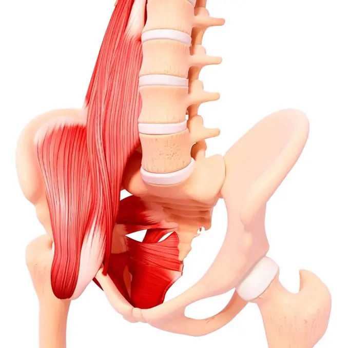
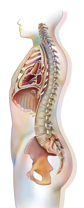
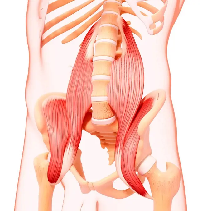
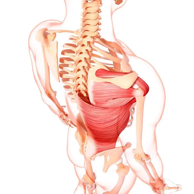
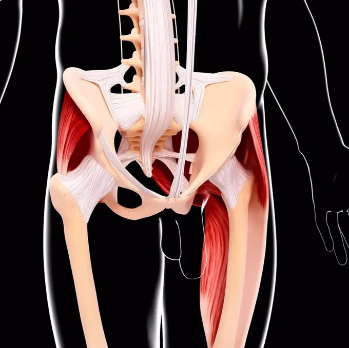
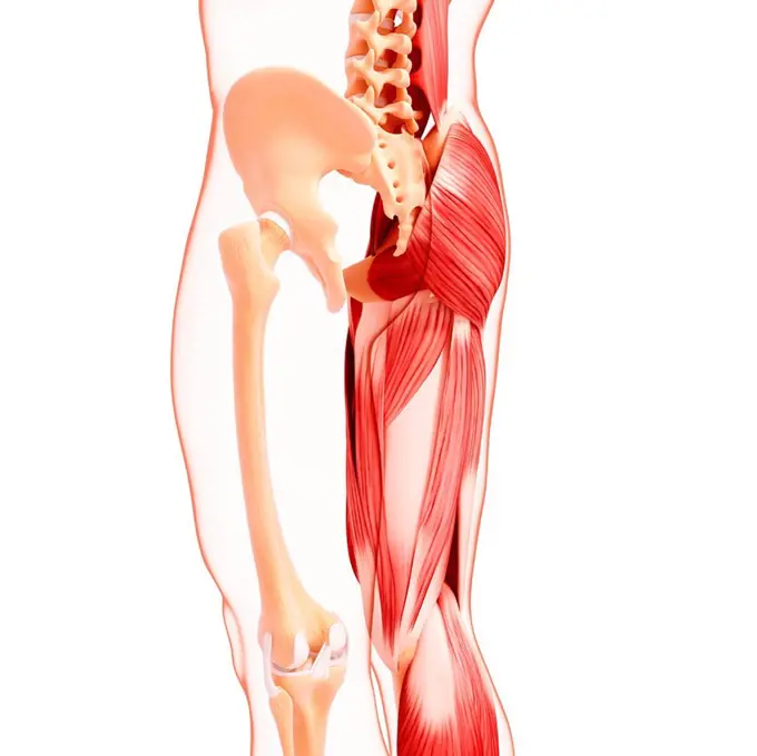
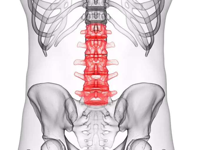
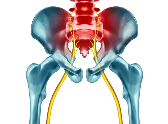
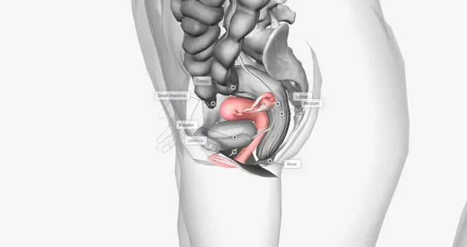
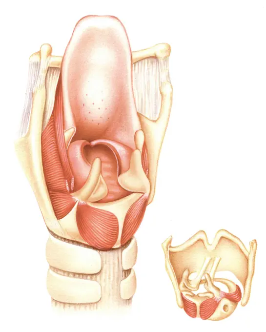
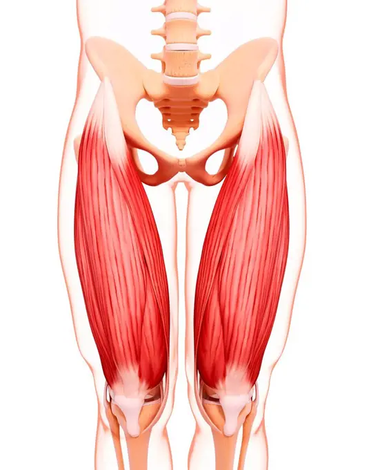
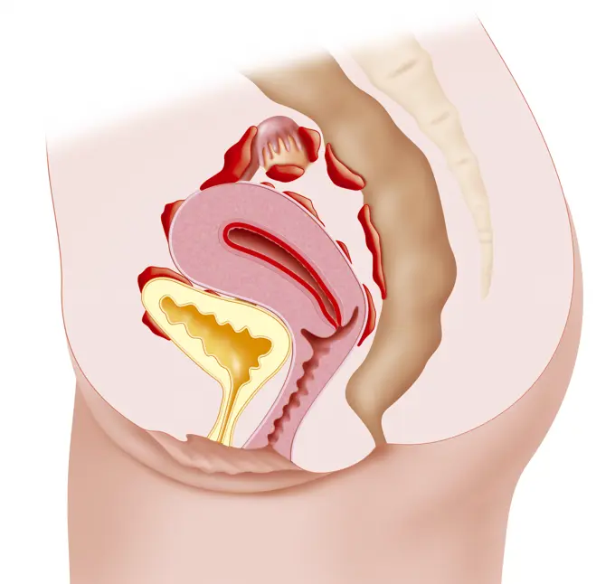
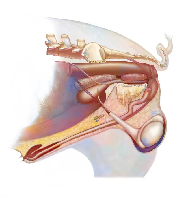
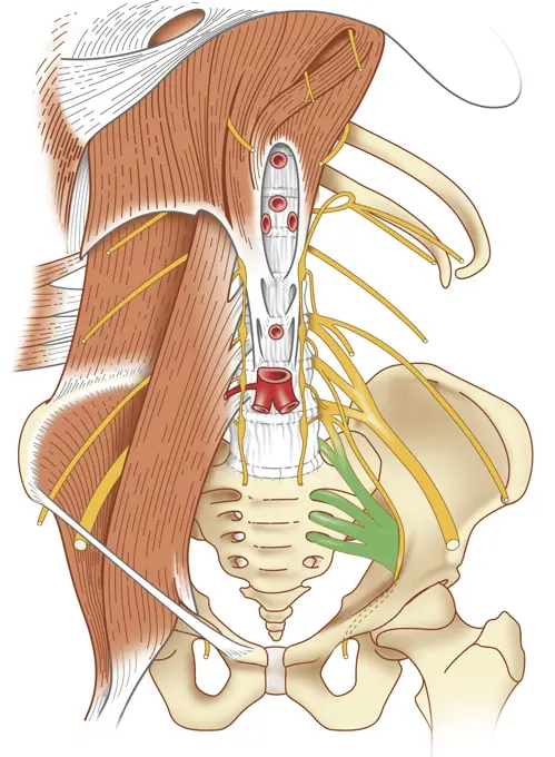
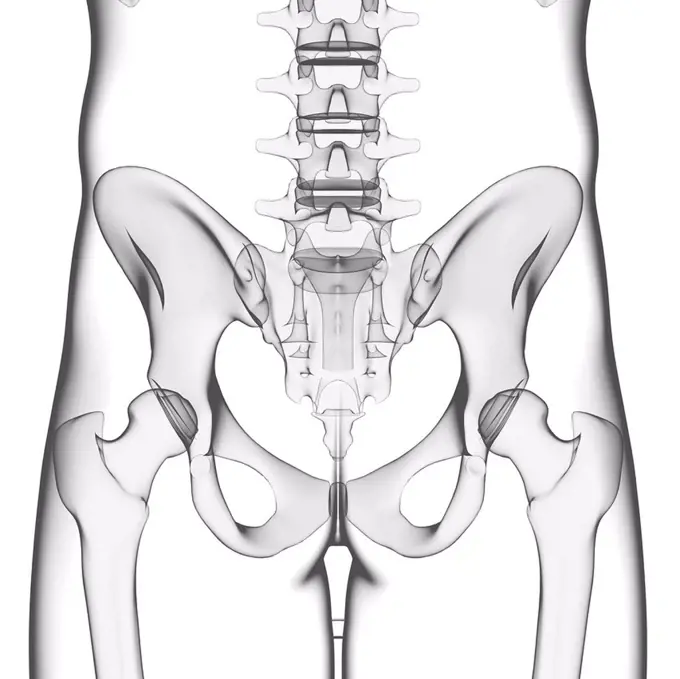
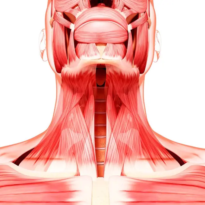
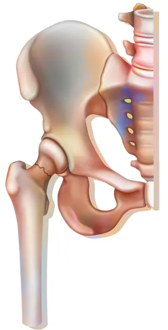
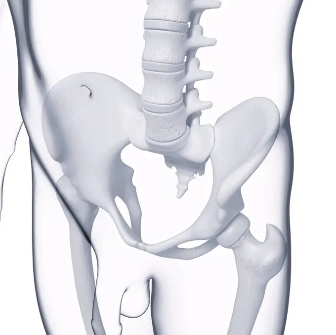
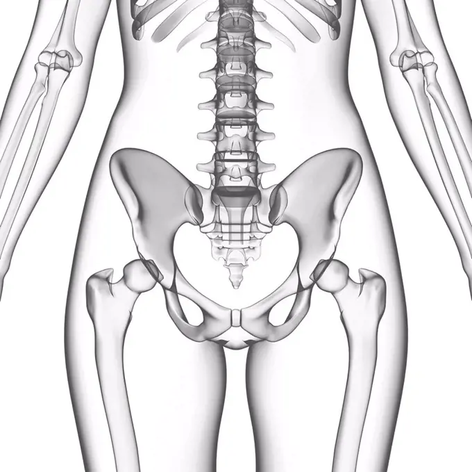
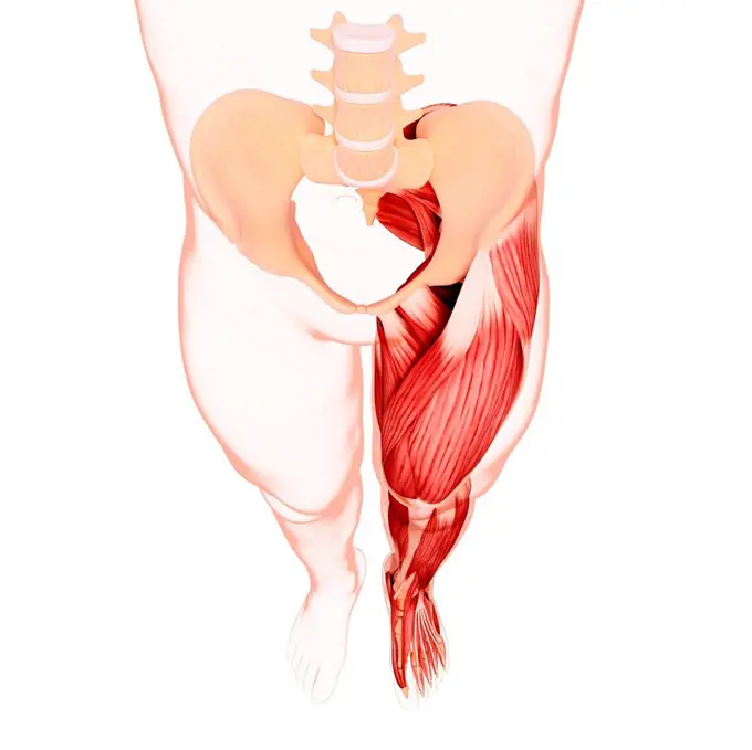
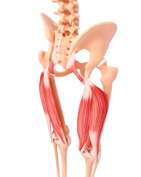
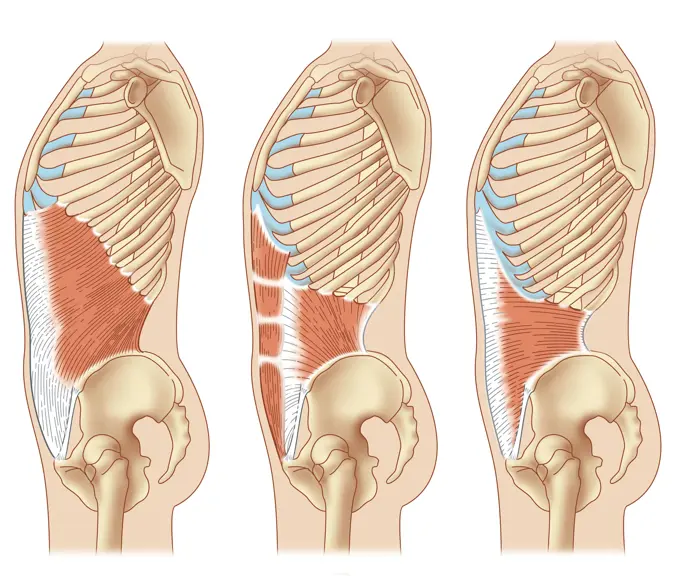
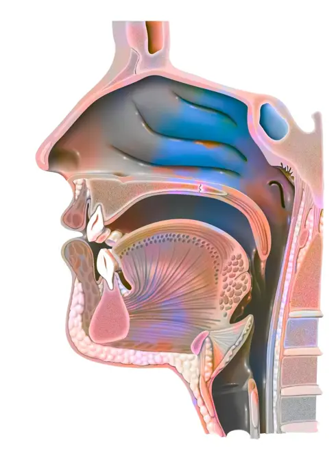
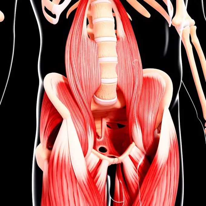
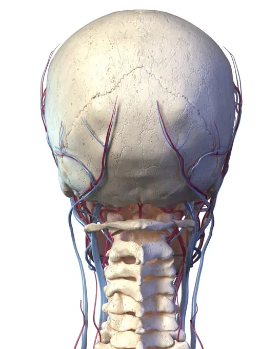
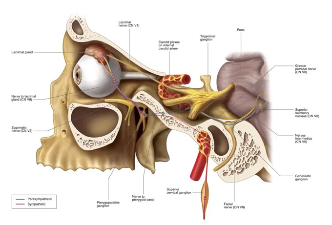
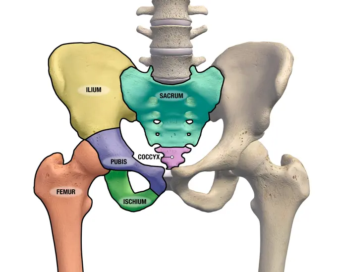
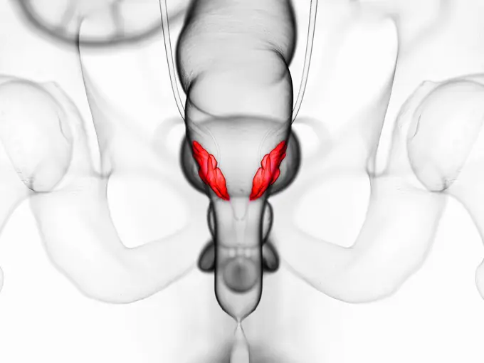
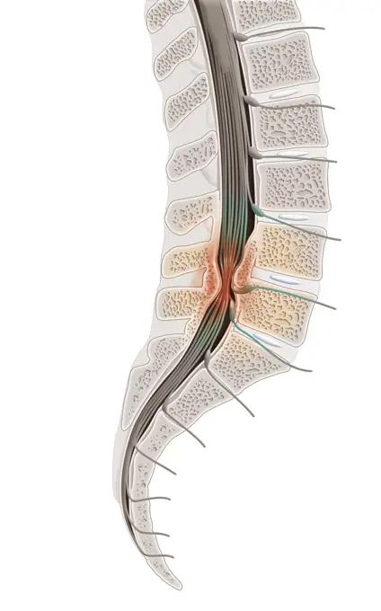
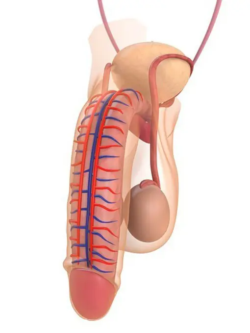
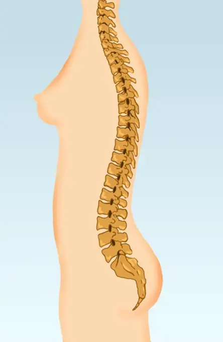
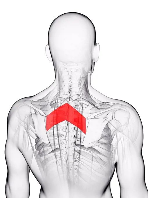
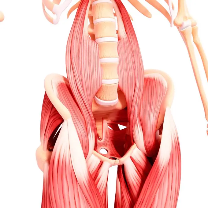
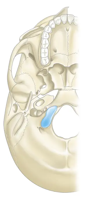
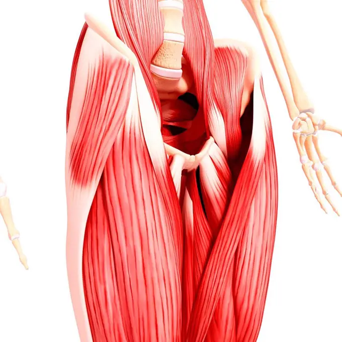
Fine ArtVintageFootageSubscriptionBoardsFree ResearchPricing
  • Fine Art
  • Vintage
  • Footage
  • Subscription
  • Boards
  • Free Research
  • Pricing

Internal Human Anatomy

Illustrations depicting various internal body structures including the spine, ribs, and musculature, emphasizing human anatomy in detail.

Human hip musculature, computer artwork.
194 assets in this story
824-63164154
Mediastinum in sagittal section with the rib cage, the sternum.
4128R-31973
Human hip musculature, computer artwork.
4128R-11288766
Male anatomy, computer artwork.
824-63195397
Illustration in the foreground of the oblique rectus muscle, covering the inferior oblique muscle, then the transverse and abdomen in the background.
4128R-37153
Human musculature, computer artwork.
824-63191332
Illustration of a herniated disc at L5 and S1, compressing S1s nerve root. The pedicles are illustrated in cross-section to show the vertebral hole and the passage of the nerve roots.
4128-28968661
Fracture of the femur neck, illustration
4128R-33412
Human musculature, computer artwork.
4128R-32845
Human hip musculature, computer artwork.
824-63164211
Upper airways showing the larynx, epiglottis.
4128R-34985
Female anatomy, computer artwork.
4128R-32921
Human arm musculature, computer artwork.
4128R-33016
Human hip musculature, computer artwork.
824-63186165
Anterior view of the abdominal lining with weak points potentially leading to inguinal hernias.
1899-53509323
Inferior view of the human skull with the upper jaw.
824-63164656
Inferior view of the human skull with the upper jaw.
4128R-13022375
Human abdominal muscles, illustration.
4128R-36546
Human pelvic bones, computer artwork.
4128R-31772
Human leg musculature, computer artwork.
4128-15665699
Lumbar spine, illustration
1525-27646751
Sciatica pain symptoms and diagnosis medical concept as a disease causing physical problems with 3D illustration elements on a white background.
4128-19248795
Facial nerve, illustration.
1525-56722374
Organs surrounding the female reproductive organ 3D rendering. Organs surrounding the female reproductive organ
1899-53513215
Posterior view of the larynx and vocal cords (bone, muscles and cartilages).
824-63163680
Anatomy of the bull's reproductive system with kidneys, bladder, ureters.
4128R-35860
Human leg musculature, computer artwork.
4128-19248798
Facial nerve, illustration.
824-63184533
Illustration of endometriosis and organs affected by this illness. Endometriosis is the presence of cells from the uterus lining outside the uterine cavity.
1899-53509008
Anatomy of the reproductive system of a male pig.
4128R-32172
Male cardiovascular system, computer artwork.
4128R-11316222
Human pelvic bones, computer artwork.
4128-19056405
Sacrum, illustration
4128R-32714
Human spine, computer artwork.
4239-18641172
Digital illustration of muscles of the human torso, anterior view (no labels).
4128R-33415
Human musculature, computer artwork.
824-63168357
Anterior view of the deep plane of the musculature of the back: diaphragm, square muscle of the loins, psoas muscle, iliac muscle.
1848-61341004
Woodcut, Opossum shrimp (Mysidacea)
4128R-11544519
Human intestine, computer illustration.
4128R-32941
Human pelvic bones, computer artwork.
4128R-11541852
Human arm muscles, computer illustration.
4128R-33437
Human neck musculature, computer artwork.
824-63163792
Bone system: fracture of the neck of the femur, linked to osteoporosis.
1525-23135397
science anatomy of human body in x-ray with glow kidneys
4128R-33137
Human musculature, computer artwork.
4128R-36888
Human skeleton, computer artwork.
824-63168377
The spinal cord, made up of gray matter surrounded by white matter, is contained in the spinal canal formed by the vertebrae and the intervertebral discs, forming the vertebral column.
4128R-12964987
Human skeletal system, Illustration
4128R-11477589
Human skeletal system, computer artwork.
4128R-36071
Male pelvic bones, computer artwork.
4128R-36999
Human skeleton, computer artwork.
4128R-22760
Male musculature, computer artwork.
4128R-34313
Human leg musculature, computer artwork.
4128R-36971
Human skeleton, computer artwork.
4128-15666195
Clavicle bone, illustration
4128R-32633
Human leg musculature, computer artwork.
824-63168358
Lateral views of the abdomen where the wall consists of three side muscles (external oblique muscle, internal oblique muscle, transverse muscle) and an anterior muscle (rectus abdominis).
1899-30609124
Anatomy of nasopharynx with nasal cavity, oral cavity.
4128R-34817
Human musculature, computer artwork.
4239-18642129
3D illustration of the vascular system of the human head, rear view on white background.
1525-56234271
The urinary bladder is surrounded by the muscles that make up the abdominal wall and pelvic floor. 3D rendering. The urinary bladder is surrounded by the muscles that make up the abdominal wall and pelvic floor.
4128R-19177
Knee anatomy, computer artwork.
4128R-36349
Human arm musculature, computer artwork.
824-63195448
Illustration of a retracted feline claw. See illustration 014337_007 to compare with an extended claw.
1899-53509405
Path traveled by information sent to the brain through the eyes.
4128R-15692
Healthy kidneys, computer artwork. Also seen are the abdominal aorta red and vena cava blue.
4128R-11316359
Male pelvic bones, computer artwork.
4239R-20482927
Anatomical pathways of innervation to the lacrimal gland.
4128R-15582350
Illustration of the human knee anatomy
1525-56722380
The last two sections of the large intestine are the sigmoid colon and rectum. 3D rendering. The last two sections of the large intestine are the sigmoid colon and rectum.
1899-53509429
Position of the mouth for the pronunciation of the 'A' in phonation.
824-63164998
Position of the mouth for the pronunciation of the 'A' in phonation.
1428-67034176
3D Rendering of Pelvis and Hip bones with major anatomical regions mapped and labeled on a white background.
824-63168360
Rectus abdominis muscle, linea alba, transverse muscle, internal oblique muscle and external oblique muscle.
4128-30416848
Seminal vesicles, illustration
1899-54027778
Illustration of spinal stenosis in L4 and L5. Spinal stenosis is a pathology located in the lumbar spinal column where the cauda equina nerves pass. It is caused by an intrinsically narrow vertebral canal or a degenerative narrow canal related to a herniated disc, an increase in the volume of rear joints and ligaments that join the vertebrae together, or an instability in the vertebrae with vertebral displacement (or degenerative spondylolisthesis), which decreases the diameter of the canal, compressing the nerves, causing lumbar pain in the lower limbs.
1525R-178743
4128-30418304
Deep muscles of the lower body, illustration
4252-4283
Vertebral column
4128R-26604
Back muscle. Computer artwork showing the rhomboid major muscle.
4128R-36857
Human musculature, computer artwork.
824-63186166
Lower left side of the skull.
4128R-34923
Human musculature, computer artwork.
4128-15665598
Seminal vesicles, illustration
4128R-13386665
Illustration of spinal cord anatomy.
824-63190783
Illustration of a paravertebral block (blue area), locoregional anaesthesia in the thoracic and lumbar regions.
1899-13244
Capsule of the wrist
1525-56722539
The bladder is supplied with oxygenated blood mainly by the superior and inferior vesical arteries 3D rendering. The bladder is supplied with oxygenated blood mainly by the superior and inferior vesical arteries
1848-49200679
Medicine, human anatomy, the circulatory system, blood circulation: section through the back of the leg, with arteries and veins shown in red and blue. Coloured lithograph by J. Maclise, 1841, 1844, Historic, digitally restored reproduction from a 19th century original
824-63163885
Sagittal section of the left kidney with the renal arteries and veins.
4128R-13586548
Illustration of lumbar vertebrae.
4128-30421600
Lumbar spine, illustration
824-63187034
Illustration of a herniated disc comparing a healthy disc to a disc that has produced a hernia. This disc is composed of a fibrous ring on the outside, and a gelatinous core on the inside. When the core becomes fissured due to a micro-blow, the cores gelatine leaks into the rachidian canal and forms a ball (hernia), that compresses the nerve roots.
4128R-12577025
Male nervous system, computer illustration.
4128R-25596
Male anatomy, computer artwork.
1525-56722376
The last two sections of the large intestine are the sigmoid colon and rectum. 3D rendering. The last two sections of the large intestine are the sigmoid colon and rectum.
4239R-20482699
Anterior neck and facial muscles of the human head (with labels).
1899-30609343
Inner ear and zoom on the macule (organ of static balance).
4239R-8066
Male muscle anatomy of the abdominal wall.
4128R-3505
Human anatomy, artwork.
824-63187030
Illustration of a herniated disc which compresses and swells the nerve root of the sciatic nerve.
PREVIOUS
of 2
NEXT
2401 S. Ervay, Suite 206
Dallas, Texas 75215
United States
Browse
CategoriesGalleriesFree Research
For Creators
License Your ContentSuperStock For StudentsFrame of Mind BlogContributor PortalNew Image SpotlightLicense AI Imagery
Resources
About UsLicensingTerms & ConditionsAPI AccessPrivacyPricing
Contact
+1 866 236 0087help@superstock.com Contact form
©2026 SuperStock. All Rights Reserved. -A
This site is protected by reCAPTCHA and Google Privacy Policy and Terms of Service apply.