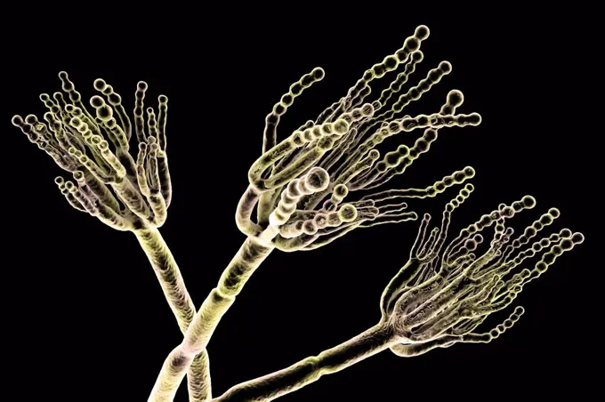
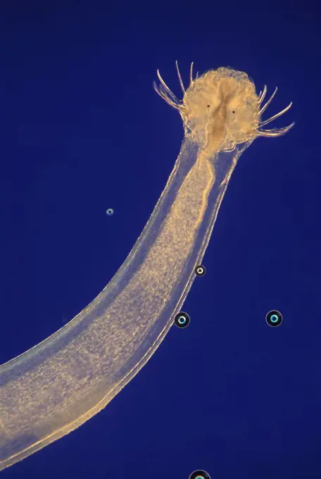
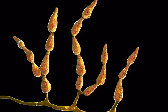
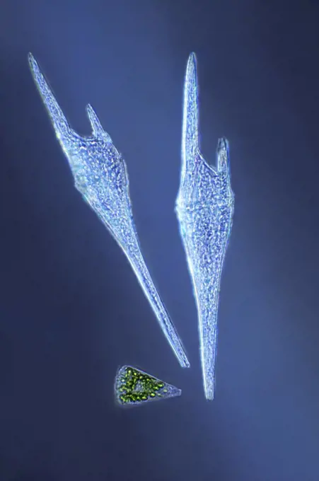
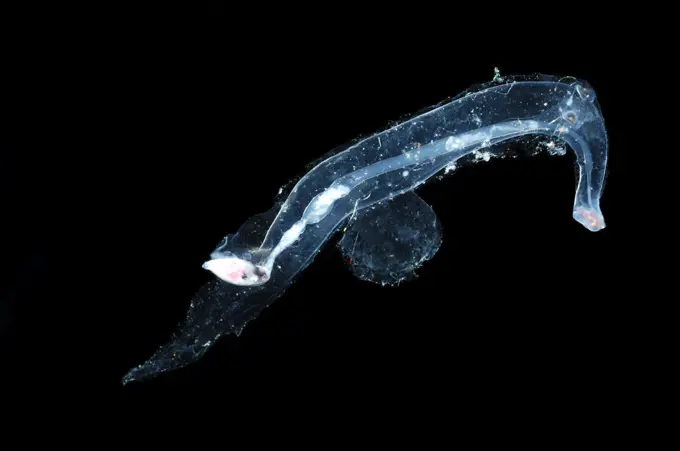
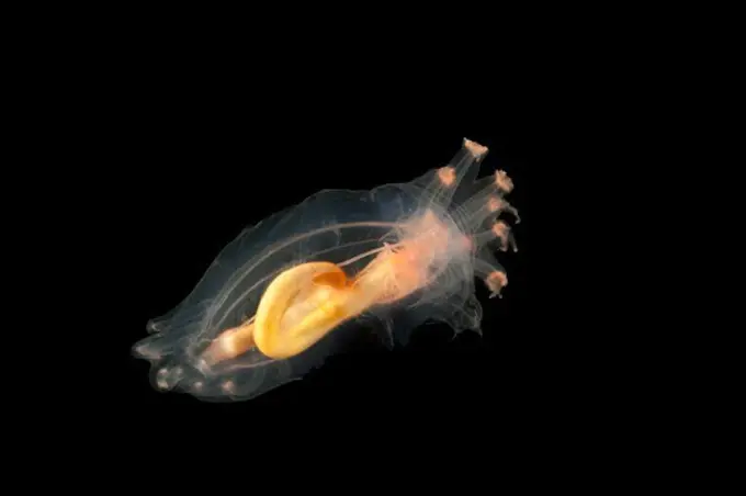
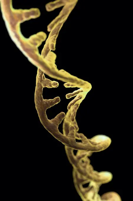
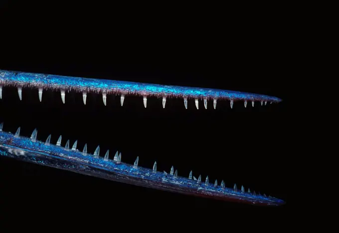
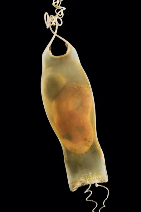
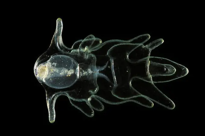
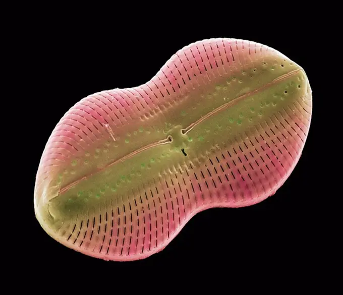
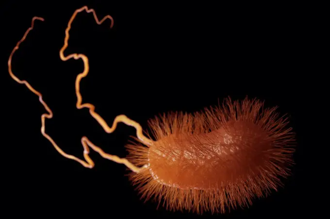
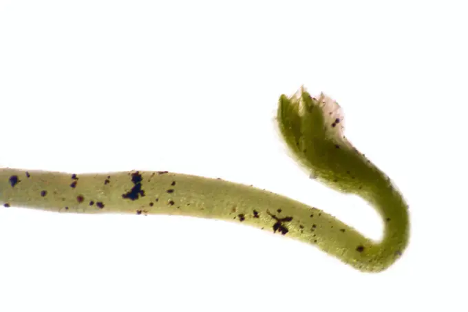
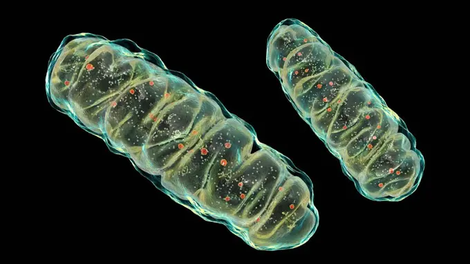
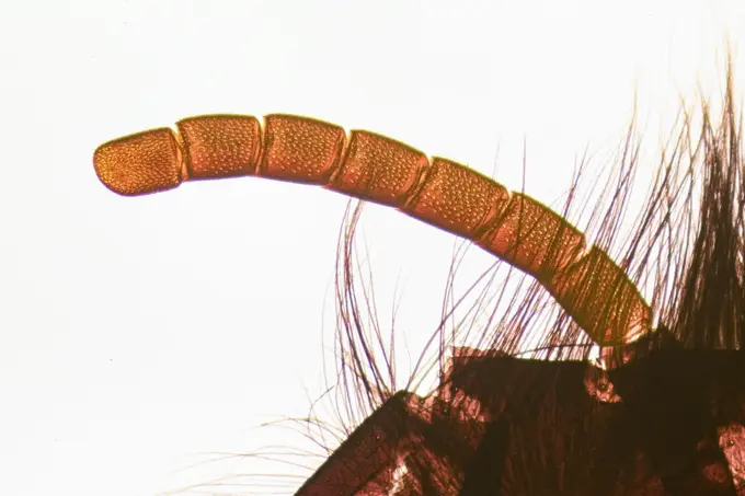
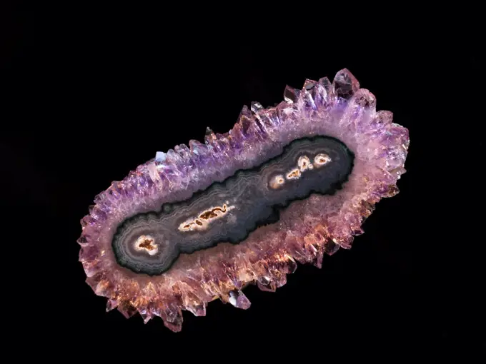
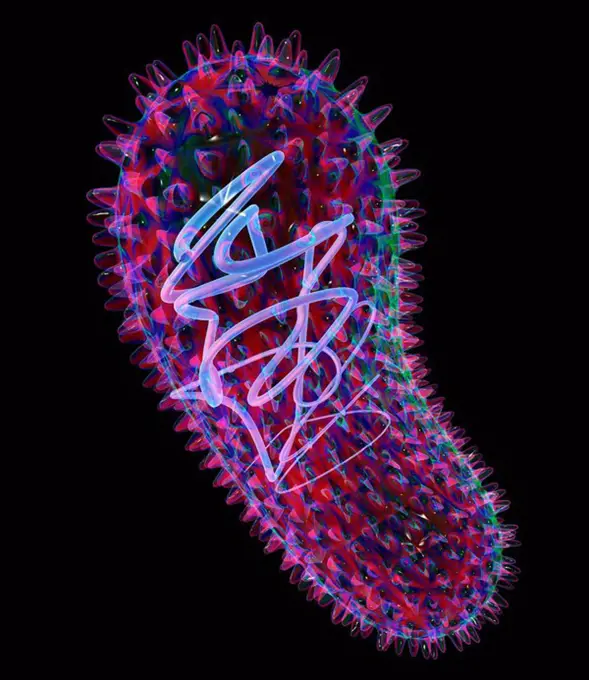
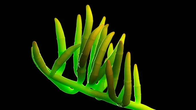
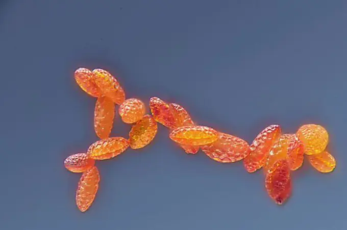
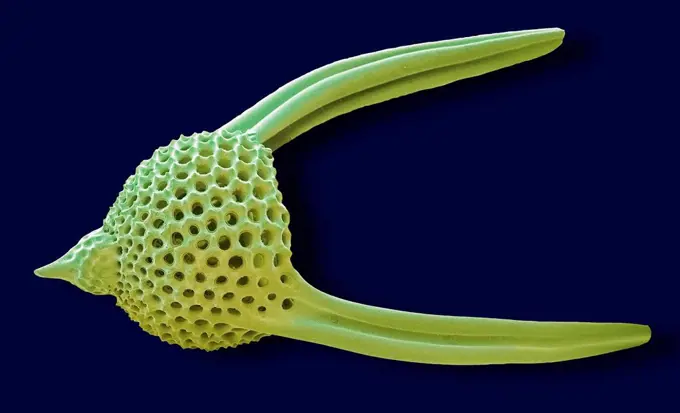
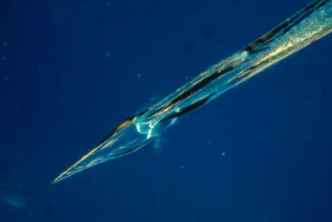
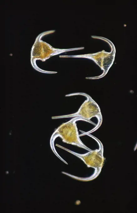
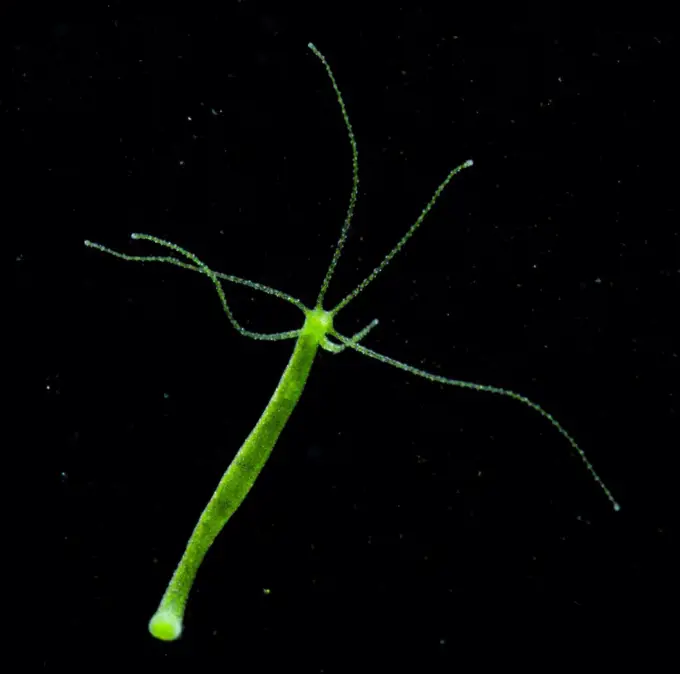
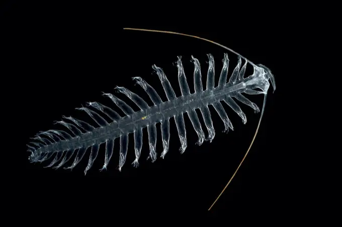
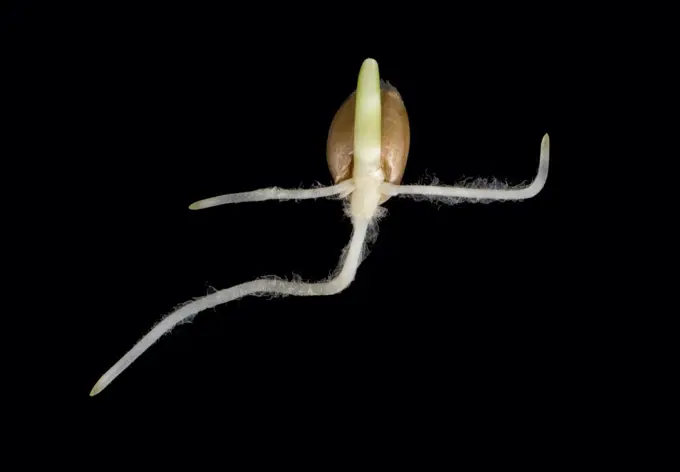
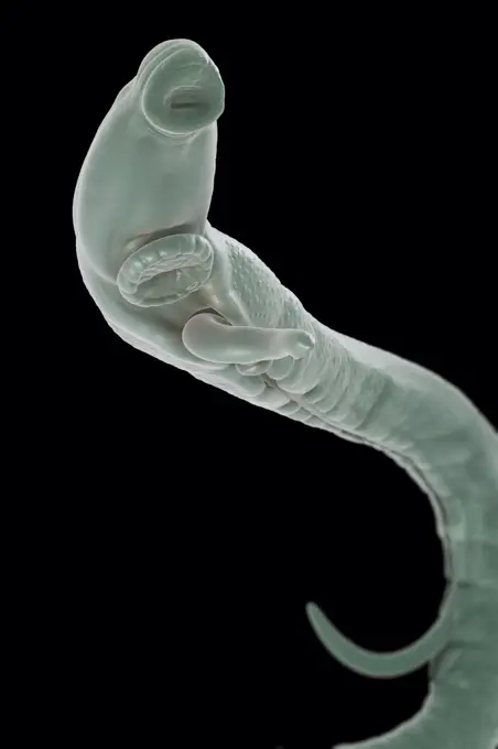
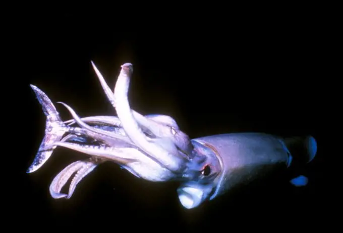
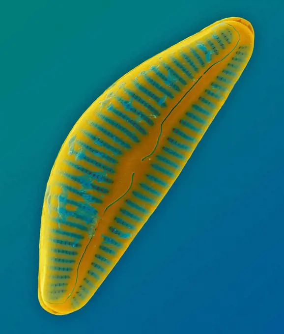
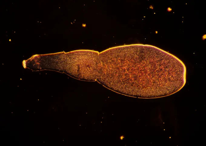
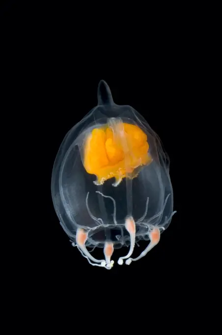
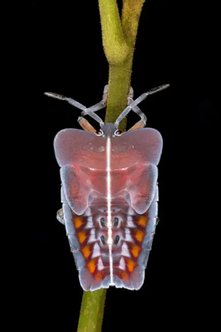
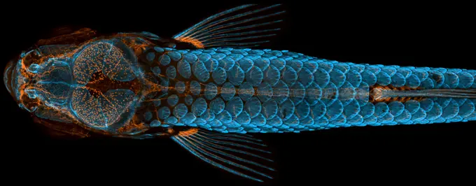
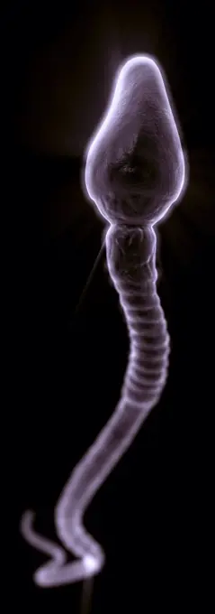
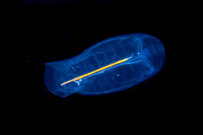
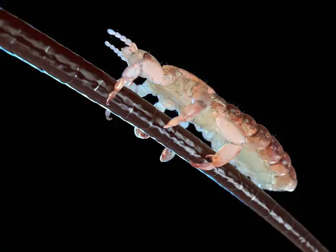
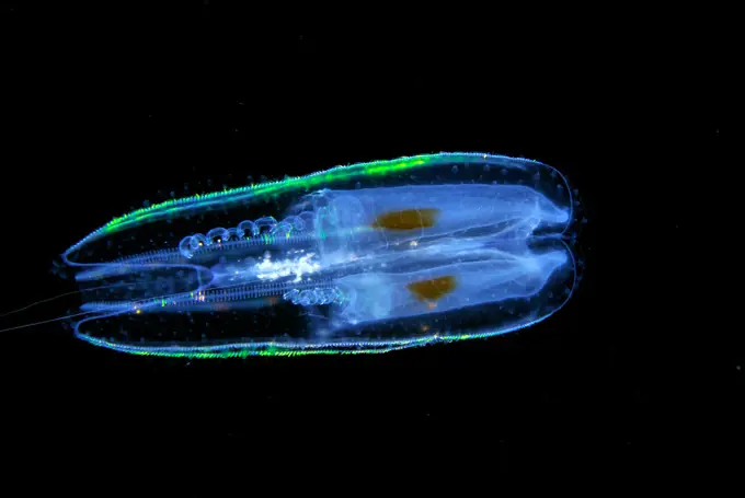
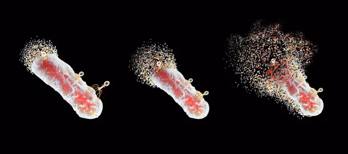
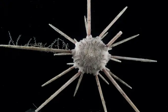
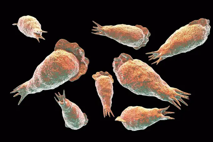
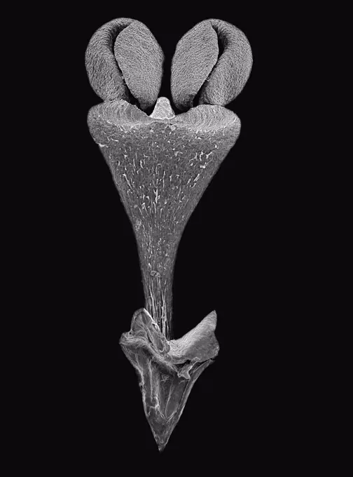
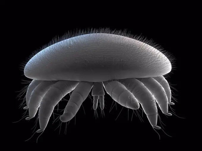
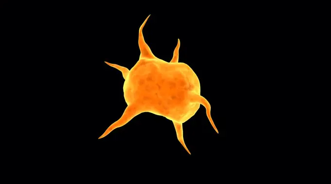
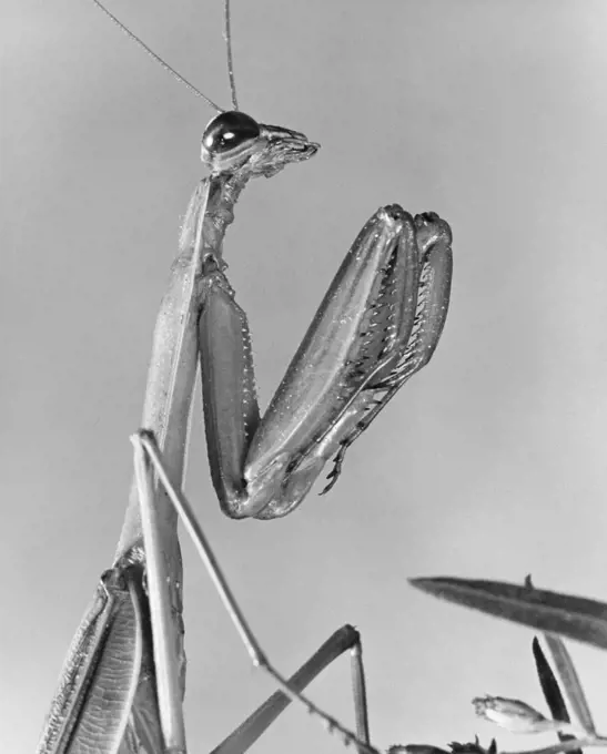
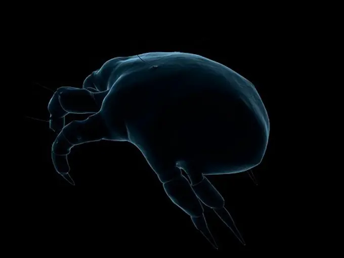
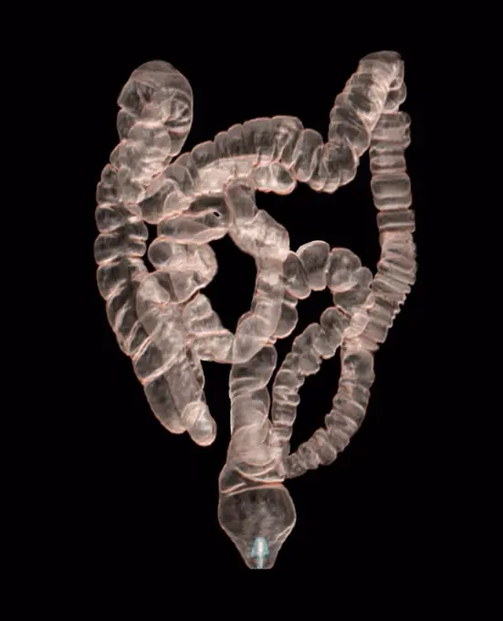
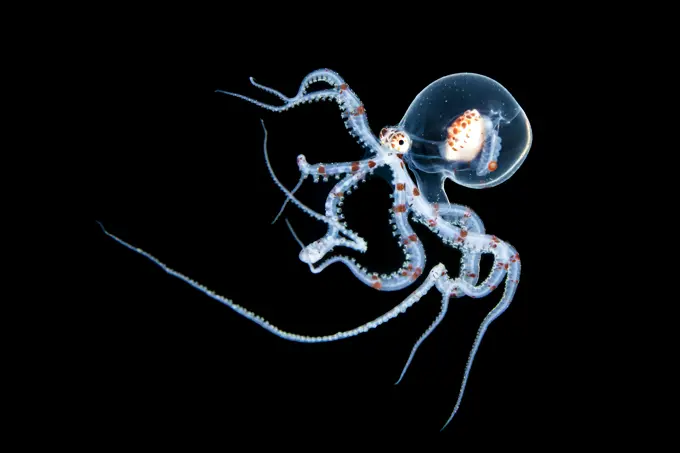
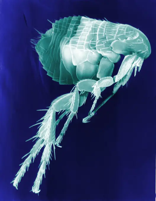
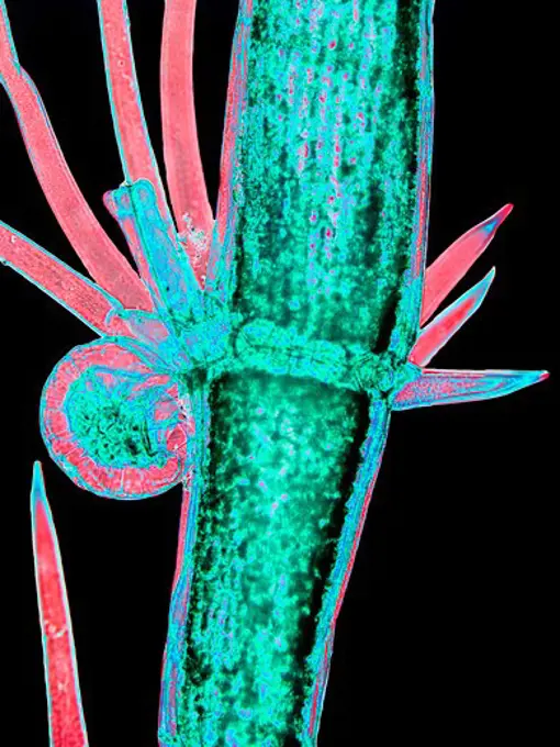
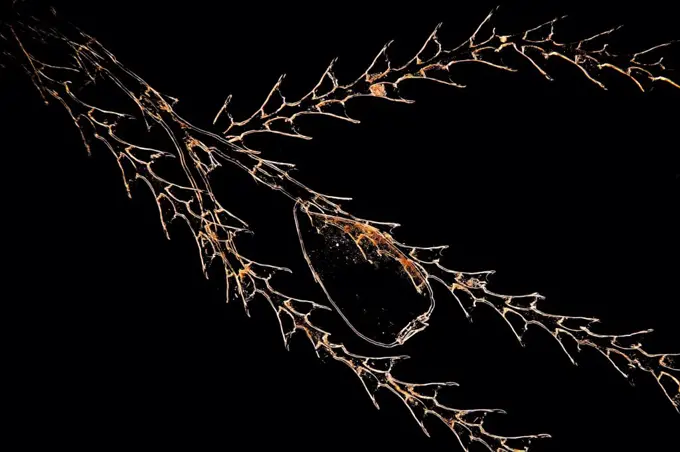
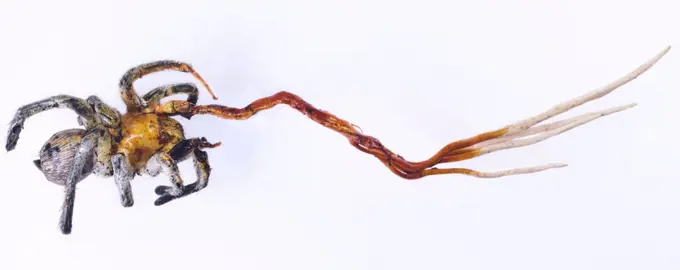
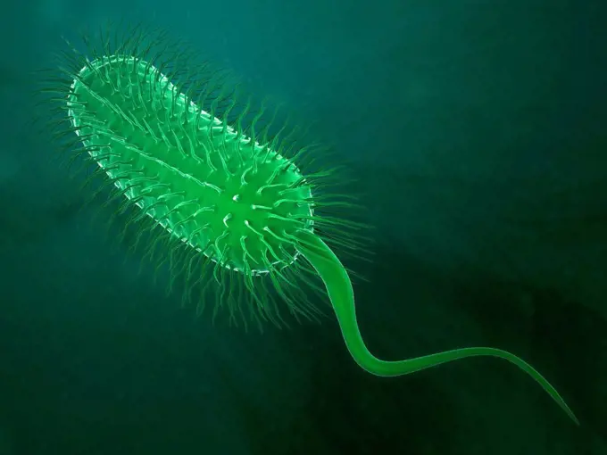
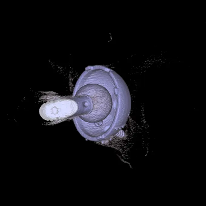
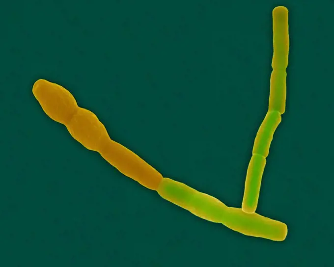
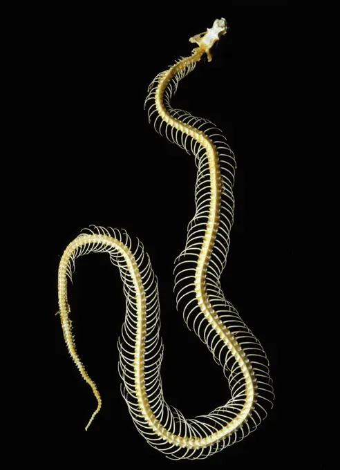
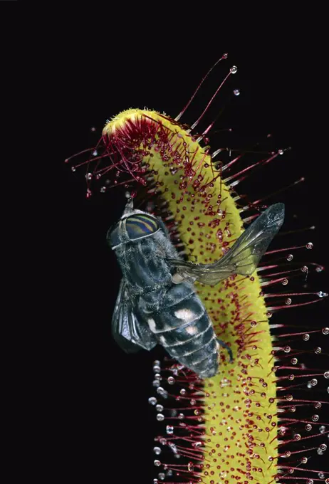
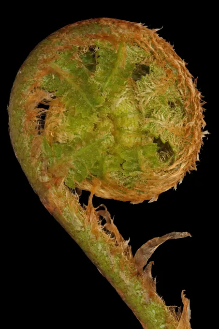
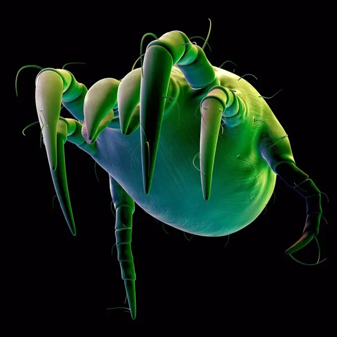
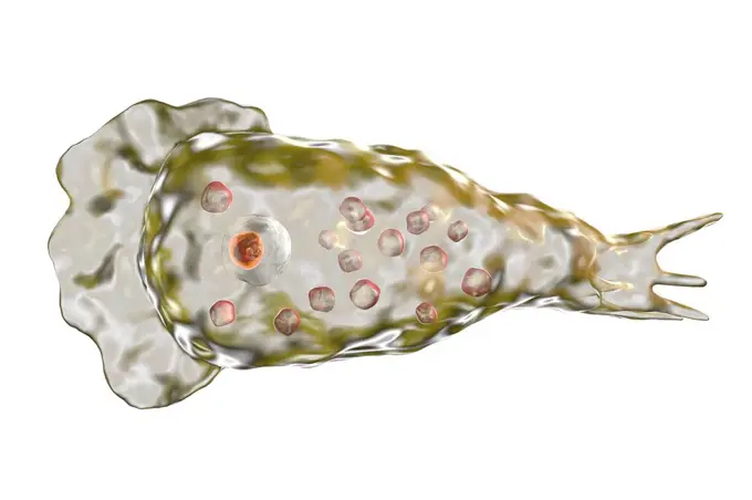
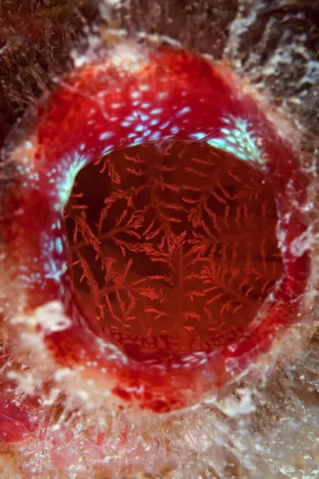
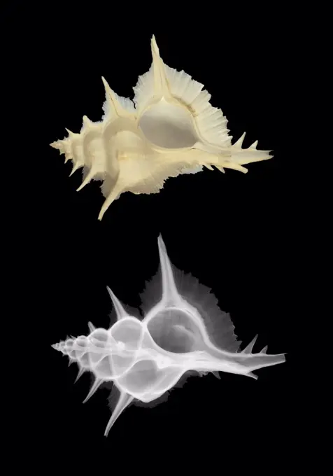
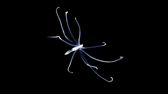
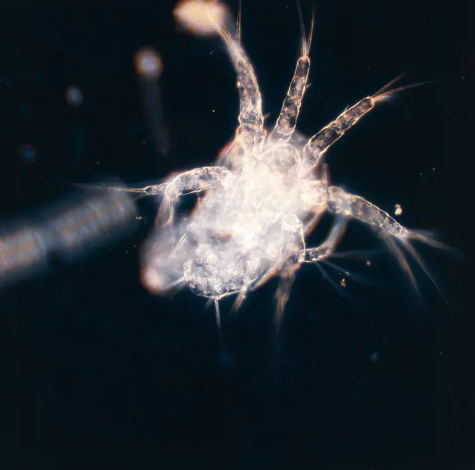
Fine ArtVintageFootageSubscriptionBoardsFree ResearchPricing
  • Fine Art
  • Vintage
  • Footage
  • Subscription
  • Boards
  • Free Research
  • Pricing

Marine Microorganisms

Magnified images of marine life including arrow worms, dinoflagellates, and unique deep-sea mollusks, showcasing intricate details and vibrant colors.

Penicillium fungus, illustration
241 assets in this story
4201-66123
Arrow Worm (Sagitta sp), Spain
4128-111612470
Filamentous allergenic fungus Alternaria alternata, computer illustration. Alternaria is a dematiaceous (phaeoid) fungus commonly isolated from plants...
4201-21586614
Dinoflagellate (Ceratium sp) group, magnified 400x, Barcelona, Spain
4070-19578733
Sea elephant, marine pelagic gastropod mollusc in the family Pterotracheoidea. Deep sea heteropod. Controlled conditions.
4115-6001
Deepsea benthic Sea cucumber (Peniagone diaphana ) from mid atlantic ridge, depth approx 2500m. Specimen caught swimming above sea floor, June 2010
4128R-14768502
Penicillium fungus, illustration
4128-V58573771
4128-V58562084
4128-V58573315
4128-17809992
Moina water flea, light micrograph
4378-1174
Stylized view of strands of human DNA or deoxyribonucleic acid.
4413-176315
Mouth of Red Sea Houndfish Red Sea
4201-61037
Small-spotted Catshark (Scyliorhinus canicula) embryo in lucent mermaid's purse, North Sea, Germany
4115-6240
Sea star / Common starfish (Asterias rubens) at brachiolaria larva stage (60 days old), digital focus stacking
4128R-2398
Diatom alga, SEM
4128-V58573603
4128-V58573373
4378-1194
Stylized view of an Escherichia coli or E.coli bacterium.
6188-54819722
Sprout, stem in particles of earth and unfolding leaves. Ultra macro Sprout, stem in particles of earth and unfolding leaves - germinant. The development of living things. Ultra macro, isolated on white Copyright: xZoonar.com/MaximilianxBuzunx 13655588
4115-5360
Giant ostracod Gigantocypris mulleri} from between 718 and 1864m, Mid-Atlantic Ridge, North Atlantic Ocean
4128-20040347
Mitochondria, illustration
4128-V58573912
4413-28973101
Honey bee (Apis mellifera) detail of an antenna seen under the microscope
4421-24385
Crab - Shore (Carcinus maenas) 1st Zoea larva lateral, X 20
4332-2404
Polished slice of amethyst crystals and agate cut from a geode mined in Artigas, Uruguay.
4141-111428966
Light micrograph whole mount of a Planarian flatworm showing the injected digestive tract, UK
4128R-4723
Bacillus bacteria, artwork.
4128-19053747
Penicillium, illustration
4128-V58573347
1848-51667540
Pollen of a lily, enlarged about 200x
4128R-11310480
Lychnocanoma radiolarian. Coloured scanning electron micrograph (SEM) of the shell of a Lychnocanoma sp. radiolarian. Radiolaria are single-celled pro...
4141-16162
textile cone shell conus textile poison dart
4201-28451
Dinoflagellates (Ceratium tripos) five specimens underwater
4201-89788
Hydra (Hydra viridis), England
4128-V58573776
4201-61038
Small-spotted Catshark (Scyliorhinus canicula) embryo in lucent mermaid's purse, North Sea, Germany
4201-80048
Polychaete (Tomopteris sp) showing parapodia, paddle-like structures, Weddell Sea, Antarctica
4128-V58573592
4421-21545231
Germinating seed of winter wheat, Triticum aestivum, with radicle, root hairs and coleoptile growth developing
4378-3534
Schistosomiasis also known as snail fever is a parasitic disease caused by the parasitic worm of the genus Schistosoma. An adult male and female are seen here.
4179-75665
Giant Squid eats Baitfish (Dosidicus gigas) Mexico
4128R-6192
Diatom, SEM
4128R-13620028
Coloured scanning electron micrograph (SEM) of marine pennate diatom frustule (Cymbella sp.). The genus Cymbella is an asymmetrical biraphid, pennate ...
4128-V58573313
4128-V58573870
4239-V53647259
4220-21334550
Light Micrograph: Tapeworm.     Date:
4201-80026
Jellyfish (Leuckartiara sp), Weddell Sea, Antarctica
1525-27028633
Tessaratomids resemble large stink bugs and are sometimes quite colorful, Amboli, India
1916-111302566
Hydromedusa, Kona, Hawaii, USA
4128-V58573581
824-63227436
This photo uses confocal microscopy and merges 350 individual images to reveal a top view of a zebrafish with a fluorescent and quottagged and quot skeleton, scales and lymphatic system. The image was awarded by NIH researcher Daniel Castranova and won 1st place in Nikon's 2020 photomicrography competition.
4378-3338
A single male sperm cell.
4128-30418625
Fusarium fungus, illustration
1889-49004380
This Pelagic tunicate or salp (Salpa aspera) is part of the Salpidae family of gelatinous animals; Yap, Federated States of Micronesia
4128-15979366
Head louse, computer illustration.
1916-111302565
Comb jelly, a pelagic ctenophore, at night, Kona, Hawaii, USA
4128R-13586603
Bacteriophages infecting bacteria, computer illustration. A bacteriophage, or phage, is a virus that infects bacteria. It consists of an icosahedral (...
4128-V58566855
4128-16224868
Sugar bush flower (Protea sp.), X-ray
1525-21054167
milk splash
4413-89028
Young Lancelet
4128R-12573047
Diatom. Coloured scanning electron micrograph (SEM) of a single diatom. Diatoms may be extremely abundant in both freshwater and marine ecosystems; it...
4201-66220
Fern (Nephrolepis sp) leaf showing clusters of spores, Venezuela
4128-V58573301
4070-21283246
Pencil urchin (Cidaroid). Collected from coral sea mount near Dragon vent field on SW Indian Ridge, Indian Ocean.
4128R-14845723
Naegleria brain-eating amoeba, illustration
4201-66331
Orchid (Dendrobium sp) SEM close-up view of stamen at 14x magnification
4128-V58573601
4128R-14511
Honeybee mite Varroa destructor, artwork, artwork.
4239R-20483441
Conceptual image of a platelet.
990-1733
Close-up of a Praying Mantis (Mantis religiosa)
1525R-178734
4128R-14901
Normal intestines. Coloured 3D computed tomography CT scan of the healthy intestines of 38 year old patient.
4128-19249831
Tick, illustration.
4070-69335070
Wunderpus (Wunderpus photogenicus) larval stage, drifting at night in open ocean as part of the plankton, North Sulawesi, Indonesia, Lembeh Strait.
824-63128608
Scanning Electron Micrograph of a Flea.
4298-1005
Chara algae showing stem with oogonium and antheridiium, magnification 40x, image enhanced
4413-174720
Hydridae by light microscopy
4201-22097127
Sac Fungus (Cordyceps caloceroides) which has infected a spider, Yasuni National Park, Ecuador
4128R-13591269
Bone cancer cell, illustration.
4128R-22291
Bacterium, computer artwork.
4269-25440
Prosthetic hip. 3D computed tomographic (CT) scan reconstruction of a ball-and-socket prosthetic joint used to replace the old hip joint. The upper 'ball' end swivels in the artificial cavity surgically implanted into the pelvis (seen here). The other end extends down into a hole drilled into the femur (thigh bone) (not seen).
6188-56079375
Golden Pancreas, 3D rendering Golden Pancreas, 3D rendering isolated on white background Copyright: xZoonar.com/alexlmxx 21252080
4128-V58557938
4128R-13620095
Coloured scanning electron micrograph (SEM) of Bacillus cereus, Gram-positive, encapsulated, rod prokaryote (vegetative stage, dividing); causes food ...
1525-25877915
Enlarge Cactus germination, Macro shot close up
4220-20119156
Snake Skeleton
4128-V58573778
4201-77650
Cape Sundew (Drosera capensis) with trapped Horse Fly (Tabanus bovinus), native to Cape Province, South Africa
6188-65083911
Straussenfarn Matteuccia struthiopteris Straußenfarn Copyright: xZoonar.com/GerdxHerrmannx 9057496
4201-53024
Mexican Axolotl (Ambystoma mexicanum) egg on day nine of development, native to Mexico, sequence 6/13
4128R-13384925
Dust mite, illustration.
6188-55585082
Moss protonemata micrograph Light photomicrograph of Moss protonemata whole mount seen through microscope Copyright: xZoonar.com/ClaudioxDiviziax 7887071
4201-80016
Copepod (Paraeuchaeta sp) female with eggs swimming, Weddell Sea, Antarctica
4128R-14845718
Naegleria brain-eating amoeba, illustration
4379-444
Abstract of an ascidian, Mabul, Sabah, Malaysia.
4070-9483
Photograph and X-ray of shell of the Alabaster murex Murex alabaster}
4239-69140954
Tube-dwelling anemone larva (Ceriantharia).
1788-111174183
Nauplius of water flea (Cyclops), copepods, seen under a microscope.
PREVIOUS
of 3
NEXT
2401 S. Ervay, Suite 206
Dallas, Texas 75215
United States
Browse
CategoriesGalleriesFree Research
For Creators
License Your ContentSuperStock For StudentsFrame of Mind BlogContributor PortalNew Image SpotlightLicense AI Imagery
Resources
About UsLicensingTerms & ConditionsAPI AccessPrivacyPricing
Contact
+1 866 236 0087help@superstock.com Contact form
©2026 SuperStock. All Rights Reserved. -A
This site is protected by reCAPTCHA and Google Privacy Policy and Terms of Service apply.