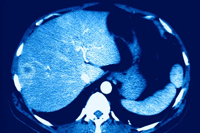
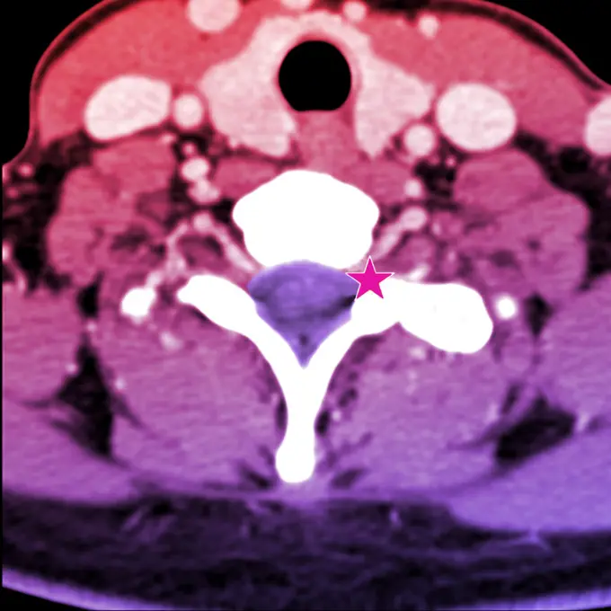
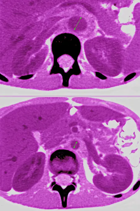
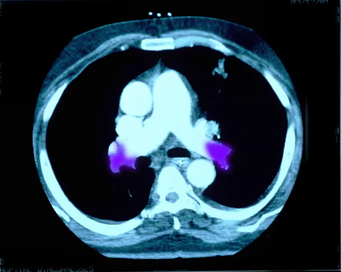
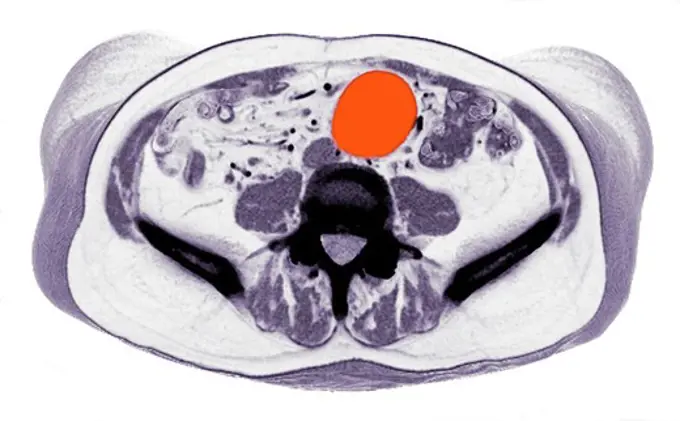
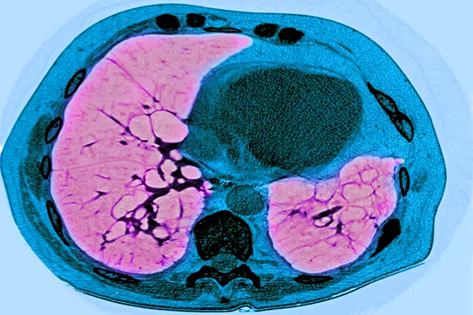
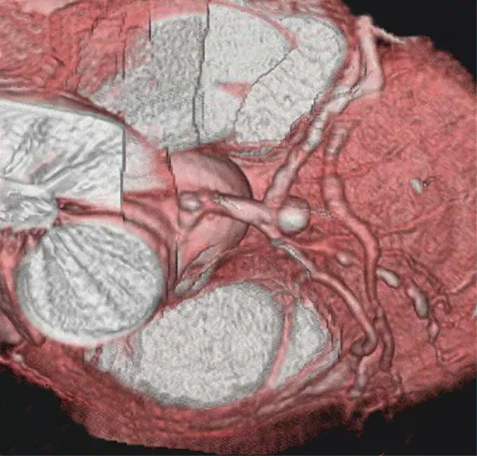
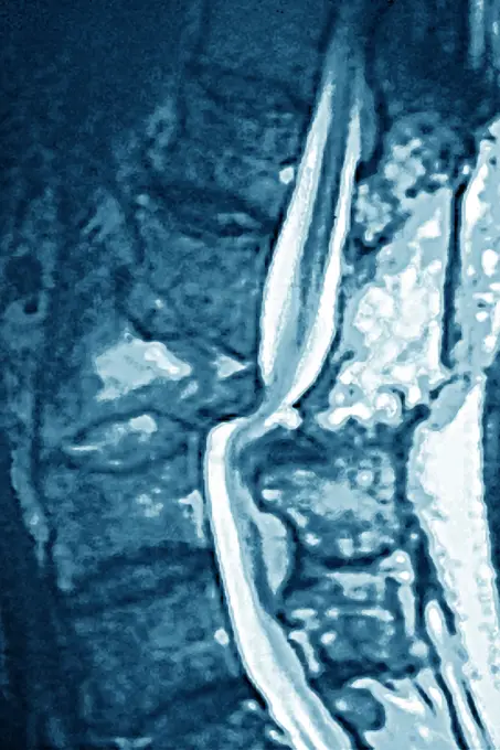
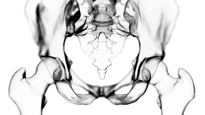
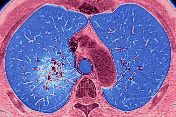
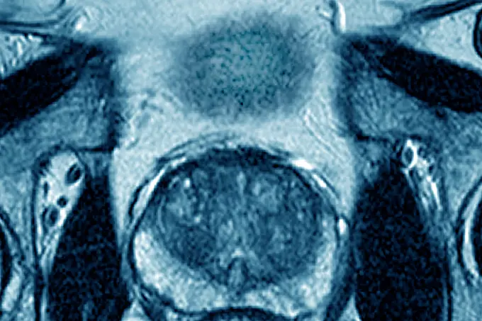
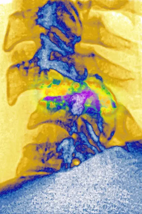
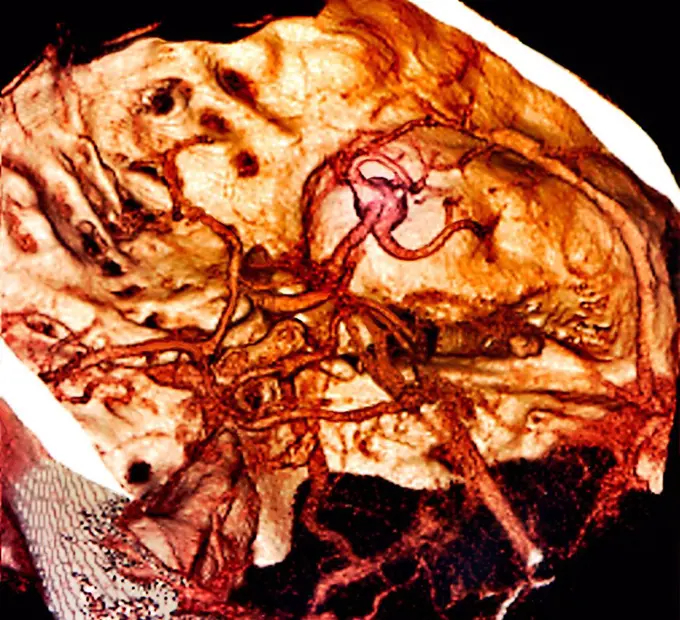
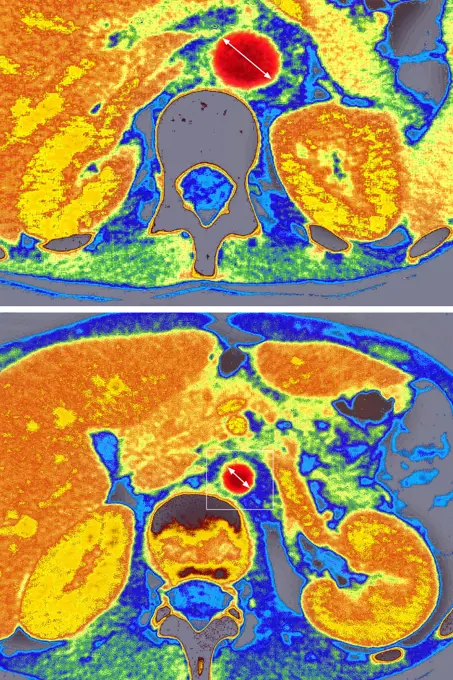
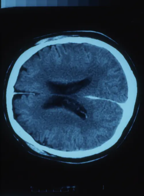
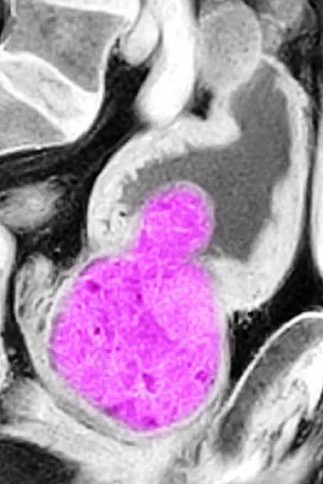
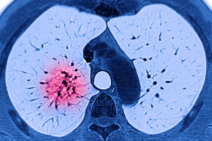
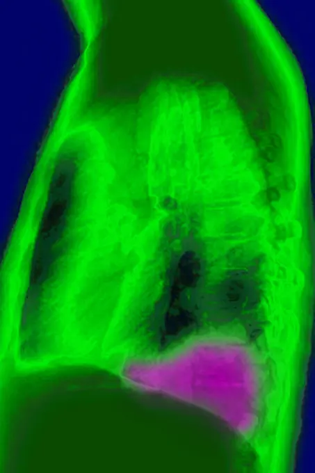
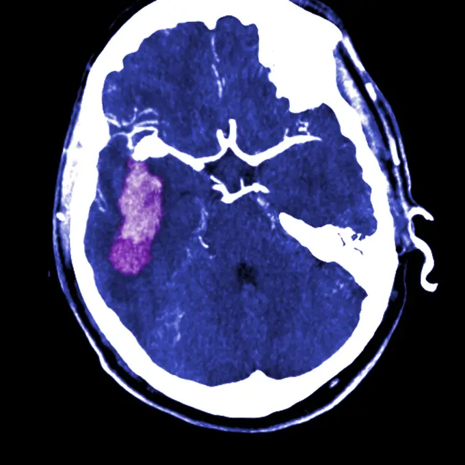
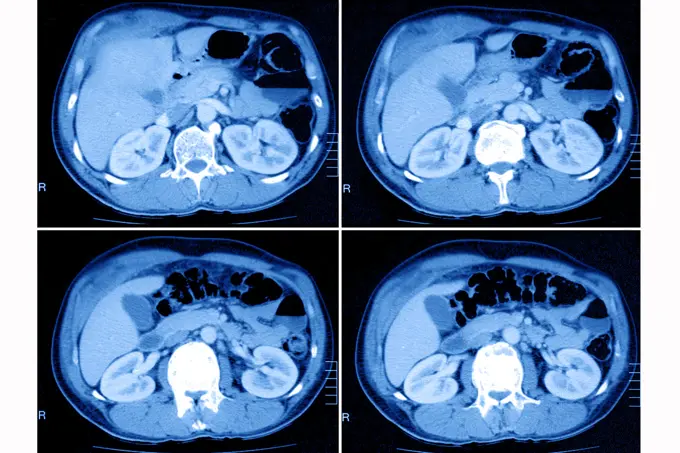
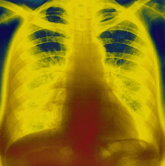
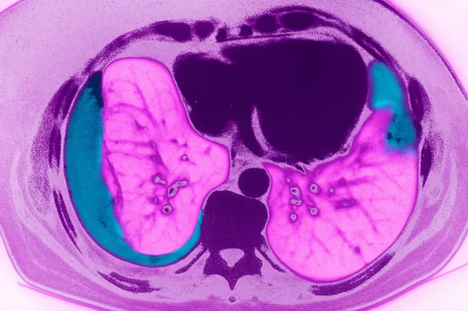
Fine ArtVintageFootageSubscriptionBoardsFree ResearchPricing
  • Fine Art
  • Vintage
  • Footage
  • Subscription
  • Boards
  • Free Research
  • Pricing

Medical Imaging and Diagnostics

Enhanced CT and MRI scans highlighting medical conditions such as stents, lung cancer, and vertebral issues, with color-coded identifiers for clarity.

Lung cancer located in the upper right lobe. Frontal chest x-ray.
207 assets in this story
4269-25389
Imroperly placed Stent prosthesis. 3D computed tomographic (CT) scan reconstruction showing a stent prosthesis that 'collapsed' on itself when inserted in the vessel.
824-63225993
Lung cancer located in the upper right lobe. Frontal chest x-ray.
4269-27230
Vertebral infiltration. Color enhanced axial Computed Tomography (CT) scan of a lumbar vertebra with arthrosis treated by infiltration of anti-inflammatory drug (corticoids) around the spinal cord and spinal nerve. The needle, which route is marked in red, will be inserted into the vertebral foramen (foraminal infiltration), but throught the vertebral canal.
4269-27218
Intestinal obstruction. Color enhanced sagittal Computed Tomography (CT) scan of the abdomen showing a volvulus of the colon responsible for a bowel obstruction. It is evidenced by the winding of the colon loops (pink circular shape) around their axis (in black at center of the loops).
1899-54028003
Ponytail syndrome (Herniated disc - medullary compression below the second lumbar vertebra). Cross section vertebral scanner.
1899-54027133
Cancer of the liver, seen in a radial cross-section abdominal scan.
4269-27493
Shoulder chondrosarcoma. Colored enhanced Axial Computed Tomography (CT) scan of the shoulder showing a chondrosarcoma of the shoulder blade. The tumor is evidenced by the presence of a heterogenous lytic area with bone calcifications. The black round shape at right is the head of the humerus inserted into the shoulder blade.
824-63205967
Adenopathy (liver lymph gland). Cross-section abdominal scan.
4269-27233
Vertebral infiltration. Color enhanced axial Computed Tomography (CT) scan of a cervical vertebra with arthrosis treated by infiltration of anti-inflammatory drug (corticoids) around the spinal cord and spinal nerve. The needle will be inserted into the vertebral foramen (foraminal infiltration). The insertion point is market with a target (purple star).
824-63206984
Takayasus arthritis, seen by 2 radial section abdominal scanners, at the descending aorta.
4269-25290
Pulmonary embolism. Coloured axial computed tomography (CT) scan of a pulmonary embolism. Two blood clots (purple) that traveled to the lungs from another part of the body block the two main pulmonary arteries.
1899-53511719
Benign prostatic hypertrophy, visualized by radial MRI of the pelvis. (Photo by: CAVALLINI JAMES/BSIP/UIG)
4269-24948
Intracranial aneurysm. Three-dimensional computed tomographic upper view of the base of a skull showing a 9 mm diameter intracranial aneurysm (red ballon shaped swelling) located on the left internal carotid artery termination showing no sign of thrombosis.
824-63204710
Frontal x-ray exmaining gastrointentinal transit.
4269-24954
Stenosis. 3D computed tomographic (CT) scan reconstruction of the left renal artery stenosis, responsible for arterial hypertension and renal ischemia.
824-63206937
Amyloidosis of the intestine, seen on a radial section CT scan.
824-63206948
Lung cancer on the left pulmonary lobe, seen on a radial section MRI scan of the chest.
4269-27566
False aneurysm of the aorta. Axial Computed Tomography (CT) scan of false aneurysm of the aorta seen as a swelling on the upper aorta, below the aortic arch. This phenomenon usually occurs after trauma to an artery, such as surgery.
4269-27553
Hepatic fracture. Axial Computed Tomography (CT) scan of the abdomen showing a fracture of the liver (dark red lesions in the middle and at right of the liver) further to a shock (road accident).
1899-54027407
Diverticulum in the bladder. Frontal pelvic x-ray. (Photo by: BSIP/UIG via Getty Images)
4269-27168
Osteoclastoma. Magnetic Resonance Image (IRM) of the knee showing a giant cell bone tumor (osteoclastoma) on the apophysis of the femur vascularized by the popliteal artery and its branches.
824-63205965
Cross-section abdomino-pelvic scan.
824-63214266
Diverticulum in the bladder. Frontal pelvic x-ray.
1899-54027161
Paget"s disease, seen in a frontal MRI scan of the L4 and L5 lumbar vertebrae.
4269-25291
Pulmonary embolism. Coloured axial computed tomography (CT) scan of a pulmonary embolism. Two blood clots (orange) that traveled to the lungs from another part of the body block the two main pulmonary arteries
4128R-15545770
Spine in ankylosing spondylitis, X-ray
1899-54027424
Ileal bladder. Frontal abdominal-pelvic x-ray. (Photo by: BSIP/UIG via Getty Images)
824-63204711
Atheromatous calcifications of the abdominal aorta and its branches. Frontal section abdominal CT scan.
1899-54027706
Pleurisy seen on a radial cross-section chest MRI scan.
824-63217260
Pleurisy seen on a radial cross-section chest MRI scan.
824-63188622
Herniated disc on a lumbar vertebra, seen on a transversal CT scan of the vertebral axis.
824-63226729
Cancer of the right kidney visualized by an abdominal scanner in radial section.
824-63217234
Lung cancer seen on an axial plane chest MRI scan.
4269-24939
Dissecting aneurysm of the aorta. 3D computed tomographic (CT) scan reconstruction of a dissecting aneurysm of the aorta.
1899-53512020
Lymphoma revealed by lymphadenopathy between the liver and the gallbladder. Abdominal scan in radial section.
824-63226752
Pulmonary metastases (secondary to kidney cancer), visualized by chest CT scan in axial section.
4297-1149
CT scan images showing an abdominal aortic aneurysm which developed between the renal arteries and the common iliac arteries
4128-30415813
Hip joint, illustration
1899-54026858
Mucoviscidosis (accumulation of mucus in the respiratory tract), seen on a radial section MRI scan.
4269-25356
Left coronary artery aneurysm. 3D computed tomographic (CT) scan reconstruction of left coronary artery aneurysm. The aneurysm appears as a swollen bulge in the center of the image. The coronary arteries feed the cardiac muscle in oxygen. An aneurysm is a bulge of a vessel due to the dilatation of its wall. A ruptured aneurysm can be fatal.
1899-53511900
Dislocation of the lumbar vertebrae by spinal trauma, between the 3rd and 4th lumbar vertebrae, visualized by MRI in sagittal section. (Photo by: CAVALLINI JAMES/BSIP/UIG)
4128-19056406
Hip bone, illustration
824-63224524
Pulmonary arteritis causing progressive occlusion of the bronchial arteries, seen by a radial CT scan of the lungs (right lung).
824-63225950
Benign prostatic hypertrophy, visualized by radial MRI of the pelvis.
4269-27507
Hemothorax. Colored enhanced Axial Computed Tomography (CT) scan of the lungs showing bilateral hemothorax : presence of blood in the pleural cavity (dark purple area in the lower parts of the lungs).
1899-53511895
Dislocation of the cervical vertebrae, with spinal trauma, between the sixth and seventh cervical vertebrae, visualized by sagittal section radiography. (Photo by: CAVALLINI JAMES/BSIP/UIG)
824-63206256
Aortic dissection in the ascending aorta, seen on a sagittal MRI scan of the thorax.
1899-54026839
Lung cancer on the left pulmonary lobe, seen on a radial section MRI scan of the chest.
4128R-12577950
Brain haemorrhage. Coloured 3D CT (computed tomography) angio scan of the brain of a 42 year old patient with a subarachnoid haemorrhage (SAH, paler a...
824-63204714
Atheromatous calcifications of the abdominal aorta and its branches. Radial section abdominal CT scan.
824-63215958
Smoking-related lung cancer, seen on a radial section chest CT scan.
1899-53511718
Benign prostatic hypertrophy, visualized by radial MRI of the pelvis. (Photo by: CAVALLINI JAMES/BSIP/UIG)
1899-54026861
Intercranial neurinoma, seen on a radial section MRI scan.
1899-54027163
Paget"s disease, seen in a frontal MRI scan of the L4 and L5 lumbar vertebrae.
824-63206986
Takayasus arthritis, seen by 2 radial section abdominal scanners, at the descending aorta.
6188-55645588
X -ray image of the human skull X -ray image of the human skull
1899-53511714
Large prostatic adenoma, visualized by frontal x-ray of the pelvis. (Photo by: CAVALLINI JAMES/BSIP/UIG)
4176-23935
Refraction on water surface in a swimming pool
824-63224522
Pulmonary arteritis causing progressive occlusion of the bronchial arteries, seen by a radial CT scan of the lungs (right lung).
824-63175201
CT scan of the abdomen, frontal view.
4269-25422
Liver cancer. 3D tomographical (CT) scan reconstruction of a liver tumor. The back of the patient is at top. The vertebraes of the backbone and the ribs are in white. The tumor appear as a white ovale spot the right lobe, at right on the photo.
824-63204733
Multiple hemorrhagic foci, in contact with the meninges. Cross-section brain scan.
4269-27545
Renal infarct. 3D Computed Tomographic (CT) reconstruction scan of the kidneys showing a partial right renal infarct (at left on the photo). The infarcted (necrotic) areas of the kidney appears in black while the healthy parts are in yellow. Renal infarct is caused by deficient blood irrigation of the kidney.
4128-30419941
Pulmonary artery, CT scan
6188-55645100
Abdomen man, man abdomen man, man, ct recording copyright: xzoonar.com/dr.xnorbertxlangeex 13790174
824-63226726
Prostate cancer seen by frontal pelvic x-ray.
824-63215955
Smoking-related lung cancer, seen on a radial section chest CT scan.
4269-24724
Lung cancer.
4128R-12577949
Brain haemorrhage. Coloured 3D CT (computed tomography) angio scan of the brain of a 42 year old patient with a subarachnoid haemorrhage (SAH, blue). ...
824-63207438
Kidney infarction seen in an abdominal angiogram.
1899-54028010
Slipped disc compression of the medulla lumbar vertebra.
4128-19248062
Abdominal vascular system, illustration.
824-29057215
Chronic colitis (inflammatory bowel disease), revealed by a frontal abdominal x-ray.
824-57656879
Spontaneous vertebral fracture due to oteoporosis (bone disease characterized by a decrease in bone mass, as well as bone deformation by compression and the risk of spontaneous fracture.). Lumbar vertebral x-ray.
4128R-30941
Chest anatomy. Coloured 3D computer tomography (CT) scan of a transverse section through the chest of a 54 year old patient. The lungs are blue. At bo...
1899-54027162
Paget"s disease, seen in a frontal MRI scan of the L4 and L5 lumbar vertebrae.
4269-25468
Breast cancer. Colored mammogram (breast X-ray) of a woman's breast showing cancerous tumor (yellow).
824-63188628
An osteolytic-shaped malignant bone tumour in a lumbar vertebra, seen on a frontal x-ray.
4269-24952
Stenosis. 3D computed tomographic (CT) scan reconstruction of the right renal artery stenosis, responsible for arterial hypertension and renal ischemia.
824-63195600
A frontal scintiscan of the entire body, showing a gastro-duodenal adenocarcinoma (cancer of the stomach and small intestine).
824-63215976
Pleurisy (right lung), seen on a radial section chest scan.
4269-27196
Sacroiliitis. Frontal colored Computed Tomography (CT) scan of the pelvis with sacroiliitis : inflammation of the sacro-iliac joint causing bone erosion (mauve) and condensation at the surface on the bone (yellow).
824-63217257
Pleurisy seen on a side chest x-ray.
4128R-13447239
Spinal cord. Light micrograph (LM) of a cross-section through the human spinal cord in the lumbar region. The spinal cord consists of a butterfly-shap...
4269-39014
Cerebral arteriovenous malformation (AVM) in the occipital lobe (bottom left), CT-Angiography.
4269-27274
Cerebral aneurysm. Computed Tomography (CT) scan of the brain showing a ruptured intracranial aneurysm with hemorrhage (stroke). The aneurysm is the white shape at left, the gemorrhage is seen in pink. An aneurysm is a bulge of a vessel due to the dilatation of its wall. A ruptured aneurysm can be fatal.
1899-53512018
Lymphoma revealed by lymphadenopathy between the liver and the gallbladder. Abdominal scan in radial section.
824-63205966
Cross-section abdomino-pelvic scan.
824-29057223
Retinal detachment of the left eye, visualized by radial section MRI.
824-63207401
Kidneys and liver in the abdomen seen on a radial cross-section scan.
4269-39007
Supra-aortic trunks (TSA) showing the collateral branches of the aorta from the aortic arch, the brachiocephalic arterial trunk, the left common carotid artery (centre), the subclavian artery, which supply blood to the upper body. Magnetic resonance angiography.
824-63207431
Heart and lungs seen in a radial cross-section MRI scan.
824-63222239
Pneumonia with radiological appearance in frosted glass, it is due to partial filling of the alveolar lumens and / or thickening of the alveolar partitions, during pneumonia with infection in an immunocompromised patient (ex COVID 19), or a demed, or hemorrhage, or lung inflammation.
824-63217259
Pleurisy seen on a radial cross-section chest MRI scan.
1899-53508426
The arterial canal carries blood from the pulmonary artery to the aorta during fetal life and normally closes within hours after birth. In certain cases, however, the canal remains open increasing blood flow into the pulmonary artery, causing increased pulmonary vascularity. 3D CT scan.
824-63165967
The arterial canal carries blood from the pulmonary artery to the aorta during fetal life and normally closes within hours after birth. In certain cases, however, the canal remains open increasing blood flow into the pulmonary artery, causing increased pulmonary vascularity. 3D CT scan.
1815R-95604
Ct scan report of head skull, close up
1899-54028005
Ponytail syndrome (Herniated disc - medullary compression below the second lumbar vertebra). Cross section vertebral scanner.
824-63165976
3D scanner of thoracic anatomy.
4269-25357
Left coronary artery aneurysm. 3D computed tomographic (CT) scan reconstruction of left coronary artery aneurysm, with views of the aneurysm from the inside at right. The aneurysm appears as a swollen bulge in the center of the image. The coronary arteries feed the cardiac muscle in oxygen. An aneurysm is a bulge of a vessel due to the dilatation of its wall. A ruptured aneurysm can be fatal.
PREVIOUS
of 3
NEXT
2401 S. Ervay, Suite 206
Dallas, Texas 75215
United States
Browse
CategoriesGalleriesFree Research
For Creators
License Your ContentSuperStock For StudentsFrame of Mind BlogContributor PortalNew Image SpotlightLicense AI Imagery
Resources
About UsLicensingTerms & ConditionsAPI AccessPrivacyPricing
Contact
+1 866 236 0087help@superstock.com Contact form
©2026 SuperStock. All Rights Reserved. -A
This site is protected by reCAPTCHA and Google Privacy Policy and Terms of Service apply.