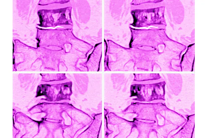
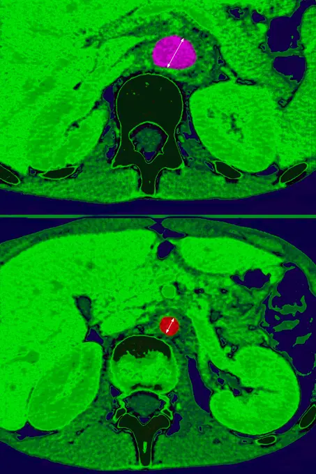
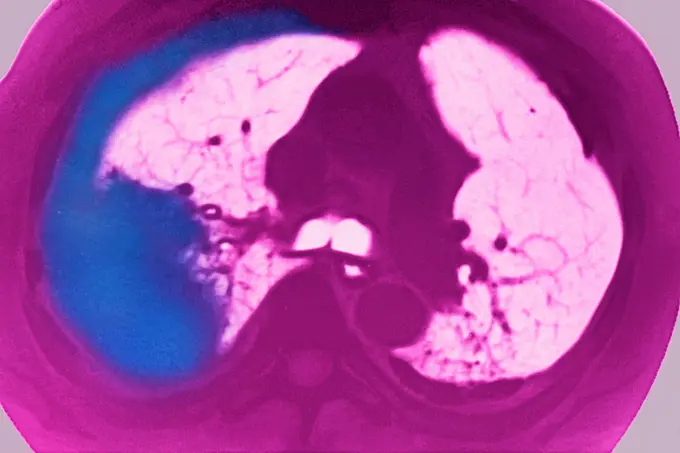
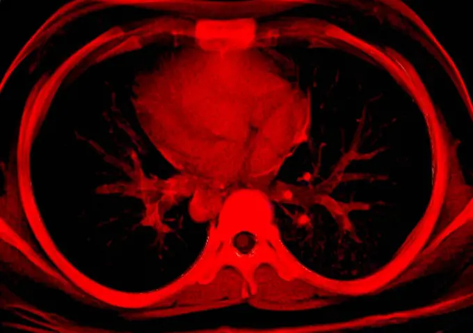
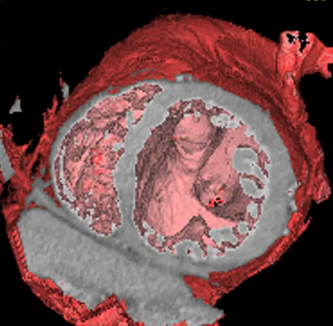
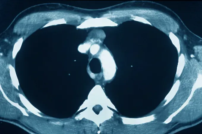
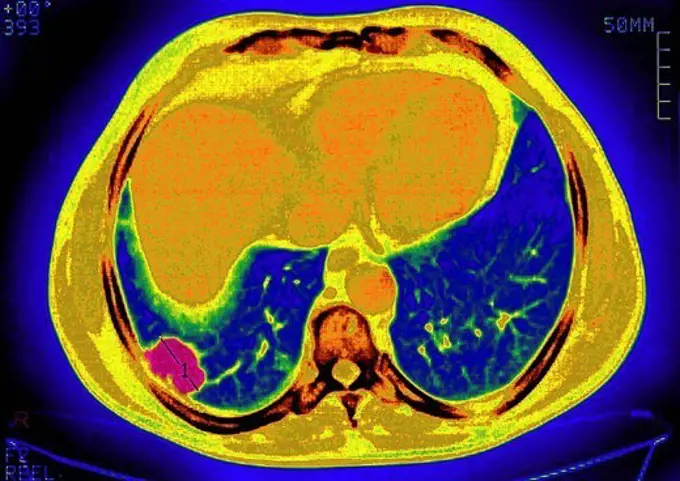
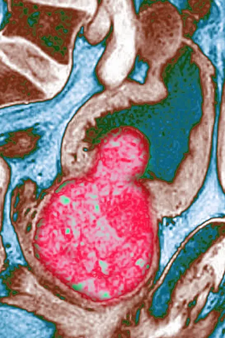
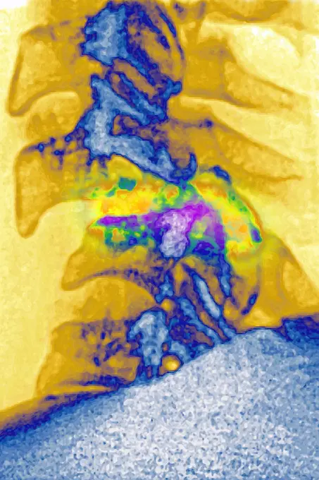
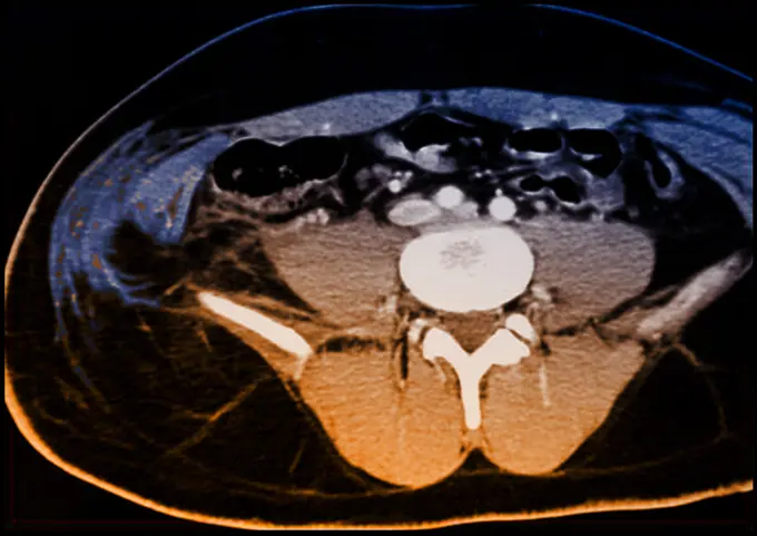
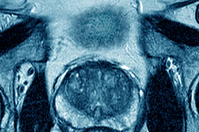
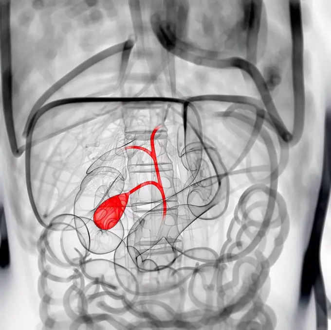
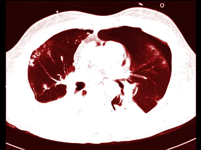
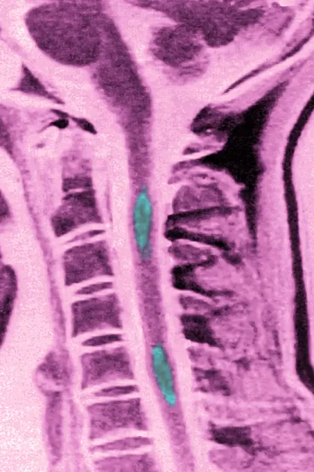
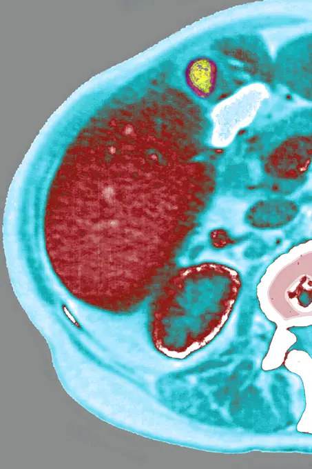
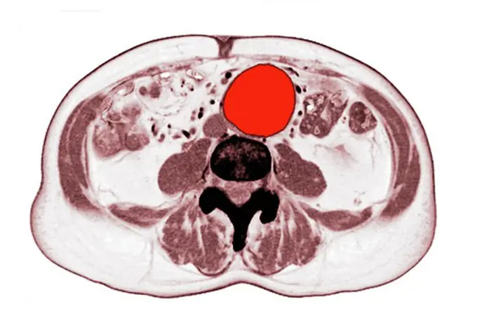
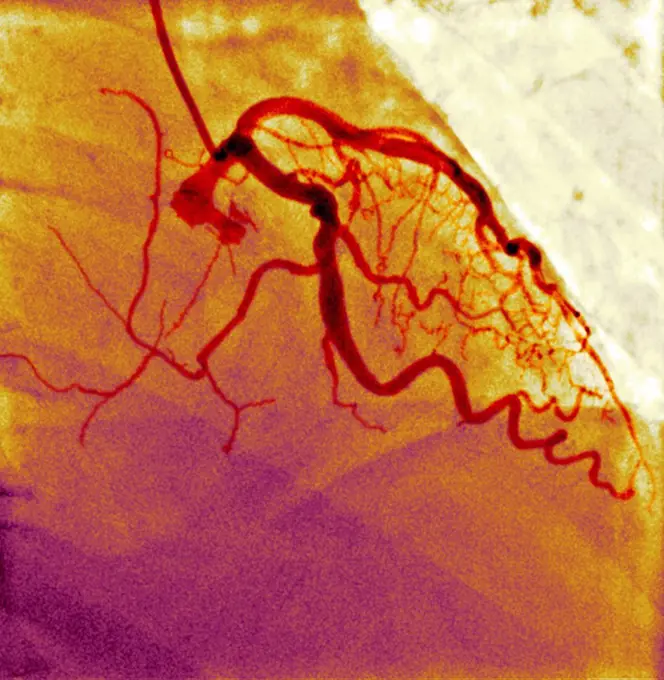
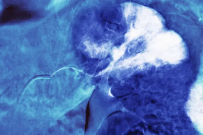
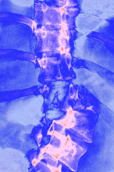
Fine ArtVintageFootageSubscriptionBoardsFree ResearchPricing
  • Fine Art
  • Vintage
  • Footage
  • Subscription
  • Boards
  • Free Research
  • Pricing

Medical Imaging Scan Visuals

Color-enhanced scans revealing various medical conditions within the human body, highlighting tools in diagnostics through vivid imagery.

Pagets disease, seen in a frontal MRI scan of the L4 and L5 lumbar vertebrae.
189 assets in this story
824-63206985
Takayasus arthritis, seen by 2 radial section abdominal scanners, at the descending aorta.
824-63214285
Vesicule calculus. Frontal pelvic x-ray.
1899-54027694
Pleurisy seen on a radial cross-section CT scan.
4269-27222
Vertebral infiltration. Color enhanced axial Computed Tomography (CT) scan of a cervical vertebra with arthrosis treated by infiltration of anti-inflammatory drug (corticoids) around the spinal cord and spinal nerve. The needle (in red at right) is inserted into the vertebral foramen (foraminal infiltration).
4128-20043085
Normal lung, CT scan
4269-26727
Asbestosis. Computer tomography (CT) scan of a horizontal slice through the chest of a patient suffering from asbestosis, a kind of pneumoconiosis caused by the inhalation of asbestos particules from the air that have damaged the lung tissus and can lead to breathing problems and heart failure. The disease generated pleural plaques (red) in the most affected parts.
824-63207445
Pagets disease, seen in a frontal MRI scan of the L4 and L5 lumbar vertebrae.
4269-25369
Myocardial infarctus. 3D CT Scan et 2D
824-63215985
Thymus cancer seen on a cross-section chest CT scan.
1899-53511514
Uterine cancer visualized by MRI in radial section.
824-33580
LUNG METASTASIS, SCAN. Pulmonary metastases secondary cancer, carcinoma. Thoracic scan in axial section.
1899-54027391
X-ray of urinary schistosomiasis. Frontal pelvic x-ray. (Photo by: BSIP/UIG via Getty Images)
824-63225949
Large prostatic adenoma, visualized by frontal x-ray of the pelvis.
4128R-1856
Coloured MRI brain scan showing an arachnoid cyst
824-29057249
Lymphoma revealed by lymphadenopathy between the liver and the gallbladder. abdominal scan in radial section.
824-63165982
Thorax: Heart, aorta, and pulmonary artery are visible, 3D CT scan.
824-63226739
Dislocation of the cervical vertebrae, with spinal trauma, between the sixth and seventh cervical vertebrae, visualized by sagittal section radiography.
824-63211061
Fracture of a dorsal vertebra, with damage to the spinal cord with a bony fragment in the neuroforamen. Cross-section scanner.
1899-54027121
Kidneys and liver in the abdomen seen on a radial cross-section scan.
4269-24830
Kidney cancer. Axial computed Tomography (CT) scan the abdomen showing a cancer of the left kidney.  The front of the patient is at top. The sections of the vertebrae and ribs are in brown-yellow, kidneys in green. The tumor is the red spot on the left kidney.
4269-27190
Intestinal obstruction. Color enhanced axial Computed Tomography (CT) scan of the abdomen showing a volvulus of the colon responsible for a bowel obstruction. It is evidenced by the winding of the colon loops around their axis (in red).
824-29057250
Lymphoma revealed by lymphadenopathy between the liver and the gallbladder. abdominal scan in radial section.
1899-54027134
Cancer of the liver, seen in a radial cross-section abdominal scan.
824-63207417
Cancer of the liver, seen in a radial cross-section abdominal scan
1899-54026840
Lung cancer on the left pulmonary lobe, seen on a radial section MRI scan of the chest.
1899-53511713
Large prostatic adenoma, visualized by frontal x-ray of the pelvis. (Photo by: CAVALLINI JAMES/BSIP/UIG)
1899-54027166
Paget"s disease, seen in a radial cross-section MRI scan of the L4 and L5 lumbar vertebrae.
4269-27504
Chondroma. Sagittal MRI of the thigh showing a chondroma of the femur (thigh bone). This benign bone tumor is formed by a calcified cartilage matrix giving the bone marrow (in red) its white spotted aspect.
1525-24460453
CT scan of an aortic aneurysm
1899-54027420
Vesicule calculus. Frontal pelvic x-ray. (Photo by: BSIP/UIG via Getty Images)
1899-61460123
Lung cancer (cancerous tumor consisting of a group of cancerous cells that invade nearby tissues and destroy them, they can also spread in metastases). Lungs visualized by thoracic scanner in radial section
1899-54027680
Lung cancer seen on an axial plane chest MRI scan.
824-32928
LUNG METASTASIS, SCAN. Pulmonary metastases secondary cancer, carcinoma. Thoracic scan in axial section.
4269-24964
Aortic aneurysm. 3D computed tomographic CT) scan reconstruction of a multiple aneurysm. The aneurysm appears as a swollen bulge (green) located on superior thoracoabdominal artery at left, and on the left common iliac artery at right . An aneurysm is a bulge of a vessel due to the dilatation of its wall. A ruptured aneurysm can be fatal.
824-29057260
Prostate (healthy prostate gland), visualized in ultrasound in frontal view.
1899-53508420
Abnormal vessel originating from the abdominal aorta and vascularizing the right lung. This anomaly, called pulmonary sequestration, can cause pulmonary hemorrhaging (hemoptysis). 3D CT scan.
4269-24969
Coarctation of the aorta. 3D computed tomographic (CT) scan reconstruction showing a coarctation of the aorta. It is a malformation of the aorta, often congenital, resulting in a narrowing of the aorta seen here just below the aortic arch.
4269-24959
Stenosis of the aorta. 3D computed tomographic (CT) scan reconstruction of an atheromatous stenosis of of the aorta, visible just before its division in 2 iliac arteries. The atheroma plaques causing the stenosis are seen in yellow.
1899-53511478
MRI of pulmonary cystic fibrosis (viscous mucus disease, cystic fibrosis affecting glandular epithelia). Radial MRI of the lungs.
4269-27505
Chondroma. Sagittal MRI of the thigh showing a chondroma of the femur (thigh bone). This benign bone tumor is formed by a calcified cartilage matrix giving the bone marrow (in red) its white spotted aspect.
4269-24989
Lungs malformation. 3D computed tomographic (CT) scan reconstruction of the left lung affected intra-lobal pulmonary sequestration, a congenital malformation of the lung caracterized by the separation of a part of bronchopulmonary tissue from its normal connexions.
4128R-1993
Carotid aneurysm, digital angiogram
4269-27508
Pneumothorax. Axial Computed Tomography (CT) scan of the lungs with pneumothorax : presence of air in the pleural cavity, in black, at top right. The whith patchy areas at left are contusions of the lung tissus (car accident).
824-63222946
Pulmonary embolism in the left lower lobe (pulmonary artery blocked by a blood clot). Thoracic angiography.
4269-27524
Ruptured abdominal wall. Axial Computed Tomography (CT) scan of the abdomen showing a ruptured right abdominal wall (at left), evidenced by the torn aspect of the muscles and the gap between the muscle (blue) and the iliac bone (white).
1899-53511717
Benign prostatic hypertrophy, visualized by radial MRI of the pelvis. (Photo by: CAVALLINI JAMES/BSIP/UIG)
4128R-21198
Healthy gallbladder, artwork
4269-27509
Pneumothorax. Axial Computed Tomography (CT) scan of the lungs with pneumothorax : presence of air in the pleural cavity, in black, at top right. The whith patchy areas at left are contusions of the lung tissus (car accident).
1899-53511925
Multiple sclerosis (MS) (autoimmune disease that affects the central nervous system by dysfunction of the immune system and leads to lesions that cause motor, sensory, cognitive, visual or even sphincter disturbances). MRI of the cervical vertebrae in sagittal section. (Photo by: CAVALLINI JAMES/BSIP/UIG)
824-63217273
Pulmonary tuberculosis seen on a radial cross-section chest CT scan.
824-63215951
Lung cancer caused by asbestosis, seen on a radial section chest CT scan.
1899-61460130
Knee fracture (caused by a violent shock, causing trauma with serious lesions to the tibial plateau and to the lower end of the femur. X-ray of the left knee from the front
4269-25416
Calcifying pericarditis. 3D computed tomographic (CT) scan reconstruction of a heart affected by calcifying pericarditis (armoured heart) seen from above. Pericarditis is the inflammation of the pericardium, a thin layer of tissue that covers the outer surfaces of the heart. Here, the pericardium is covered by a fibrous calcarious rigid bag (white coat) and can not complete its natural mouvements.
4269-27490
Shoulder chondrosarcoma. Colored enhanced Axial Computed Tomography (CT) scan of the shoulder showing a chondrosarcoma of the shoulder blade. The tumor is evidenced by the presence of a heterogenous lytic area with bone calcifications. The black round shape at right is the head of the humerus inserted into the shoulder blade.
824-63225947
Large prostatic adenoma, visualized by frontal x-ray of the pelvis.
1899-53511472
MRI of pulmonary cystic fibrosis (viscous mucus disease, cystic fibrosis affecting glandular epithelia). Radial MRI of the lungs.
824-63224705
MRI of pulmonary cystic fibrosis (viscous mucus disease, cystic fibrosis affecting glandular epithelia). Radial MRI of the lungs.
824-63207451
Pagets disease, seen in a radial cross-section MRI scan of the L4 and L5 lumbar vertebrae.
824-63204746
Pulmonary infarction seen on a cross-section CT scan of the chest.
824-63206966
Mucoviscidosis (accumulation of mucus in the respiratory tract), seen on a radial section MRI scan.
824-63212829
Colon cancer (tumor on the caecum and ileocolic valve). Visualisation on a radial CT scan.
824-29057251
Lymphoma revealed by lymphadenopathy between the liver and the gallbladder. abdominal scan in radial section.
4269-25334
Stenosis. 3D computed tomographic (CT) scan reconstruction of a stenosis of the brachiocephalic trunk. The narrowed section of the artery is visible just above the aortic arch.
4297-1150
CT scan images showing an abdominal aortic aneurysm which developed between the renal arteries and the common iliac arteries
1899-54027679
Lung cancer seen on an axial plane chest MRI scan.
4128R-12922514
Normal blood vessels, X-ray
4269-25420
Atherosclerosis. 3D computed tomographic (CT) scan (virtual endoscopy) showing atheromatous plaques (in pink) inside an artery. This fatty deposit narrows the vessel and restricts the blood flow (atherosclerosis).
6188-55645088
Abdomen man, man abdomen man, man, ct recording copyright: xzoonar.com/dr.xnorberbertxlangeex 13790159
1899-54027154
Kidney infarction seen in an abdominal angiogram.
4269-26719
Aortic aneurysm. Axial coloured Computed Tomography (CT) scan of an abdominal aortic aneurysm. The face of the body is downwards. Spinebone is in blue. At centre is the severely enlarged, rounded artery (yellow) with its central lumen visible purple). An aneurysm is a bulge of a vessel due to the dilatation of its wall. A ruptured aneurysm can be fatal.
1899-53511732
Iliac artery stenosis before and after percutaneous angioplasty. (Photo by: CAVALLINI JAMES/BSIP/UIG)
4269-25345
Stenosis. Virtual endoscopic view of the left internal carotid artery showing a stenosis (narrowing) and atheromatous plaques in pink.
824-63226747
Pulmonary metastases (secondary to kidney cancer), visualized by chest CT scan in axial section.
1899-53511891
Rotational dislocation at the junction of the thoracic and lumbar vertebrae, visualized by an AP chest radiograph. (Photo by: CAVALLINI JAMES/BSIP/UIG)
824-63226735
Rotational dislocation at the junction of the thoracic and lumbar vertebrae, visualized by an AP chest radiograph.
4128R-4193
Normal brain, CT scan
4297R-1896
CT scan showing aortic aneurysm. The enlarged and dilated aortic aneurysm is highlighted in red.
1899-61460138
Spontaneous vertebral fracture due to oteoporosis (bone disease characterized by a decrease in bone mass, as well as bone deformation by compression and the risk of spontaneous fracture.). Lumbar vertebral x-ray
4128-30416837
Parathyroid glands, illustration
824-63222948
Pulmonary embolism in the left lower lobe (pulmonary artery blocked by a blood clot). Thoracic angiography.
4128-V58573943
824-63128413
Stenosis of the abdominal aorta.
4269-24976
Rupture of prosthesis. 3D computed tomography (CT) scan showing the rupture of a prosthesis on the left external iliac artery (at right), due to the rupture of the threads maintaining the prosthesis (anastomic rupture), and causing an  internal heamorrhage (in purple).
824-63206021
Acute pneumonia, seen on a cross-section thoracic scan.
824-65830283
Ischemic mitral insufficiency (MI) - Pathology of the left ventricle (mitral ring and subvalvular apparatus). transthoracic echocardiography (TTE).
1899-53508458
3D CT scan of coronary bypasses.
824-63224770
Uterine cancer visualized by frontal MRI.
1899-61460116
Arteriography or angiography (radiological examination of the arteries of a lower limb seen in profile)
824-63203248
Ensethopathy of the supeficial plantar fascia. Sagittal section MRI of the foot.
824-63204748
X-ray exam of the digestive tract, seen in 3 saggital-plane views.
4269-25347
Bi-iliac aneurysm. Three-dimensional computed tomographic (CT) scan reconstruction of a bi-iliac aneurysm. The aneurysms appear as a swollen bulge located on the beginning of the common iliac arteries that irrigate the lower limbs. An aneurysm is a bulge of a vessel due to the dilatation of its wall. A ruptured aneurysm can be fatal.
4269-25349
Abdominal aortic aneurysm. Three-dimensional computed tomographic (CT) scan reconstruction of an abdominal aorta aneurysm. It appears as swollen bulge  located under the renal arteries and before its division in the two comon iliac arteries. An aneurysm is a bulge of a vessel due to the dilatation of its wall. A ruptured aneurysm can be fatal.
6188-55645096
Abdomen man, man abdomen man, man, ct recording copyright: xzoonar.com/dr.xnorberbertxlangeex 13790170
1899-54027392
X-ray of urinary schistosomiasis. Frontal pelvic x-ray. (Photo by: BSIP/UIG via Getty Images)
4269-25812
Peptic ulcer. Barium X-ray of a stomach affected by gastric or peptic ulcer (yellow). An ulcer is the damage of the lining membrane of the stomach, often related to diet, stress, alcohol, and drug consumption and sometime associated with the bacterium Helicobacter pilori.
1899-53511740
Osteoarthritis of the left hip (coxarthrosis) visualized by a pelvic x-ray of the front. (Photo by: CAVALLINI JAMES/BSIP/UIG)
1899-54027118
Kidneys and liver in the abdomen seen on a radial cross-section scan.
824-63188664
Secondary malignant tumour on a lumbar vertebra, seen on a saggital plane MRI scan.
1899-53511477
MRI of pulmonary cystic fibrosis (viscous mucus disease, cystic fibrosis affecting glandular epithelia). Radial MRI of the lungs.
824-63215992
Pulmonary tuberculosis, seen on a frontal chest x-ray.
PREVIOUS
of 2
NEXT
2401 S. Ervay, Suite 206
Dallas, Texas 75215
United States
Browse
CategoriesGalleriesFree Research
For Creators
License Your ContentSuperStock For StudentsFrame of Mind BlogContributor PortalNew Image SpotlightLicense AI Imagery
Resources
About UsLicensingTerms & ConditionsAPI AccessPrivacyPricing
Contact
+1 866 236 0087help@superstock.com Contact form
©2026 SuperStock. All Rights Reserved. -A
This site is protected by reCAPTCHA and Google Privacy Policy and Terms of Service apply.