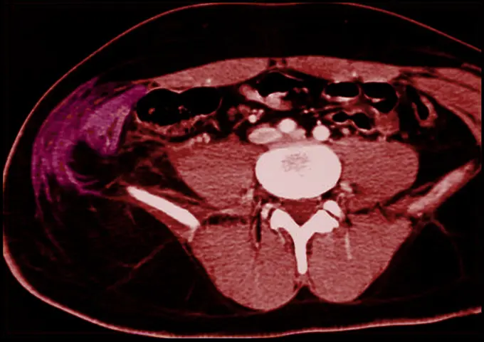
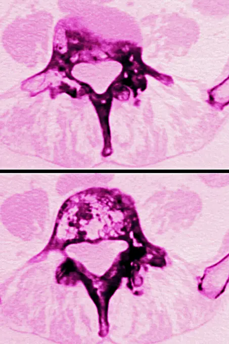
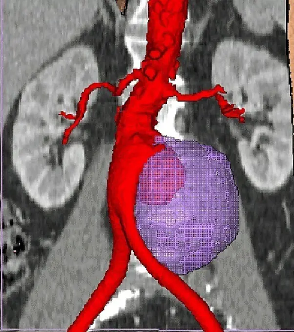
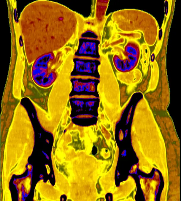
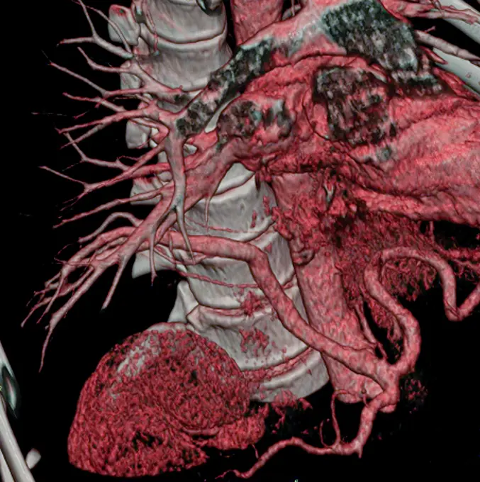
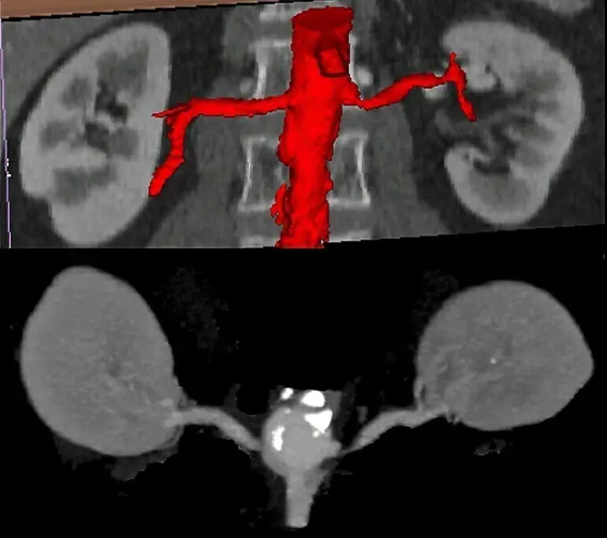
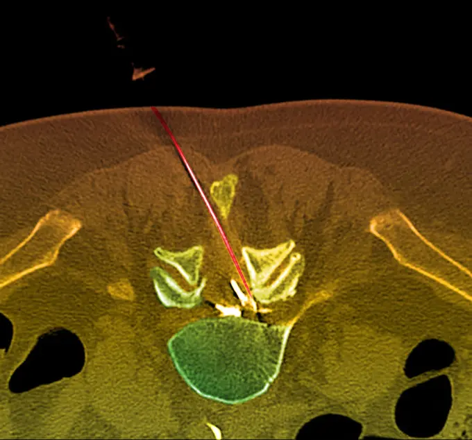
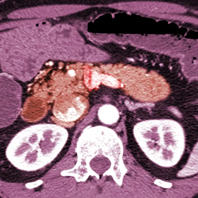
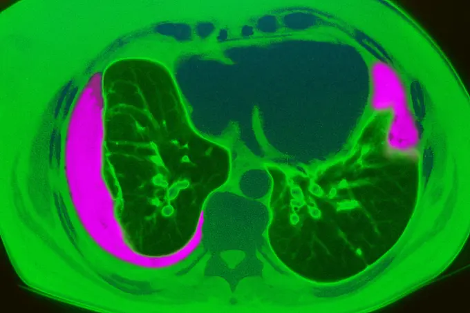
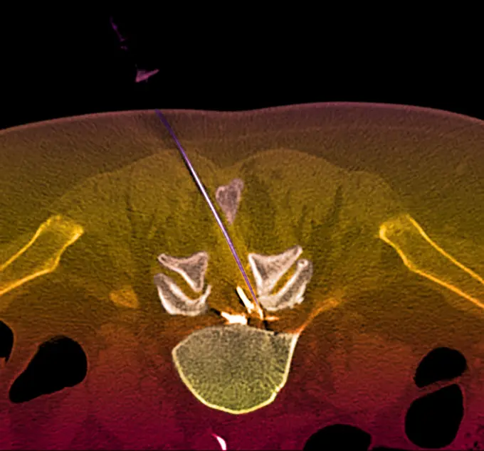
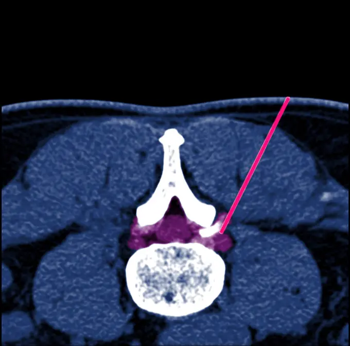
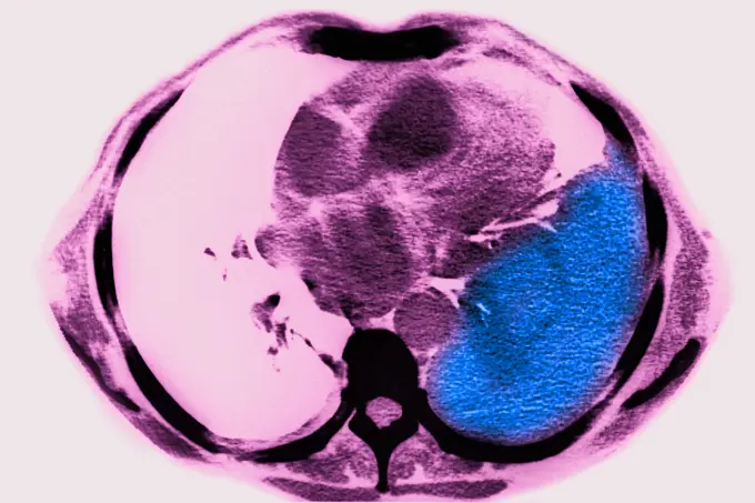
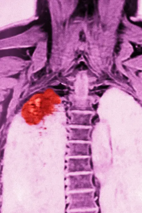
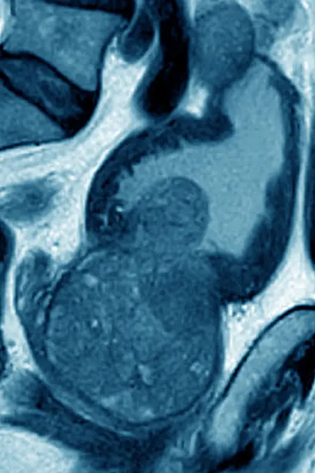
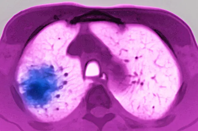
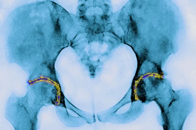
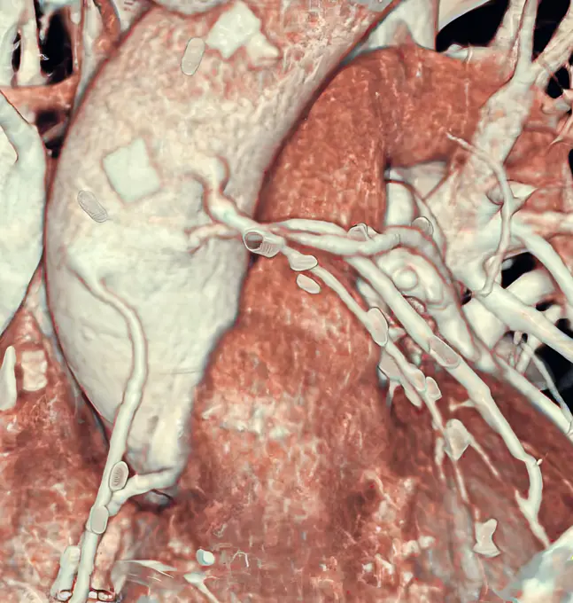
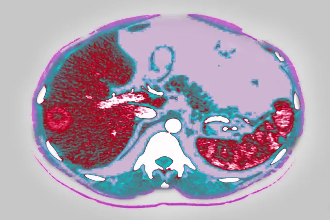
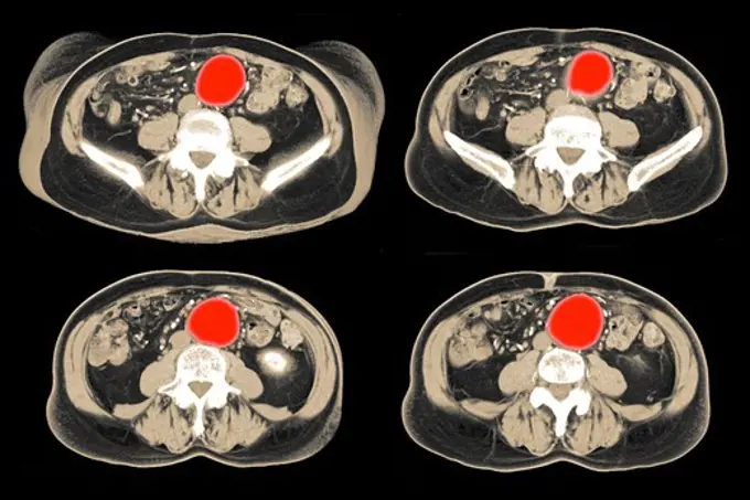
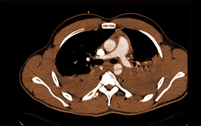
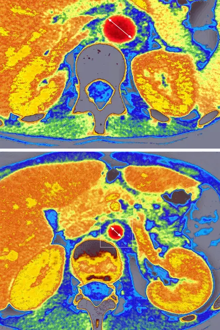
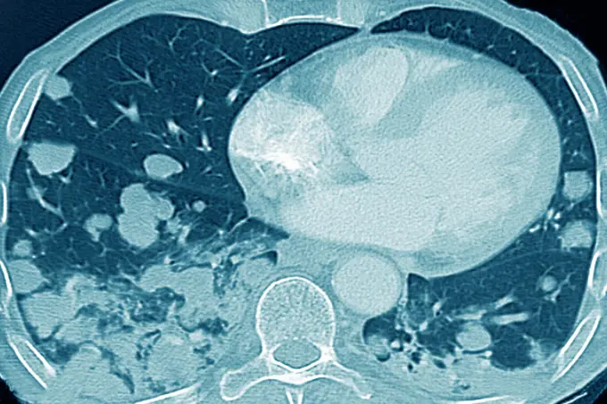
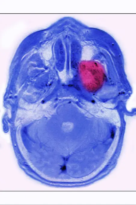
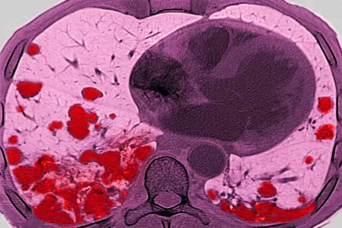
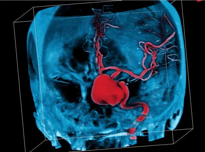
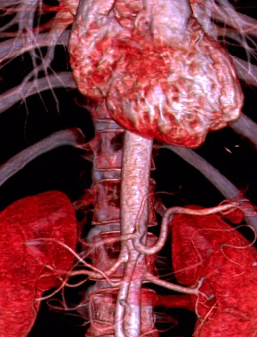
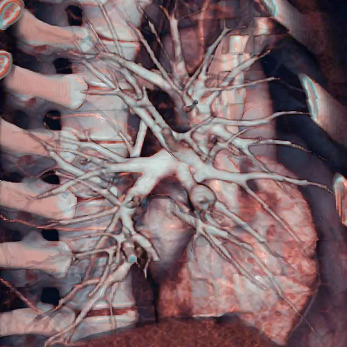
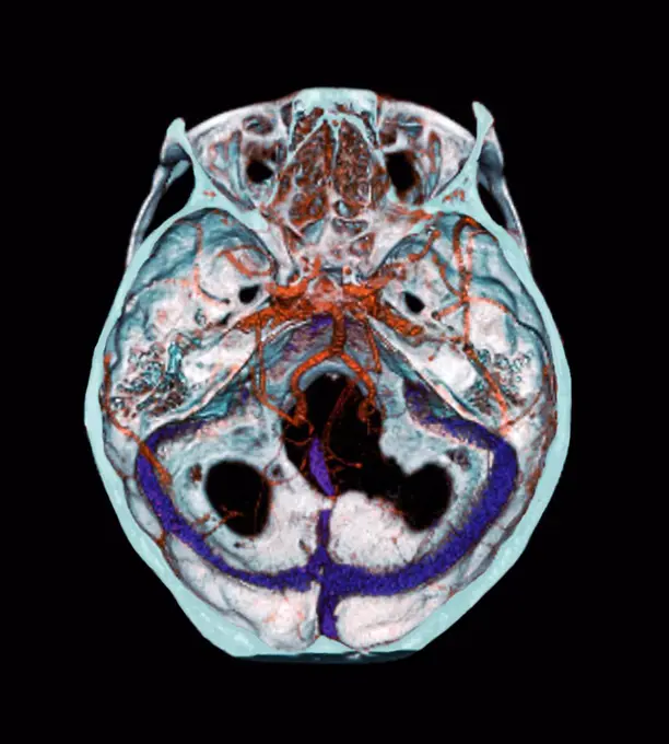
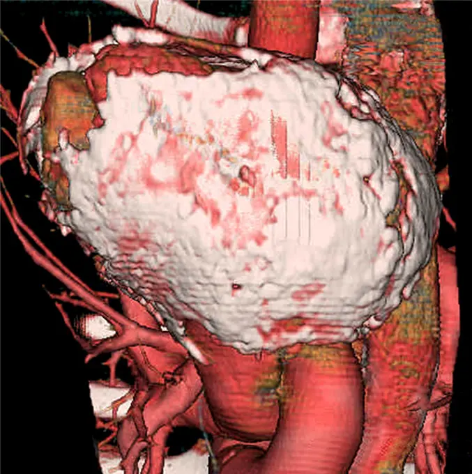
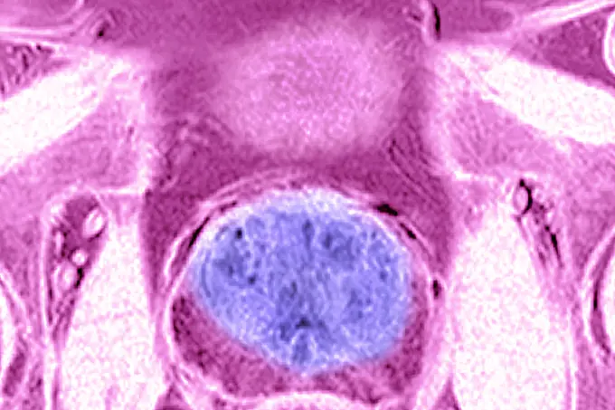
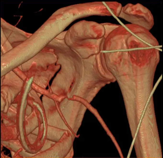
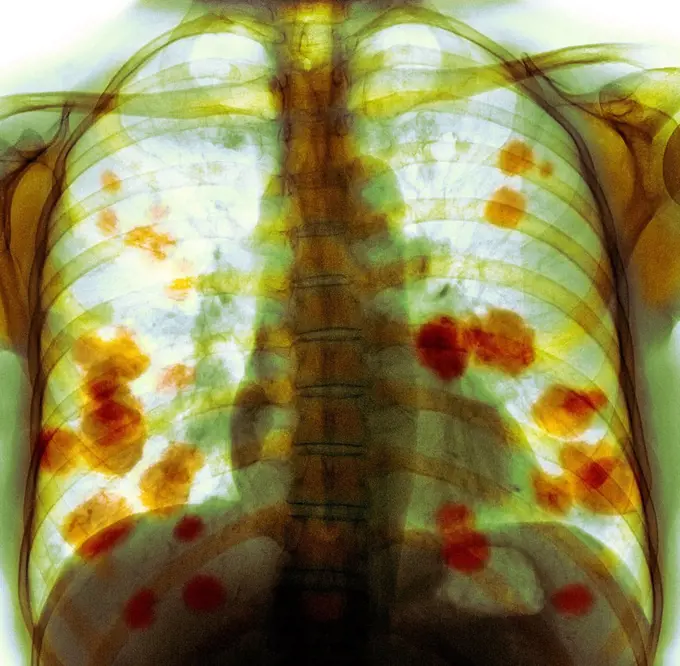
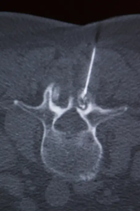
Fine ArtVintageFootageSubscriptionBoardsFree ResearchPricing
  • Fine Art
  • Vintage
  • Footage
  • Subscription
  • Boards
  • Free Research
  • Pricing

Medical Imaging Scans

Detailed medical imaging scans highlighting various abdominal conditions, showcasing cross-sectional views of organs and tissues.

Pleurisy (left lung), seen on a radial section chest scan.
135 assets in this story
824-63205971
Adenopathy (spleen lymph gland). Cross-section abdominal scan.
4269-27232
Vertebral infiltration. Color enhanced axial Computed Tomography (CT) scan of a cervical vertebra with arthrosis treated by infiltration of anti-inflammatory drug (corticoids) around the spinal cord and spinal nerve. The needle will be inserted into the vertebral foramen (foraminal infiltration). The insertion point is market with a target (red star).
1899-53511878
Suppurative adenopathy in the liver and spleen, due to cat scratch disease (benign lymphoreticulosis). Abdominal scanner in axial section. (Photo by: CAVALLINI JAMES/BSIP/UIG)
824-63207416
Cancer of the liver, seen in a radial cross-section abdominal scan
4269-27124
Ileo caecal intussusception. Color enhanced axial computed tomography (CT) scan of the abdomen showing ileo caecal intussusception.
1899-53511474
MRI of pulmonary cystic fibrosis (viscous mucus disease, cystic fibrosis affecting glandular epithelia). Radial MRI of the lungs.
824-63215969
Pleurisy (left lung), seen on a radial section chest scan.
4269-27525
Ruptured abdominal wall. Axial Computed Tomography (CT) scan of the abdomen showing a ruptured right abdominal wall (at left), evidenced by the torn aspect of the muscles and the gap between the muscle (red) and the iliac bone (white).
824-63204742
Spleen infarction seen on a radial section CT scan of the abdomen.
1899-54027167
Paget"s disease, seen in a radial cross-section MRI scan of the L4 and L5 lumbar vertebrae.
4269-25003
Aortic aneurysm. Three-dimensional computed tomographic (CT) scan reconstruction of an abdominal aorta aneurysm.
824-63175202
CT scan of the abdomen, frontal view.
824-63188651
Silicosis, seen on a transversal CT scan of the thorax.
824-63184388
Cystic fibrosis. Cross-section thoracic scan.
1899-54026829
Amyloidosis of the intestine, seen on a radial section CT scan.
4269-24973
Dissecting aneurysm of the aorta and its complications. 3D computed tomographic (CT) scan reconstruction of a dissecting aneurysm of the aorta (left side view).
1899-53511458
Angiography of myocardial infarction caused by atheromatous plaque in the left coronary artery. Angiographic visualization is produced by injection of contrast product.
1899-53511742
Osteoarthritis of the hips (coxarthrosis) visualized by a pelvic x-ray of the front. (Photo by: CAVALLINI JAMES/BSIP/UIG)
824-63225975
Osteoarthritis of the hips (coxarthrosis) visualized by a pelvic x-ray of the front.
824-63206258
Aortic dissection in the ascending aorta, seen on a sagittal MRI scan of the thorax.
824-63224711
MRI of pulmonary cystic fibrosis (viscous mucus disease, cystic fibrosis affecting glandular epithelia). Radial MRI of the lungs.
4269-27862
Liver lymphoma. Colored CT (computed Tomography) scan of the liver (brown, left) of a patient with HIV with a lymphoma (violet, upper left).
824-63165961
Abnormal vessel originating from the abdominal aorta and vascularizing the right lung. This anomaly, called pulmonary sequestration, can cause pulmonary hemorrhaging (hemoptysis). 3D CT scan.
4269-24955
Stenosis. 3D computed tomographic (CT) scan reconstruction of the left renal artery stenosis, responsible for arterial hypertension and renal ischemia.
4269-27228
Vertebral infiltration. Color enhanced axial Computed Tomography (CT) scan of a lumbar vertebra with arthrosis treated by infiltration of anti-inflammatory drug (corticoids) around the spinal cord and spinal nerve. The needle is inserted into the vertebral foramen (foraminal infiltration), but throught the vertebral canal.
1899-53511759
Lung cancer located in the upper right lobe. Frontal chest x-ray. (Photo by: CAVALLINI JAMES/BSIP/UIG)
4269-27865
Pancreas tumor. Colored Computed Tomography (CT) scan of the abdomen showing a tumor of the pancreas (center).
824-63184389
Cystic fibrosis. Cross-section thoracic scan.
1899-53511447
Angiography of myocardial infarction caused by atheromatous plaque in the left coronary artery. Angiographic visualization is produced by injection of contrast product.
824-63217261
Pleurisy seen on a radial cross-section chest MRI scan.
4269-24943
Iliac artery aneurysm. 3D computed tomographic (CT) scan view of internal iliac artery aneurysm treated with a Stent. The aneurysm appears as a swollen bulge (left, in red).
824-63195627
Abdominal CT scan showing the adrenal glands.
4269-24970
Trauma of the aorta. 3D computed tomographic (CT) scan reconstruction of a traumatic rupture of the thoracic aorta further to a car accident. The treatment involves surgery.
4269-27229
Vertebral infiltration. Color enhanced axial Computed Tomography (CT) scan of a lumbar vertebra with arthrosis treated by infiltration of anti-inflammatory drug (corticoids) around the spinal cord and spinal nerve. The needle is inserted into the vertebral foramen (foraminal infiltration), but throught the vertebral canal.
1899-53511885
Cancer of the right kidney visualized by an abdominal scanner in radial section. (Photo by: CAVALLINI JAMES/BSIP/UIG)
1899-53511324
Pulmonary arteritis causing progressive occlusion of the bronchial arteries, seen by a radial CT scan of the lungs (right lung).
4269-27225
Vertebral infiltration. Color enhanced axial Computed Tomography (CT) scan of a lumbar vertebra with arthrosis treated by infiltration of anti-inflammatory drug (corticoids) around the spinal cord and spinal nerve. The needle will be inserted into the vertebral foramen (foraminal infiltration). The route of the needle is marked by the pink line.
824-63215973
Pleurisy (left lung), seen on a radial section chest scan.
824-63225991
Lung cancer located in the upper right lobe. Frontal chest x-ray.
824-63225946
Large prostatic adenoma, visualized by frontal x-ray of the pelvis.
824-63198908
Sinusitis in the left maxillary sinus, seen on a frontal, cross-section MRI scan.
1899-54027678
Lung cancer seen on an axial plane chest MRI scan.
824-63225977
Osteoarthritis of the hips (coxarthrosis) visualized by a pelvic x-ray of the front.
824-63165996
3D CT scan of coronary bypasses.
824-63226724
Suppurative adenopathy in the liver and spleen, due to cat scratch disease (benign lymphoreticulosis). Abdominal scanner in axial section.
4297-1146
CT scan images showing an abdominal aortic aneurysm which developed between the renal arteries and the common iliac arteries
4269-24941
Aortic aneurysm. Three-dimensional (left) and plane (right) computed tomographic(CT) scan reconstruction of an abdominal aorta aneurysm.
4269-27567
False aneurysm of the aorta. Axial Computed Tomography (CT) scan of false aneurysm of the aorta seen as a swelling on the upper aorta, below the aortic arch. This phenomenon usually occurs after trauma to an artery, such as surgery.
1899-54026878
Takayasu"s arthritis, seen by 2 radial section abdominal scanners, at the descending aorta.
824-63206022
Acute pneumonia, seen on a cross-section thoracic scan.
1899-53511904
Pulmonary metastases (secondary to kidney cancer), visualized by chest CT scan in axial section. (Photo by: CAVALLINI JAMES/BSIP/UIG)
4269-25388
Improperly placed Stent prosthesis. 3D computed tomographic (CT) scan reconstruction showing a stent prosthesis into the aortic arch that 'collapsed' on itself.
4269-27861
Gastric tumor. Three-dimensional computed tomographic reconstruction scan of the stomach (gray) showing a massive exophytic tumor in yellow (GIST, Gastrointestinal Stromal Tumor). The liver is seen behind the liver in red ocre.
824-63206969
Intercranial neurinoma, seen on a radial section MRI scan.
4269-27547
Renal infarct. Oblique sagittal Compted Tomography (CT) scan of the the trunc showing a partial right renal infarct. The infarcted (necrotic) areas of the kidney appears in dark ocre (upper part of the kidney) while the healthy part is in white (lower part). Renal infarct is caused by deficient blood irrigation of the kidney.
824-63226749
Pulmonary metastases (secondary to kidney cancer), visualized by chest CT scan in axial section.
1899-53511765
Lung cancer located in the upper right lobe. Frontal chest x-ray. (Photo by: CAVALLINI JAMES/BSIP/UIG)
824-63198871
Cerebral atrophy. Parietal cortico-sub-cortical atrophies (aftereffects of left, frontoparietal parenchymal ischemia), seen on a sagittal plane cross-section brain CT scan.
6188-55645082
Abdomen man, man abdomen man, man, ct recording copyright: xzoonar.com/dr.xnorbertxlangeex 13790139
4269-27665
Cerebral aneurysm. Three-dimensional computed tomographic (CT) scan reconstruction of a skull showing an intracranial aneurysm (red ballon shaped swelling) located in the arterial circle of Willis  (where the basilar artery and internal carotid arteries meet). An aneurysm is a bulge of a vessel due to the dilatation of its wall. A ruptured aneurysm can be fatal.
4128R-30916
Abdominal aorta. Coloured 3D computed tomography (CT) angiogram of the abdominal aorta in a 42 year old patient. The aorta is the body's main artery. ...
824-63224521
Pulmonary arteritis causing progressive occlusion of the bronchial arteries, seen by a radial CT scan of the lungs (right lung).
4269-27863
Liver abscess. Colored CT (computed Tomography) scan of abdomen showing liver abces (green spots at upper center of the liver).
1899-53508421
Pulmonary artery (PA) aneurysm. Pulmonary aneurysm is associated with pulmonary artery dilation, with a risk of intra-pulmonary hemorrhaging and rupture. 3D CT scan.
1899-53508436
Thorax of newborn : Mediastinum, heart, aorta, and mammary arteries are visible. 3D CT scan.
4269-25011
Goitre. 3D computed tomographic (CT) scan reconstruction of the throat showing a voluminous thyroid goitre (8cm x 6cm x 5cm) causing a compresion of the veins (blue) and arteries (red ) of the neck.
4269-24968
Coarctation of the aorta. 3D computed tomographic (CT) scan reconstruction showing a coarctation of the aorta. It is a malformation of the aorta, often congenital, resulting in a narrowing of the aorta seen here just below the aortic arch.
824-63203258
Calcified atheromatous of the aortic arch. Frontal thoracic x-ray.
4269-27855
Small intestine. MRI (Magnetic Resonance Imaging) scan of the abdomen showing  the small intestine. Are also seen the liver at top (pale gray), the stomach at right (black), part of the large intestine at left, and the baldder at bottom (black).
4269-25273
Heart. 3D computed tomographic (CT) scan reconstruction of a normal heart seen from the side. At left : the backbone. The branched structure at left of the heart is the lung vascular system.
4128R-12664
Arteries of the brain. 3D computed tomography CT scan of normal arteries in the brain of a 35 year old.
4269-25413
Calcifying pericarditis. 3D computed tomographic (CT) scan reconstruction of a heart affected by calcifying pericarditis (armoured heart) seen from above. Pericarditis is the inflammation of the pericardium, a thin layer of tissue that covers the outer surfaces of the heart. Here, the pericardium is covered by a fibrous calcarious rigid bag (white coat) and can not complete its natural mouvements.
824-63225951
Benign prostatic hypertrophy, visualized by radial MRI of the pelvis.
4269-27520
Axillary artery. 3D Computed Tomographic (CT) reconstruction scan of the shoulder showing a post-traumatic stenosis of the left axillary artery.
4269-25355
Inferior abdominal aortic aneurysm. Left side 3D computed tomographic (CT) scan reconstruction of  inferior abdominal aortic aneurysm. The aneurysm appears as a swollen double bulge located on the descending aorta, just after the aortic arch. An aneurysm is a bulge of a vessel due to the dilatation of its wall. A ruptured aneurysm can be fatal.
4269-25419
Heart and pulmonary vascular system. Thorax horizontal view 3D CT Scan
1899-54027155
Kidney infarction seen in an abdominal angiogram.
824-63188621
Herniated disc on a lumbar vertebra, seen on a transversal CT scan of the vertebral axis.
4269-27565
False aneurysm of the aorta. Sagittal Computed Tomography (CT) scan of false aneurysm of the aorta seen as a swelling on the upper aorta (pink), below the aortic arch. This phenomenon usually occurs after trauma to an artery, such as surgery.
4128R-11287903
Secondary lung cancer. Coloured X-ray of the chest of a 52 year old female patient with metastatic (secondary) lung cancer (orange).
824-63211591
Acute pneumonia, seen on a cross-section thoracic CT-scan.
4128-16073310
Multiple scans of shoulder joint, MRI
824-63224709
MRI of pulmonary cystic fibrosis (viscous mucus disease, cystic fibrosis affecting glandular epithelia). Radial MRI of the lungs.
1899-53512015
Normal knee visualized by MRI in sagittal section.
4269-24656
Lesion of the trachea. 3D computed tomographic (CT) scan reconstruction (virtual endoscopy) showing a lesion on the trachea viewed from under.
824-63173383
Lumbar vertebra, infiltration
6145-44626921
Iapetus' New Year's Flyby
4269-25266
Heart and left coronary. 3D computed tomographic (CT) scan reconstruction of a normal heart (left side) seen from above, and the left coronary arteries. At top are seen the sections of the pulmonary artery (oval) and the aorta (round).
824-66065395
A revealing look at the stroke brain: This image combines pre- and post-treatment scans from the same patient. Analysis of the two scans revealed that the area and size of post-treatment bleeding corresponded to blood-brain barrier disruption (shown in green, yellow and red) prior to therapy.
4128-30419381
Prostate, illustration
1899-54027695
Pleurisy seen on a radial cross-section CT scan.
824-63225995
Lung cancer located in the upper right lobe. Frontal chest x-ray.
4269-27278
Cerebral aneurysm. 3D Computed Tomography (CT) scan reconstruction of an aneurysm of the right vertebral artery. An aneurysm is a bulge of a vessel due to the dilatation of its wall. A ruptured aneurysm can be fatal.
824-57656900
Vertebral osteitis (inflammation of bone tissue in a dorsal vertebra, caused by a bacterial infection). radial x-ray of a dorsal vertebra.
6114-50976749
C.T. scan of comminuted fractures of calcaneums - heel bones. Computed tomography is an x-ray technique that produces a film representing a detailed cross section of tissue.
824-63128365
Gastric pains. Stomach of a patient having swallowed pebbles.
4269-27184
Arterious system. Colored Computed Tomography (CT) scan of the whole arterious system. Is seen at center the aorta, the main artery of he body. At top, the aortic arch branching in the brachiocephalic artery, the left carotid artery and the left subclavian artery. Going down : the renal arteries, and the division in 2 iliac arteries and their branches. The black spots on the aorta are atheromathous plaques.
1899-53511517
Uterine cancer visualized by sagittal MRI.
1899-54027150
Heart and lungs seen in a radial cross-section MRI scan.
824-29057206
Thrombosis of the cerebral venous sinuses of the dura mater, visualized by cranial MRI angiography.
PREVIOUS
of 2
NEXT
2401 S. Ervay, Suite 206
Dallas, Texas 75215
United States
Browse
CategoriesGalleriesFree Research
For Creators
License Your ContentSuperStock For StudentsFrame of Mind BlogContributor PortalNew Image SpotlightLicense AI Imagery
Resources
About UsLicensingTerms & ConditionsAPI AccessPrivacyPricing
Contact
+1 866 236 0087help@superstock.com Contact form
©2026 SuperStock. All Rights Reserved. -A
This site is protected by reCAPTCHA and Google Privacy Policy and Terms of Service apply.