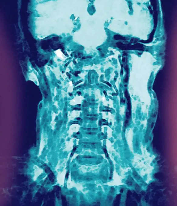
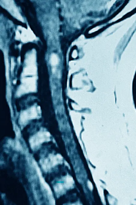
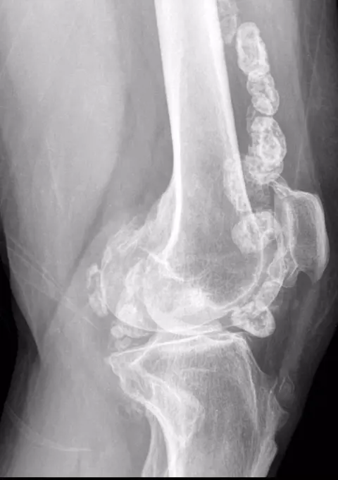
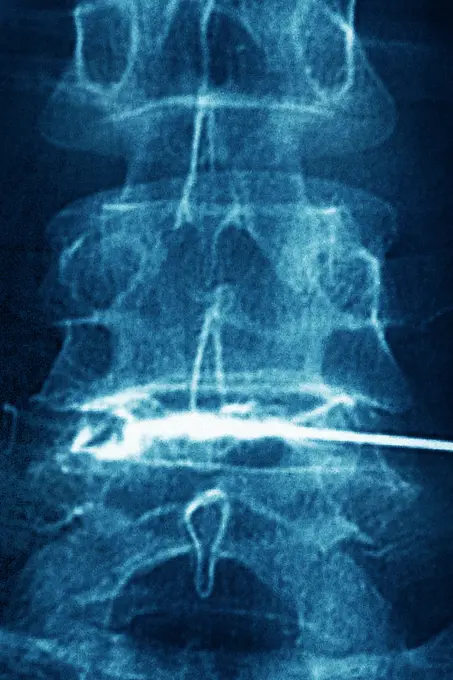
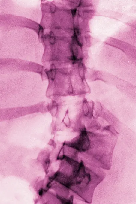
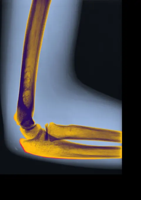
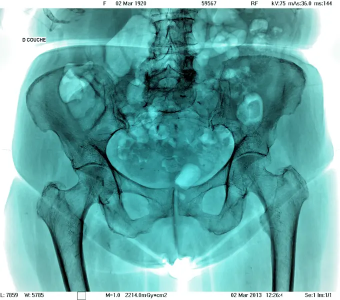
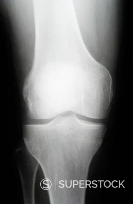
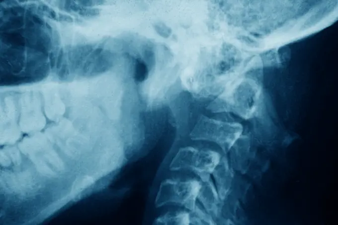
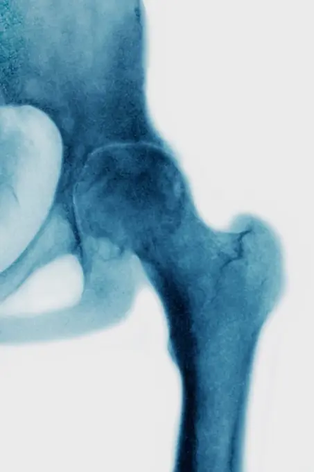
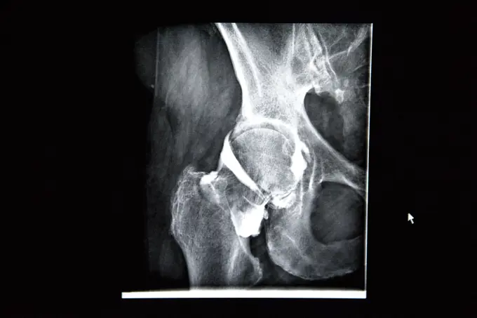
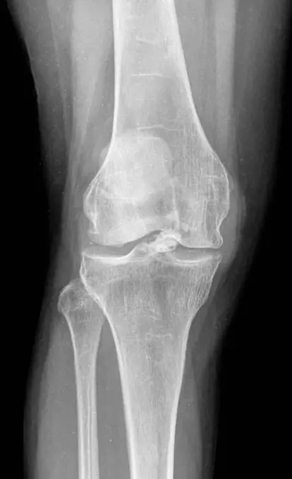
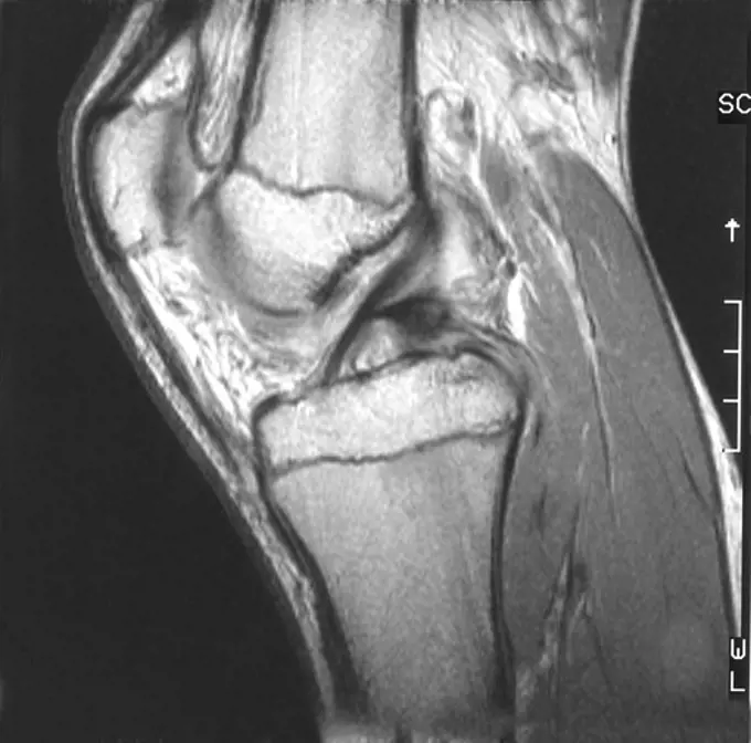
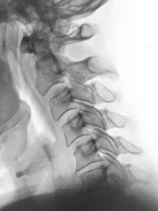
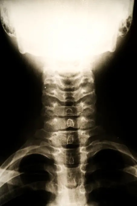
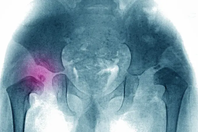
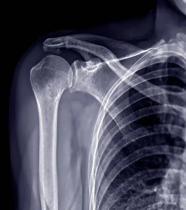
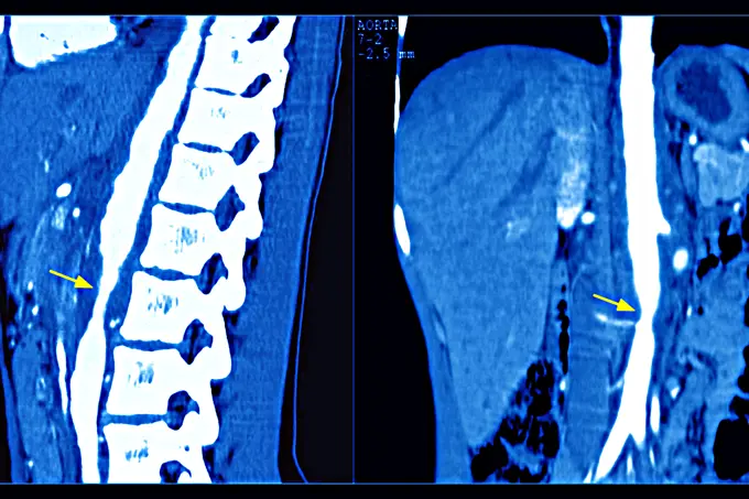
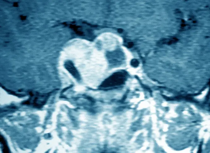
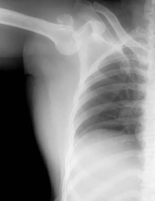
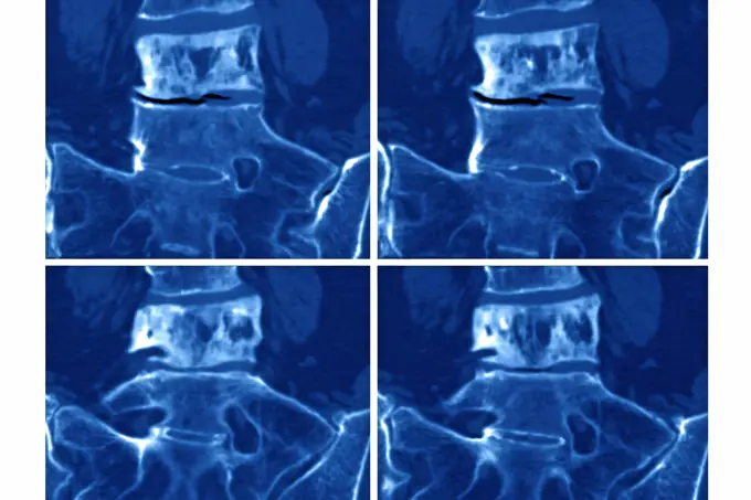
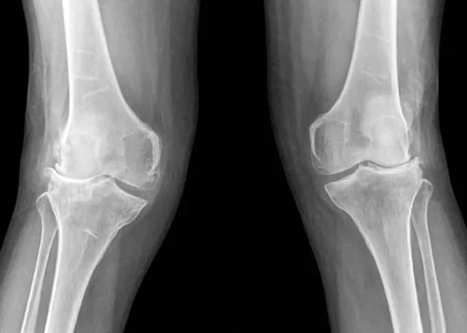
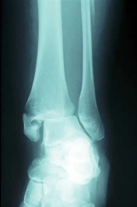
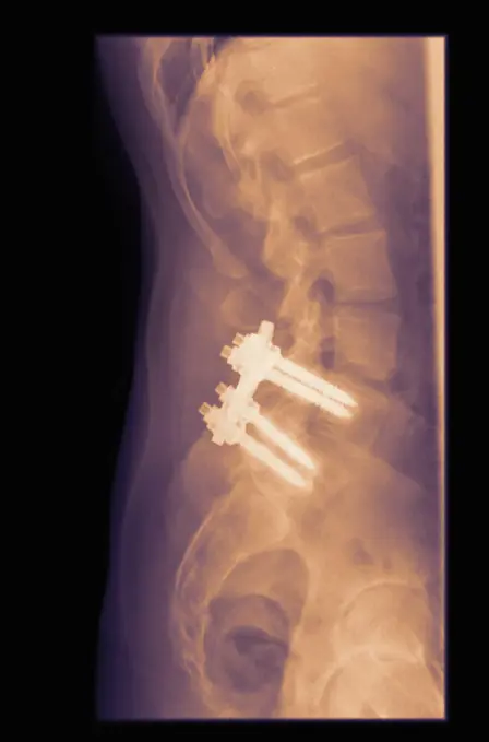
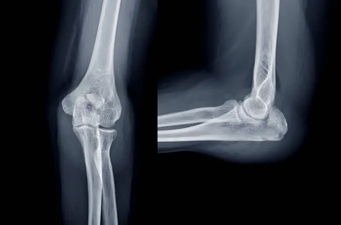
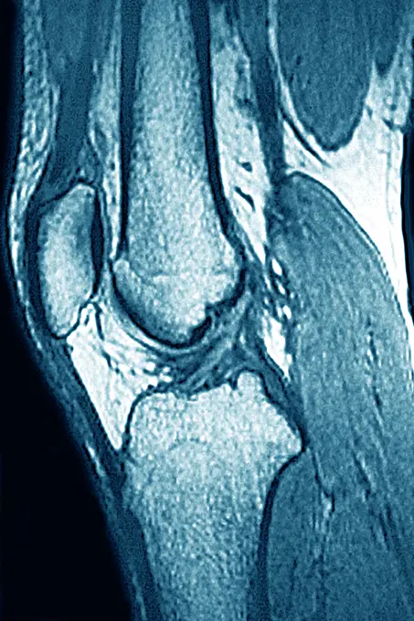
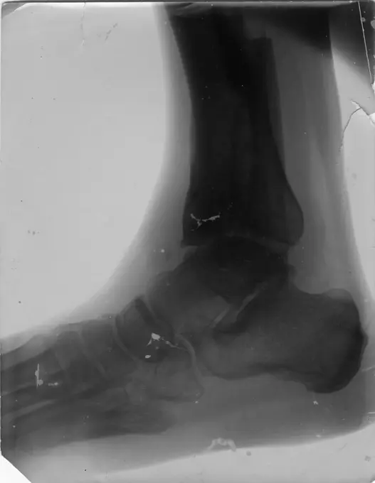
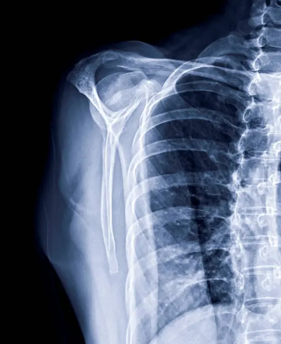
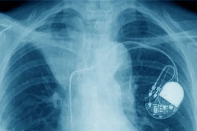
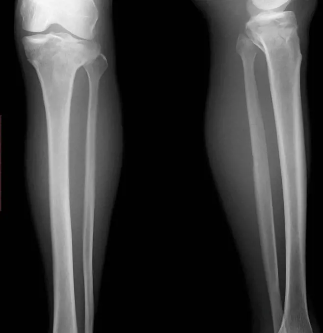
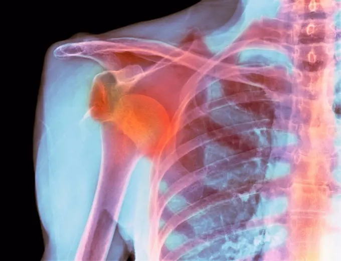
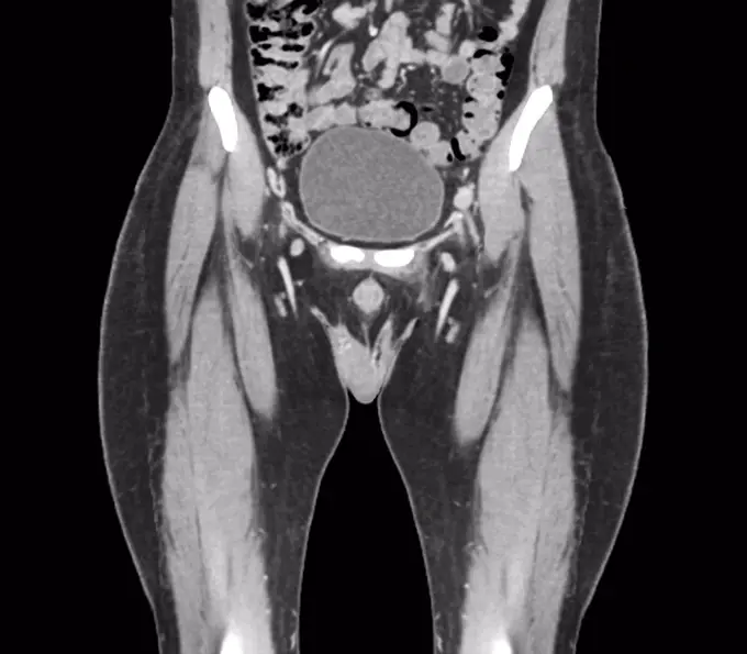
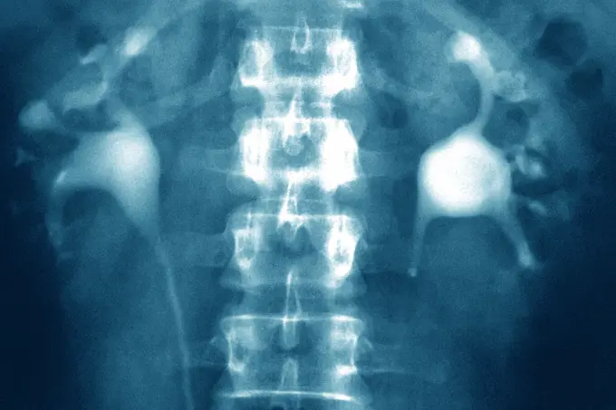
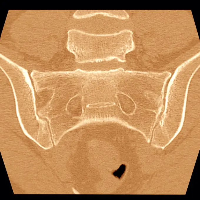
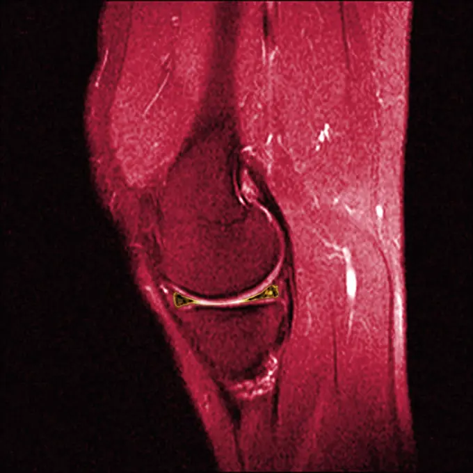
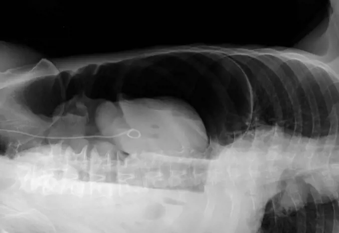
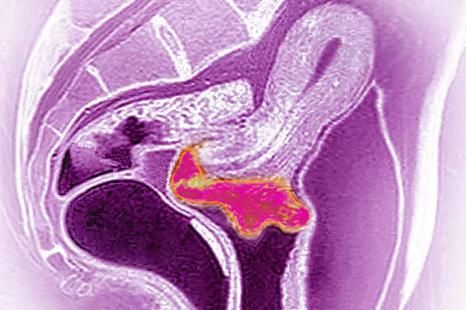
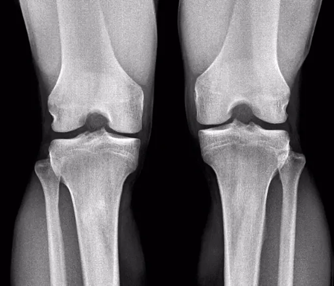
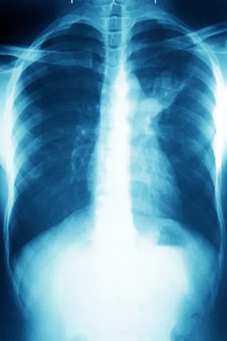
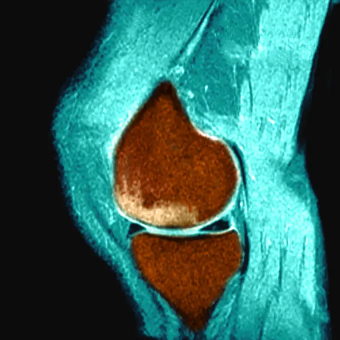
Fine ArtVintageFootageSubscriptionBoardsFree ResearchPricing
  • Fine Art
  • Vintage
  • Footage
  • Subscription
  • Boards
  • Free Research
  • Pricing

Medical Imaging Techniques

X-ray and MRI scans displaying various conditions such as spinal issues and joint placements. Highlights diagnostic imaging in healthcare.

Normal head and neck, MRI scan
291 assets in this story
824-63218245
Spinal neuromas (schwannomas) in cervical vertebrae, seen on a sagittal section MRI scan.
4128-18680265
Synovial chondromatosis, X-ray
824-29055707
X-ray of a hip prosthesis.
4128R-3276
Normal head and neck, MRI scan
1899-53511619
Corticosteroid infiltration between the fourth and fifth lumbar vertebrae, done under radiographic control.
824-63226733
Rotational dislocation at the junction of the thoracic and lumbar vertebrae, visualized by an AP chest radiograph.
824-63178633
Calculation of the bone age with a radiography. 12 years old child elbow radiography. Normal
824-63194017
Pelvic x-ray on a 93-year old woman, showing widespread bone demineralisation.
1815R-98306
X ray of knee, close up
824-63206302
Ruptured Achilles tendon, seen on a sagittal section MRI scan of the ankle.
4128R-12577979
Coloured profile X-ray showing the left knee of a 46 year old obese patient with arthritis.
4128R-12780
Normal hip. Coloured X_ray of the hip of a 90 year old man.
1848-61113910
Cervical spine
1899-53511366
Fracture of the foot on the wedge bone. Foot scanner.
4269-27500
Chondroma. Sagittal MRI of the knee showing a chondroma of the femur (thigh bone). This benign bone tumor is formed by a calcified cartilage matrix giving the bone marrow its white spotted aspect.
4128-19489554
Broken knee cap, X-ray
4297-1071
Normal knee x-ray of a 58 year old man
1899-54027400
Nephritic colic caused by a stenosis on the left ureter. Frontal abdominal-pelvic x-ray. (Photo by: BSIP/UIG via Getty Images)
824-63207410
Transluminal agioplasty of the right, subclavian artery, seen on a frontal chest x-ray.
824-63206989
Right and left ureter, seen on two frontal abdominal x-rays.
824-63203280
Osseous edema on the knee. Sub-cortical lesion of the condyle, seen on a sagittal section MRI.
4128R-12657
Arthritis of the knee. Frontal X_ray showing the knee of a 59 year old patient with arthritis.
824-63218241
Dislocated cervical vertebrae (traumatic lesion of cervical vertebrae C1-C2). X-ray in profile.
824-63211558
Osteoarthritis of the hip seen on a frontal x-ray.
1525-28468527
X ray film for human hands and legs
824-29055568
In a radiology center, an x-ray of the hip with osteoarthritis.
1848-53980273
Screwed
4128-19357557
X-ray of a knee, taken from a patient with knee pain, showing a calcified mass within Hoffa's fat pad. This is suggestive Hoffa's fat pad syndrome.
4128-18680252
Broken knee cap, X-ray
1296R-224
Scan of the human knee
824-63224753
Cervical cancer visualized by sagittal MRI, papillomavirus infection is often the cause.
4297-1021
Normal cervical (neck) x-ray of a 51 year old woman who was seen in the Emergency Room after a car accident complaining of neck pain
6019-29996485
Neck and head x-ray film vertical
824-63194016
Pelvic x-ray on a 93-year old woman, showing widespread bone demineralisation.
1899-54026849
Congential dislocation of the right hip in a 2 and a half-year old child, seen on a frontal pelvic x-ray.
1525-22286596
VDM
4128-28769185
Fractured shoulder, X-ray
1899-54026879
Takayasu"s arthritis, seen by 2 adominal scanners (sagittal section on the left and frontal on the right), at the descending aorta.
1525-25210478
hand on x-ray negative film
5514-67623065
MRI in Ventura CA
824-63175206
Hypophyseal tumor is an abnormal growth in the pituitary gland which may cause endocrine disorders. Axial CT scan of the brain.
4128-24795645
Dislocated shoulder, X-ray
4128-19357532
Contrast barium enema X-ray of a 40-year-old man with recurrent abdominal pain showing reverse intestinal malrotation and colon malrotation. The sigmo...
4128R-12655
Normal child´s knee. Frontal X_ray of the knee of a 10 year old child.
4269-27210
Meniscus. Sagittal Magnetic Resonance Image (MRI) of the knee showing the meniscus (two red triangles). The patella or knee cap (above) and the proximal tibia (below) are in purple.
1899-54028017
Screw and plate osteosynthesis on a fracture of the fibula. Frontal x-ray: tibia, fibula, malleolus and astragalus.
1899-54027157
Paget"s disease, seen in a frontal MRI scan of the L4 and L5 lumbar vertebrae.
4128-24795474
Knee osteoarthritis, X-ray
4128-19489812
Tibial plateau fracture, X-ray
6114-50976706
An x-ray of an ankle pott's fracture. The common low boot injury. It is a fracture of the fibula near the ankle. It is also called Dupuytren's fracture.
4128-19499664
Broken leg, x-ray
1899-53508071
In a radiology center, an x-ray of the hip with osteoarthritis.
1899-53511504
Significant degenerative lumbar disc disease in a 75-year-old woman, frontal x-ray.
1439-57940802
Xray showing spinal fusion hardware in place
1525-22721013
X-ray image film detail of neck and red zone pain.
824-63204754
Complete hip prostheses (treatment for the destruction of hip cartilage through arthritis). Frontal pelvic x-ray.
6145-59431628
Few hands, anonymous, c. 1896 - c. 1915 photograph  Netherlands paper x-ray photograph / gelatin silver print
6188-55645106
Hand, Mann Röntgenbild, Mann, Hand Copyright: xZoonar.com/Dr.xNorbertxLangex 13793153
4128-28766899
Fractured ulna, X-rays
824-29057240
Normal knee visualized by MRI in sagittal section.
6145-46703649
X-ray photography of kneeed taken by Oscar Karlowitch Larsson at Åminne, Mount or Sockholm.
4128-28769188
Fractured shoulder, X-ray
1899-54027685
Pacemaker implanted in the chest. Frontal chest x-ray.
1848-55997740
X-ray image of broken hip in a dog
824-63178616
Renal lithiasis, x-ray
1899-54027402
Diverticulum in the bladder. Frontal pelvic x-ray. (Photo by: BSIP/UIG via Getty Images)
4128-19489785
Tibial plateau fracture, X-ray
824-63204758
Complete hip prostheses (treatment for the destruction of hip cartilage through arthritis). Frontal pelvic CT scan.
4128R-11287602
Dislocated shoulder. Coloured X-ray of a 77 year old female patient with a dislocated shoulder.
4128R-12577957
Computed tomography (CT) scan of the healthy full bladder (oval) of a 45 year old patient.
4128-30419935
Healthy aortic valve, CT scan
6188-55508963
X-ray elbow Two elbow x-rays on a black background Copyright: xZoonar.com/NatalliaxYaumenenkax 3626529
1841R-111793
1525-22453794
X-ray picture
1660R-55738
Male hands holding xrays
4128-19489769
Supracondylar fracture, X-ray
824-63206942
Asbestosis (pulmonary fibrosis) seen on an axial section chest scan.
4128-18929464
Fractured leg, X-ray
824-63170767
Post-traumatic wrist arthritis. X-ray of right hand.
824-63225970
Osteoarthritis of the left hip (coxarthrosis) visualized by a pelvic x-ray of the front.
4269-27847
Crohn's disease. Colored MRI (Magnetic Resonance Imaging) of the small intestine of a 10 years old child with Crohn's disease. The walls of the ileum (inferior terminal part in the small intestine, at left in gray) have a thicker and ragged aspect.
4128R-15200
Tendinitis of the shoulder. Coloured X_ray of the shoulder of a patient showing calcification grainy patches around the rotator cuff tendon upper left...
4128-28769210
Healthy spine, X-ray
1899-53511531
Uterine cancer visualized by MRI in radial section.
824-63224696
X-ray of renal colic on the left kidney (very intense pain in the lumbar region and genitals). This pain results from an obstruction of the upper urinary tract, most often secondary to a kidney stone (urolithiasis) between the kidney and the bladder. Abdominal x-ray.
824-63188661
Secondary malignant tumour at the top of the right femur, seen on a frontal MRI scan.
1899-86076
Scanner Sacroiliac Joint
4269-27209
Meniscus. Sagittal Magnetic Resonance Image (MRI) of the knee showing the meniscus (two yellow triangles). The patella or knee cap (above) and the proximal tibia (below) are in dark red.
4128-19358213
Lateral left decubitus (LLD) abdominal X-ray of a patient with abdominal pain showing a crescent shape radiolucent lesion above the liver suggestive o...
1899-53511523
Cervical cancer visualized by sagittal MRI, papillomavirus infection is often the cause.
4128R-12577962
Normal knees. Frontal X-ray of the healthy knees of a 28 year old.
824-63215989
Pulmonary tuberculosis, seen on a frontal chest x-ray.
824-63178482
Severe osteonecrosis of the knee of a 78 years old patient. RMN
824-63178665
X-ray of the right hand and fourth finger of a 10-year-old boy. No visible recent bone lesion.
4128-19489614
Broken ankle, X-ray
1525-25279085
hand holds radiograph
824-63176420
Reportage in the Osteoarticular Infections Referral Centre (CRIOA) at Diaconesses Croix Saint Simon hospital in Paris. Septic surgery, removing an internal fixation in the ankle following an infection. X-ray of the patient's ankle.
824-63224950
Dental panoramic of a 70 year old man with an implant and many crowns, the crosses visualizing the teeth that will need care. Paris, Ile-de-France, France.
4128R-6042
Normal elbow and wrist joints, X_ray
824-63128447
Here total bilateral prostheses. Frontal x-ray of the hips. See image n0622705 on the site.
PREVIOUS
of 3
NEXT
2401 S. Ervay, Suite 206
Dallas, Texas 75215
United States
Browse
CategoriesGalleriesFree Research
For Creators
License Your ContentSuperStock For StudentsFrame of Mind BlogContributor PortalNew Image SpotlightLicense AI Imagery
Resources
About UsLicensingTerms & ConditionsAPI AccessPrivacyPricing
Contact
+1 866 236 0087help@superstock.com Contact form
©2026 SuperStock. All Rights Reserved. -A
This site is protected by reCAPTCHA and Google Privacy Policy and Terms of Service apply.