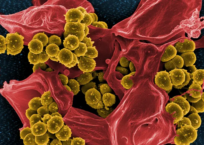
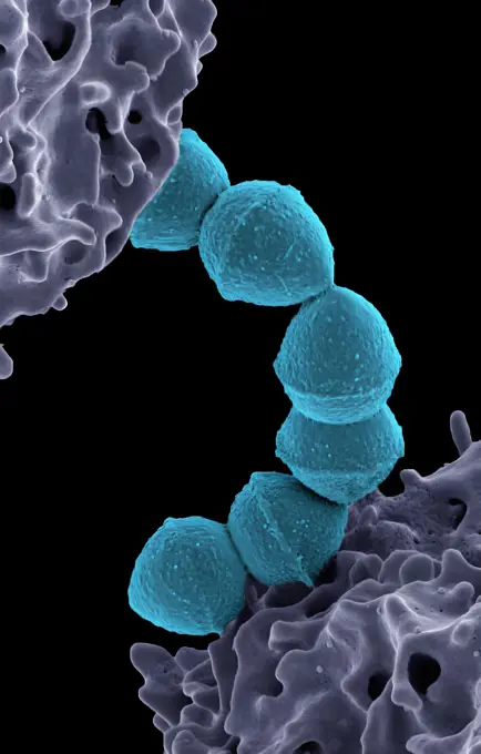
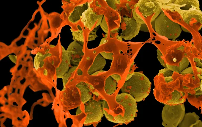
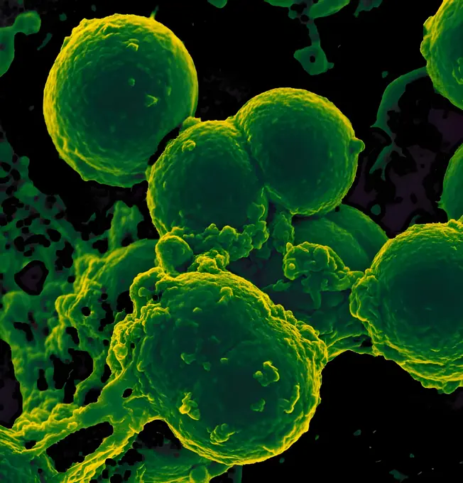
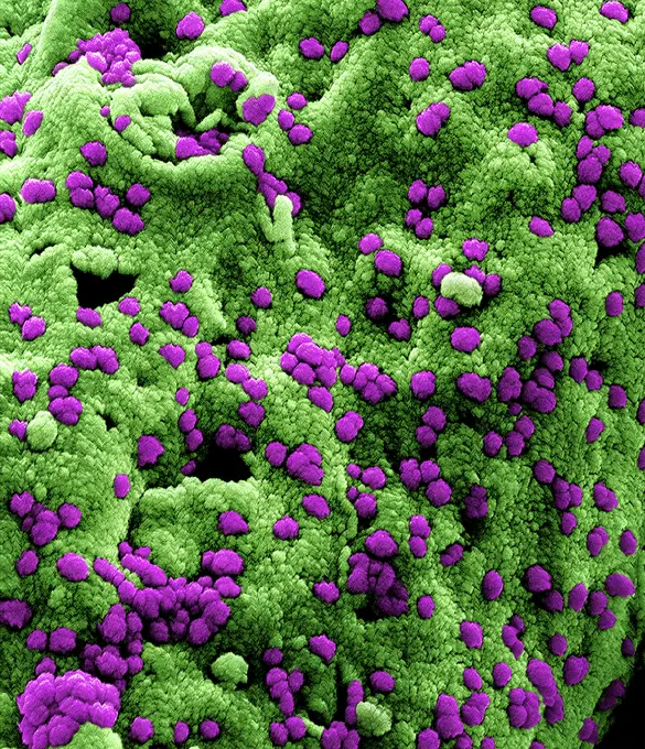
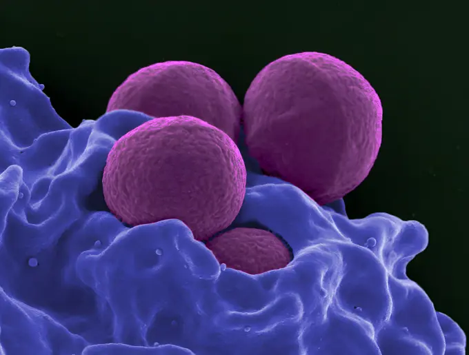
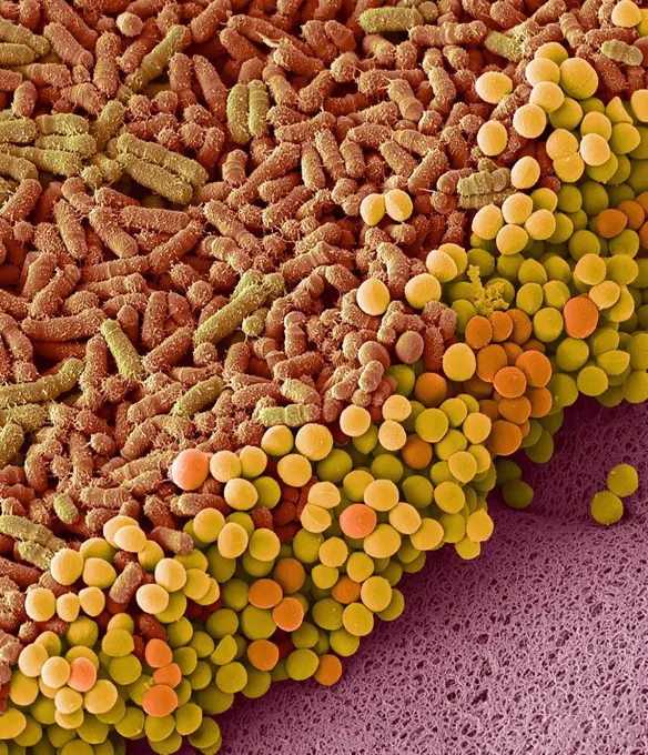
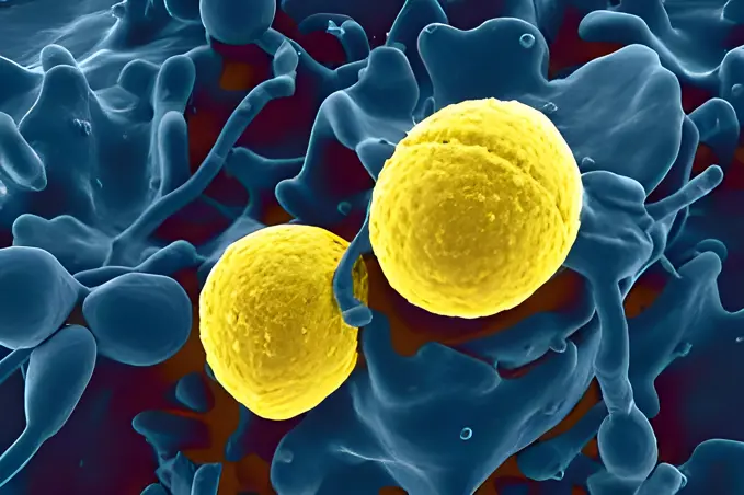
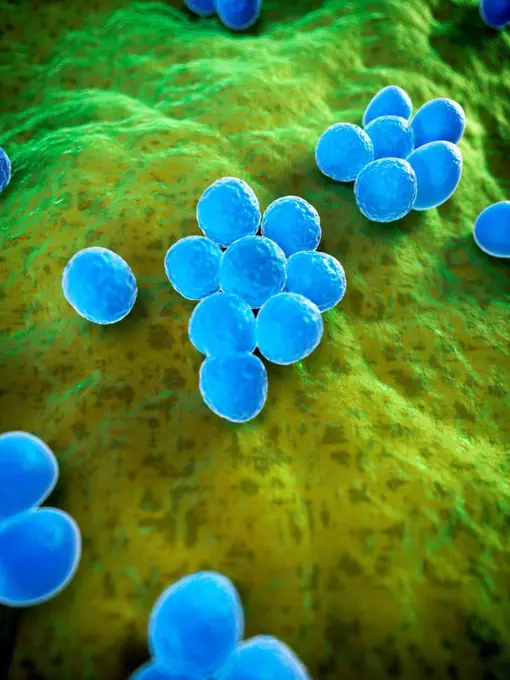
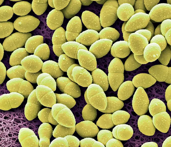
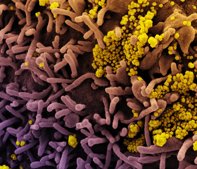
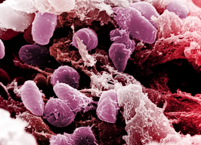
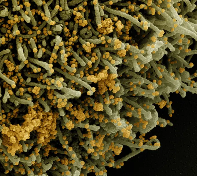
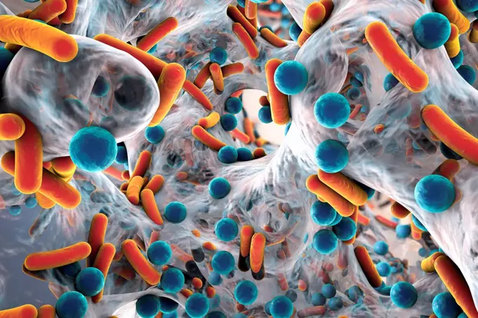
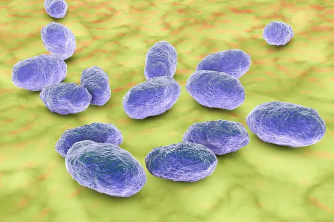
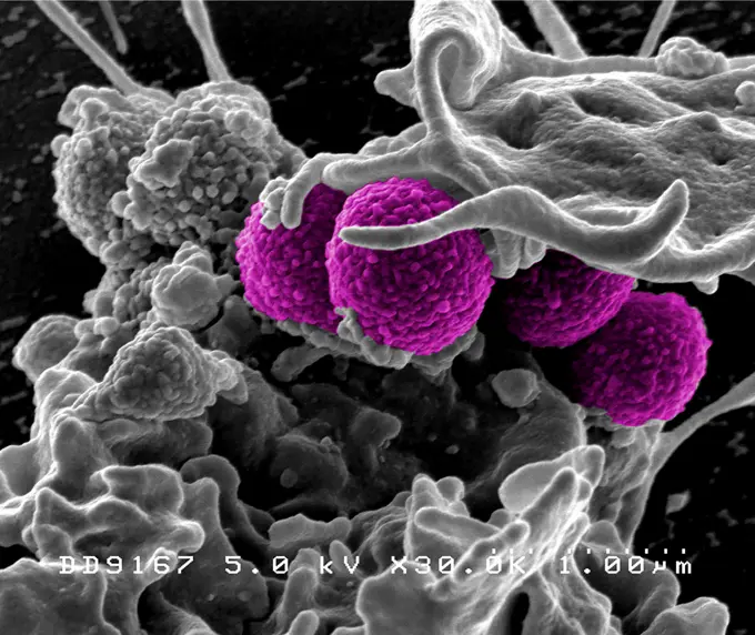
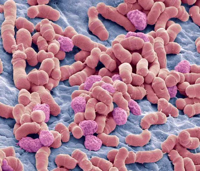
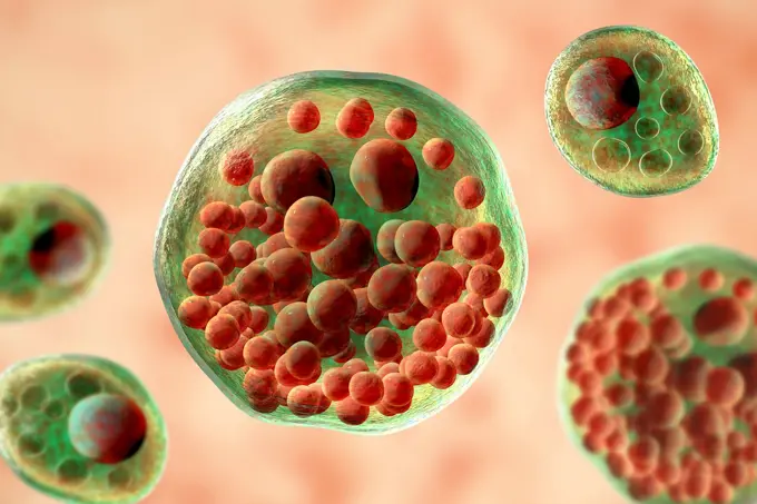
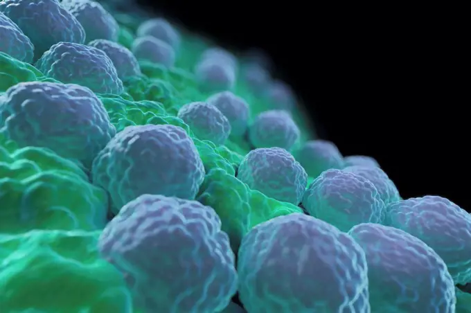
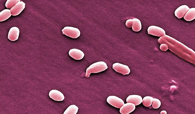
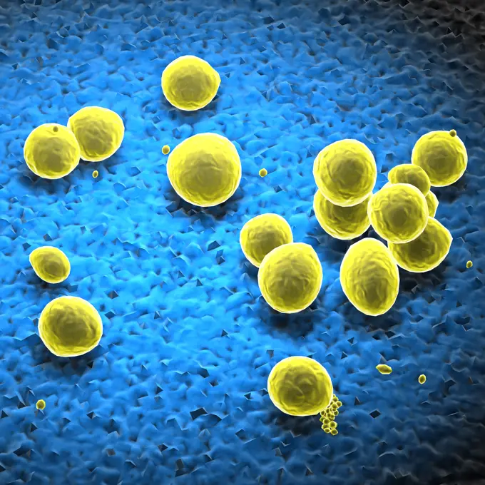
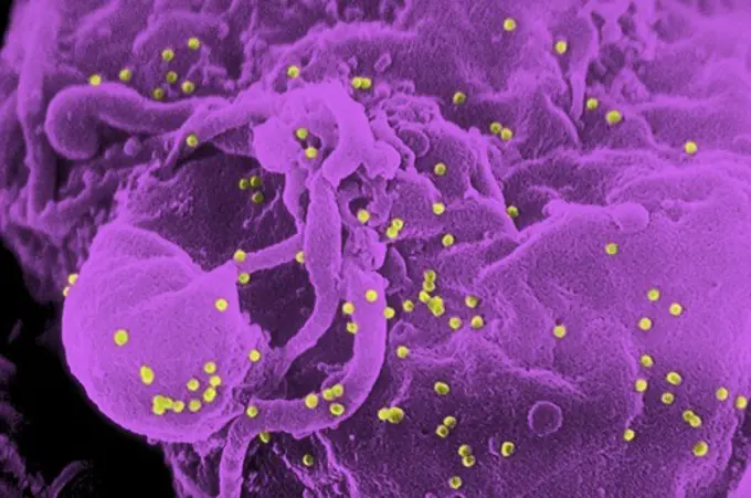
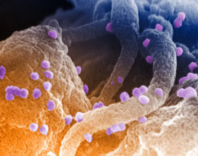
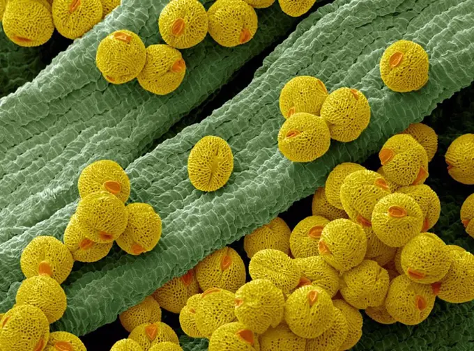
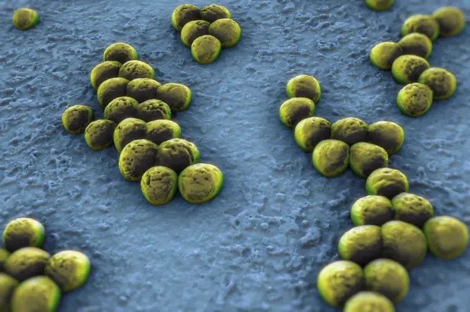
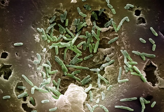
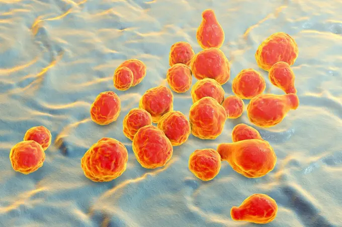
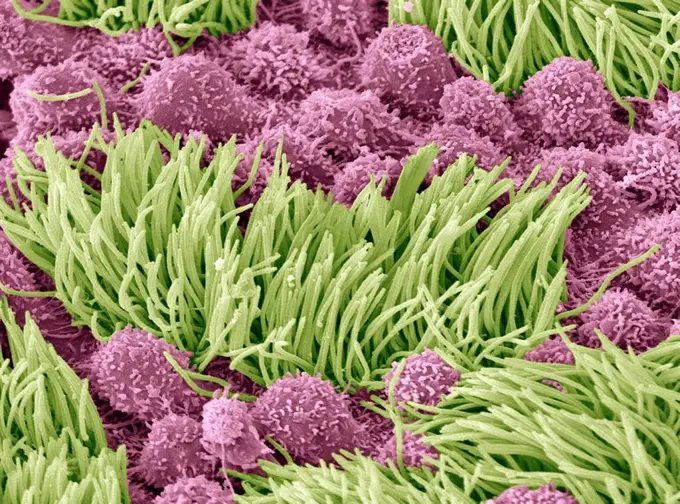
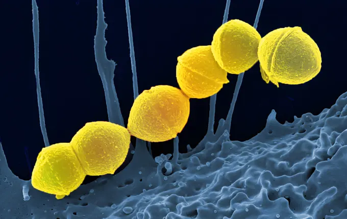
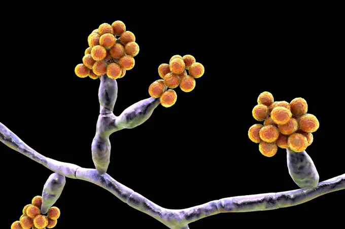
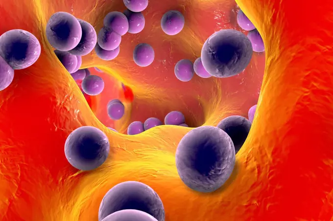
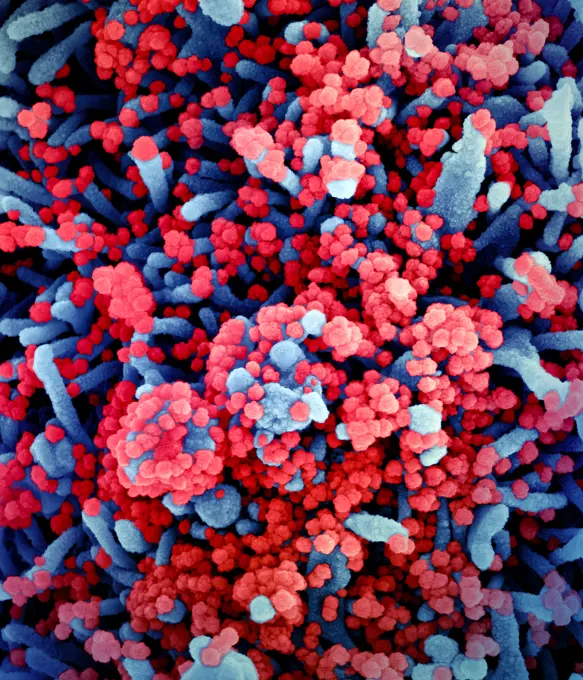
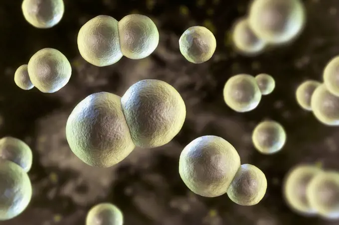
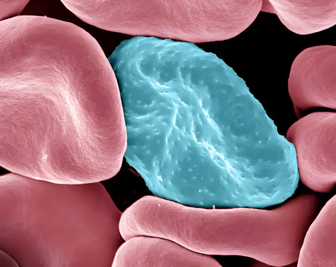
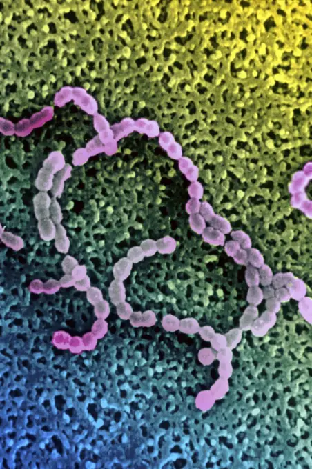
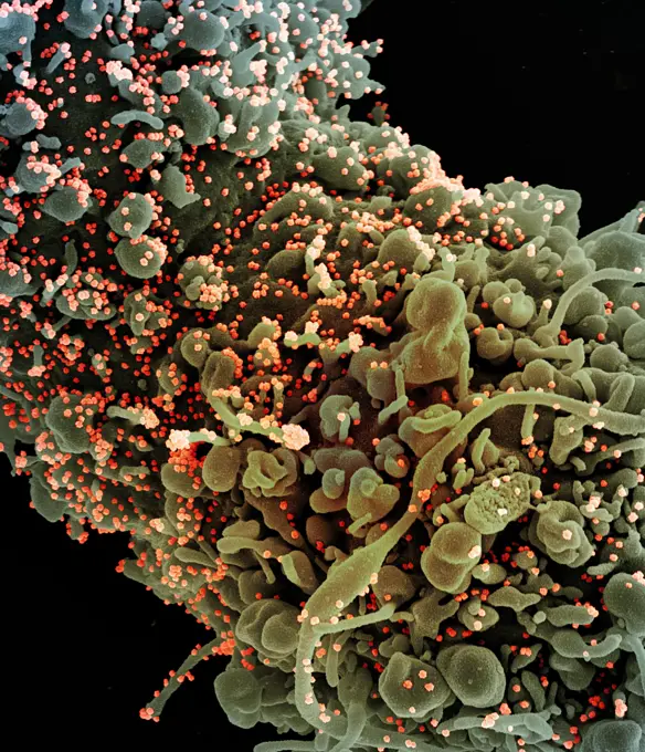
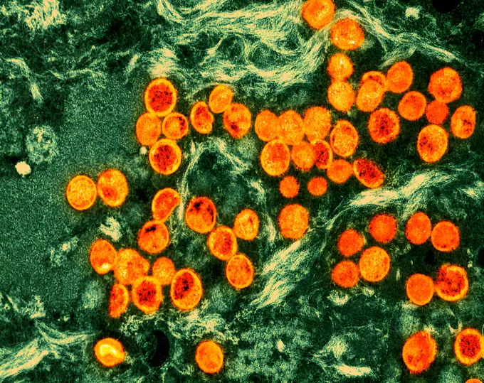
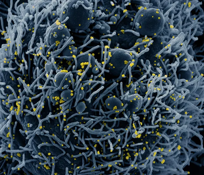
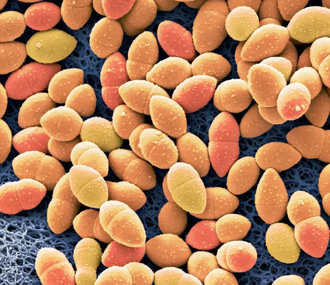
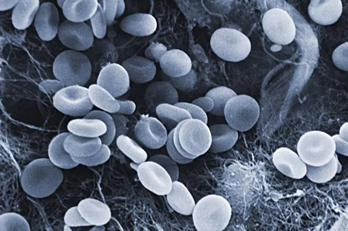
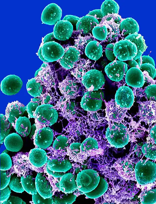
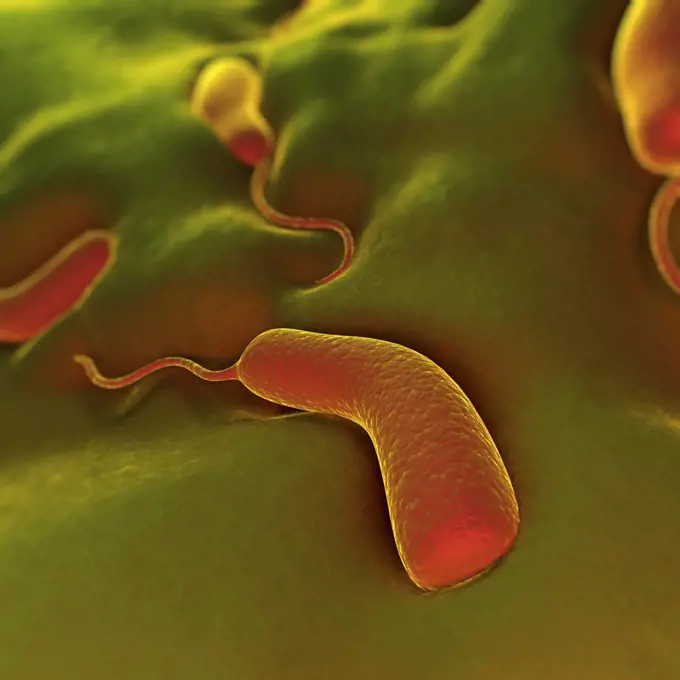
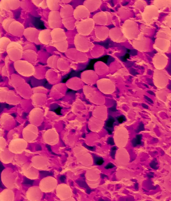
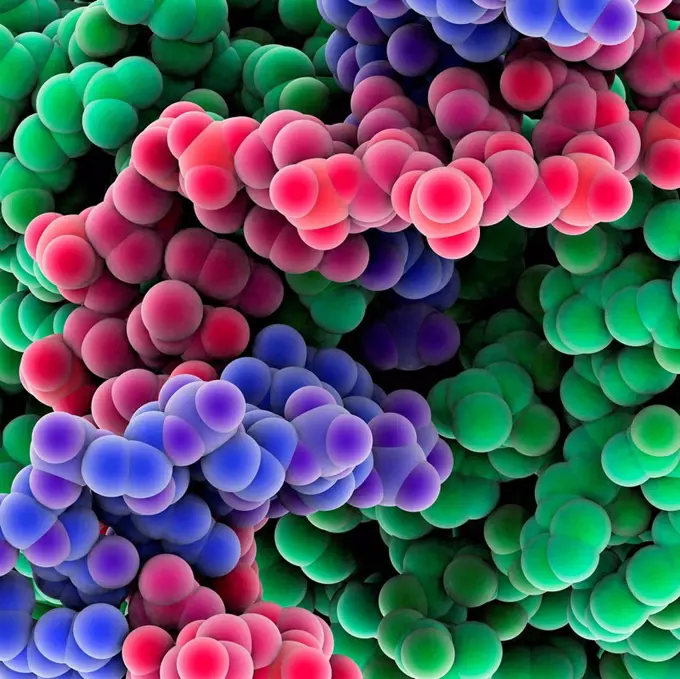
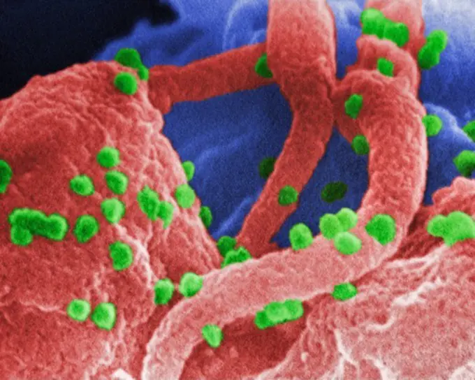
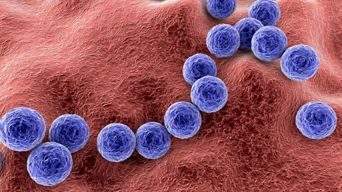
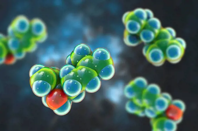
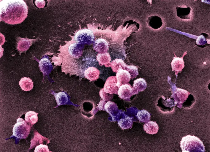
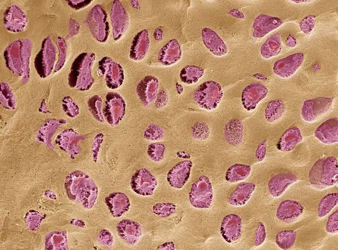
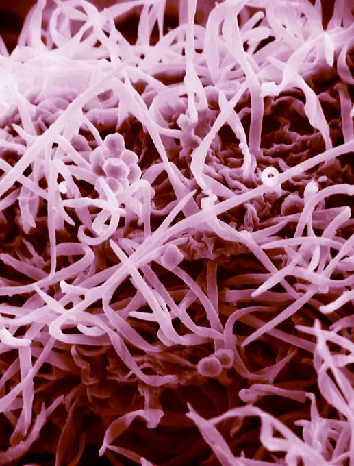
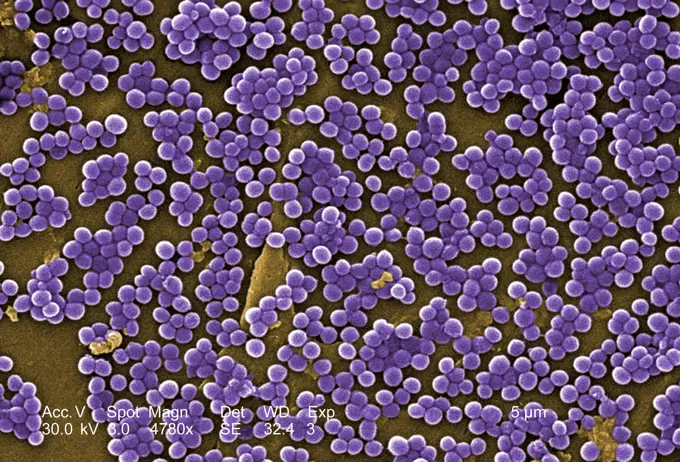
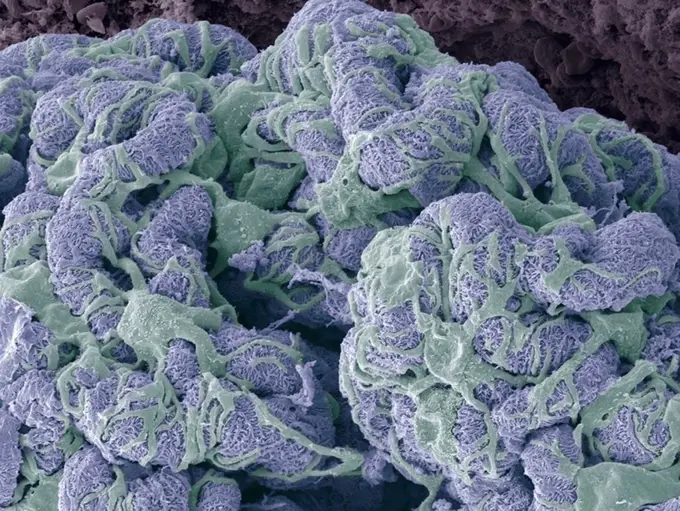
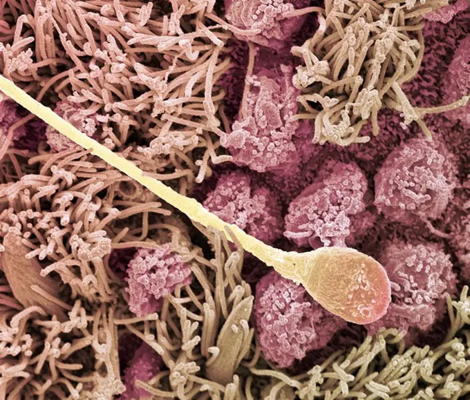
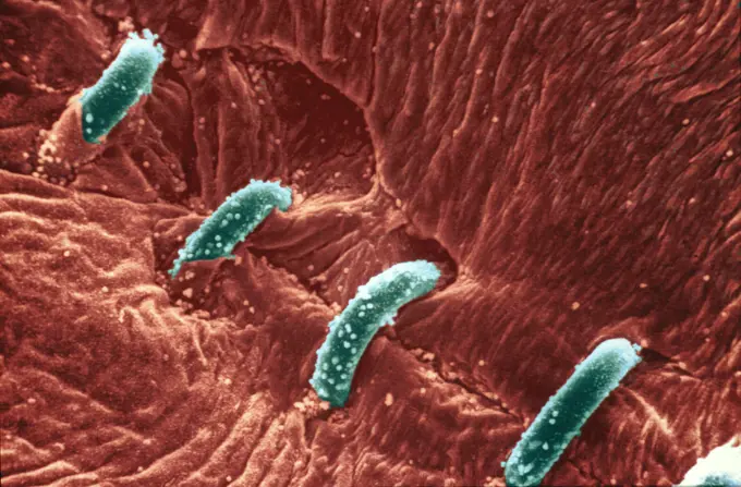
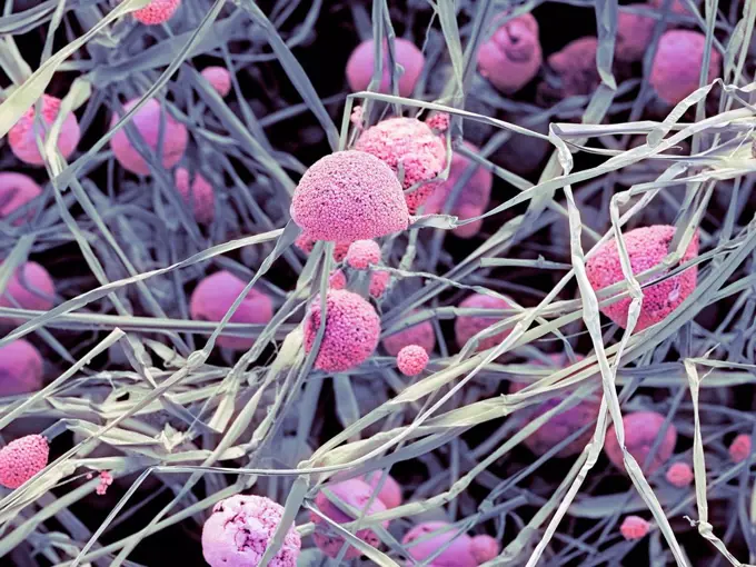
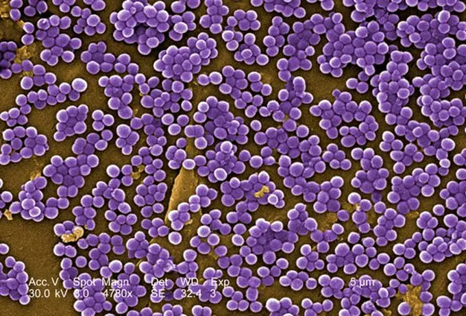
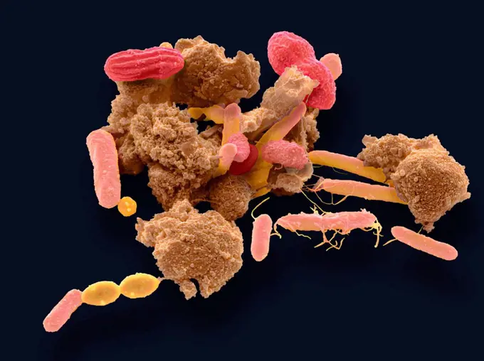
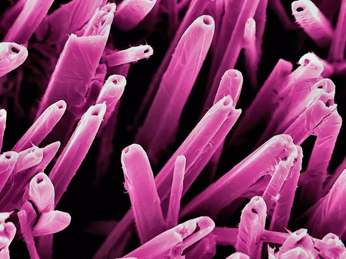
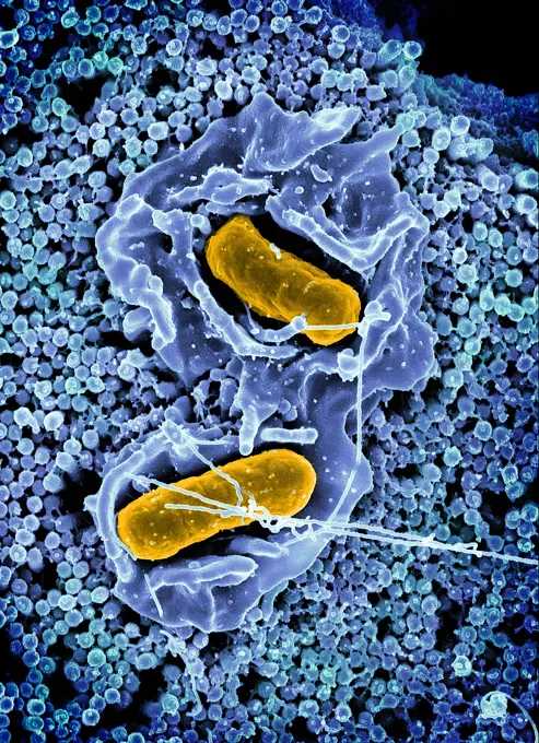
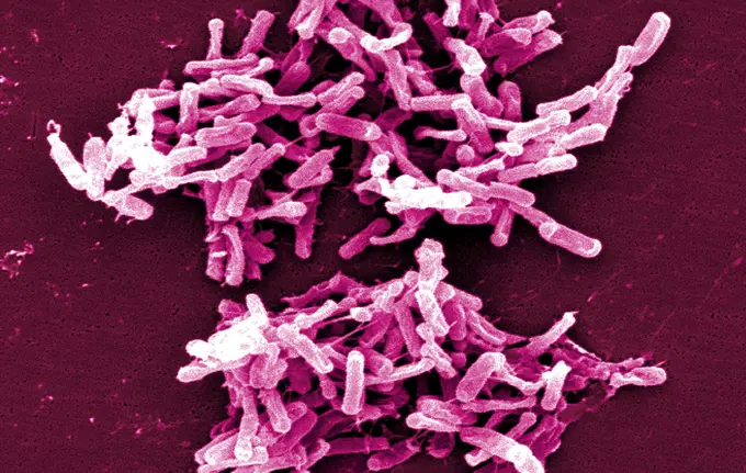
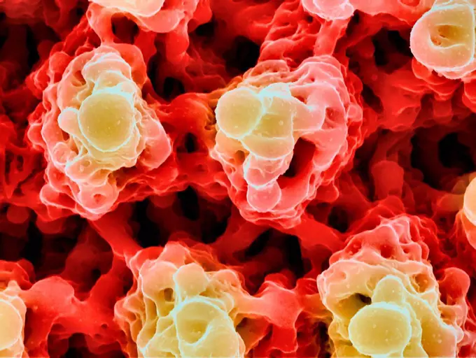
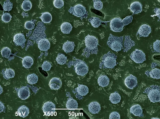
Fine ArtVintageFootageSubscriptionBoardsFree ResearchPricing
  • Fine Art
  • Vintage
  • Footage
  • Subscription
  • Boards
  • Free Research
  • Pricing

Microbial Structures under Microscope

Colorized scanning electron micrographs revealing intricate details of cells and bacteria, showcasing infections and immune responses.

Foot bacteria, SEM
Foot bacteria, SEM
179 assets in this story
824-65831032
Colorized scanning electron micrograph of a cell (pink) infected with the Omicron strain of SARS-CoV-2 virus particles (yellow), isolated from a patient sample.
824-63223195
Scanning electron micrograph of methicillin-resistant Staphylococcus aureus and a dead human neutrophil. Credit: NIAID.
1899-65662514
Colorized scanning electron micrograph of Group A Streptococcus (Streptococcus pyogenes) bacteria (teal) and a human neutrophil (purple).
824-63225816
Scanning electron micrograph of methicillin-resistant Staphylococcus aureus (MRSA, yellow) surrounded by cellular debris. MRSA resists treatment with many antibiotics.
824-63225819
Scanning electron micrograph of neutrophil ingesting methicillin-resistant Staphylococcus aureus bacteria.
4269-27385
Methicillin-Resistant Staphylococcus Aureus. Scanning electron micrograph (SEM) of methicillin-resistant Staphylococcus aureus bacteria (MRSA); Magnification 9560x. This strain of Gram-positif bacterium has developed wide-ranging antibiotic resistance such as methicillin, and are hospital pathogens.
824-63194667
Colorized scanning electron micrograph of Middle Eastern Respiratory Syndrome virus particles attached to the surface of an infected VERO E6 cell.
4128-V58562045
4128R-15545732
Foot bacteria, SEM
824-63191238
Scanning electron micrograph of neutrophil ingesting methicillin-resistant Staphylococcus aureus bacteria.
824-63227278
Scanning electron micrograph of a human neutrophil ingesting MRSA (purple).
4128R-15465084
Fecal bacteria, SEM
824-65830700
Transmission electron micrograph of cytomegalovirus (CMV) particles (pink) found within intracellular vesicles of an infected macrophage (purple).
4128R-11476221
Protein molecules, computer artwork.
4269-27444
Escherichia coli. This scanning electron micrograph (SEM) of a Escherichia coli bacterium ; Magnification 3607x. E. coli are rod-shaped Gram-negative bacteria that normally colonize the digestive tract in humans and other animals. However, some strains can produce a toxin that leads to severe illness or even death.
824-63194665
Colorized scanning electron micrograph of Middle Eastern Respiratory Syndrome virus particles attached to the surface of an infected VERO E6 cell.
4128-V58557888
824-63225943
Scanning electron micrograph of Staphylococcus aureus bacteria (gold) outside a white blood cell (blue).
4128-111453073
Bacteria found on mobile phone, SEM
4128R-26273
MRSA bacteria. Computer artwork of methicillin-resistant Staphylococcus aureus (MRSA) bacteria.
4128-20041809
Kefir bacteria, SEM
824-63225882
Colorized scanning electron micrograph of a cell (tan) infected with SARS-CoV-2 virus particles (yellow), isolated from a patient sample. Image captured at the NIAID Integrated Research Facility (IRF) in Fort Detrick, Maryland.
4269-6710
Scanning electron micrograph (SEM) depicting a mass of Yersinia pestis bacteria (the cause of bubonic plague) in the foregut of the flea vector.
824-63225922
Colorized scanning electron micrograph of a cell (olive green) infected with a variant strain of SARS-CoV-2 virus particles (orange), isolated from a patient sample. Image captured at the NIAID Integrated Research Facility (IRF) in Fort Detrick, Maryland.
824-65830672
Scanning electron micrograph of Neisseria gonorrhoeae bacteria, which causes gonorrhea.
4128R-13048913
Spherical bacteria and rod-shaped bacteria inside biofilm, illustration. A biofilm is a colony of bacteria that forms a coating on a surface. Common p...
4128R-13763914
Computer illustration of Haemophilus influenzae, coccobacillus bacteria. These bacteria cause a number of disease including meningitis in children, pn...
4128-V58562106
824-63191260
Interaction of MRSA with a human white cell. The bacteria shown is strain MRSA252, a leading cause of hospital-associated infections in the United States and United Kingdom.
4128-V58557921
4128R-30463
Athlete's foot fungus. Coloured scanning electron micrograph (SEM) of Malassezia sp. yeast cells on the skin of a human foot. This fungus can cause th...
4128-28767321
Rhinosporidium seeberi parasite, illustration
4378-3774
Rubella virus particles on a cellular surface. Rubella disease, commonly known as German measles, is the cause of congenital rubella syndrome when infection occurs during the first weeks of pregnancy.
4269-27650
Anthrax bacteria. Electron micrograph of spores of Bacillus anthracis, a Gram-positive species of bacteria which is highly pathogenic and causes the disease anthrax in man.
824-63225876
Colorized scanning electron micrograph of a cell (purple) infected with SARS-CoV-2 virus particles (green), isolated from a patient sample. Image captured at the NIAID Integrated Research Facility (IRF) in Fort Detrick, Maryland.
4292-76712
Staphylococcus bacterium
4269-27652
Anthrax bacteria. Electron micrograph of spores of Bacillus anthracis, a Gram-positive species of bacteria which is highly pathogenic and causes the disease anthrax in man.
4297-1420
Scanning electron micrograph of HIV-1 budding from cultured lymphocyte. Multiple round bumps on cell surface represent sites of assembly and budding of virions
4269-27247
AIDS virus. Scanning electron micrograph (SEM) of human immunodeficiency virus (HIV-1) appearing as small spheres on the surface of a lymphocyte.
4128R-639
Bellflower pollen, SEM
4378-2959
Clusters of 'superbug' (MRSA) bacteria.
4269-27748
Mycobacterium chelonae. Scanning Electron Micrograph (SEM)of Mycobacterium chelonae, a bacterium causing postoperative wound infections in soft tissue and bone.
4128R-14060503
Cryptococcus neoformans fungus, illustration
4128R-2951
Fallopian tube, SEM
824-63227205
Image of group A streptococcus bacteria.
4128-19054666
Madurella fungi, illustration
824-63227075
This scanning electron microscope image shows SARS-CoV-2 (round gold particles) emerging from the surface of a cell cultured in the laboratory. SARS-CoV-2, also known as 2019-nCoV, is the virus that causes COVID-19. Image captured and colorized at Rocky Mountain Laboratories in Hamilton, Montana. Credit: NIAID
4128R-11476189
Protein molecules, computer artwork.
4128R-13023011
Staphylococcus aureus bacteria infected bone (osteomyelitis), computer illustration. Ostemoyelitis is an infection of bone or bone marrow, often cause...
824-63227334
Colorized scanning electron micrograph of a cell (blue) heavily infected with SARS-CoV-2 virus particles (red), isolated from a patient sample. Image requested from the NIAID Integrated Research Facility (IRF) in Fort Detrick, Maryland.
4128-19489924
Blastomyces fungus, illustration
1899-65662315
Colorized scanning electron micrograph of red blood cell infected with malaria parasites (teal). The small bumps on the infected cell show how the parasite remodels its host cell by forming protrusions called 'knobs' on the surface, enabling it to avoid destruction and cause inflammation. Uninfected cells (pink) have smoother surfaces.
4128R-11476203
Protein molecules, computer artwork.
824-63223238
Colorized scanning electron micrograph of a VERO E6 cell (purple) exhibiting elongated cell projections and signs of apoptosis, after infection with SARS-COV-2 virus particles (pink), which were isolated from a patient sample. Image captured at the NIAID Integrated Research Facility (IRF) in Fort Detrick, Maryland. Credit: NIAID.
824-201
Streptococcus Pyogenes
4269-24929
Progesterone. SEM (Scanning Electron Microscope)
824-63223236
Colorized scanning electron micrograph of a cell showing morphological signs of apoptosis, infected with SARS-COV-2 virus particles (orange), isolated from a patient sample. Image captured at the NIAID Integrated Research Facility (IRF) in Fort Detrick, Maryland. Credit: NIAID.
1899-61460459
Colorized transmission electron micrograph of monkeypox virus particles (orange) found within an infected cell (green), cultured in the laboratory
824-63223231
Colorized scanning electron micrograph of an apoptotic cell (blue) infected with SARS-COV-2 virus particles (yellow), isolated from a patient sample. Image captured at the NIAID Integrated Research Facility (IRF) in Fort Detrick, Maryland. Credit: NIAID.
4128-20041755
Kefir bacteria, SEM
1439R-995003
Red blood cells
824-63194641
Scanning electron micrograph of a clump of Staphylococcus epidermidis bacteria in the extracellular matrix, which connects cells and tissue.
4378-2540
Group of vibrio cholerae bacteria which causes cholera.
4128-V58557862
4128R-13620042
Coloured scanning electron micrograph (SEM) of Oral bacterium, Streptococcus mutans with surrounding dextran polysaccharide mucilage (glucan). S. muta...
4128R-11476183
Protein molecules, computer artwork.
4384-241
Scanning Electron Micrograph of HIV Virions
4128-17927939
Streptococcus pyogenes bacteria, illustration
4128-20044483
Linalool molecule, illustration
4269-6696
Scanning electron micrograph (SEM) of malignant B-cell lymphocytes seen in Burkitt's lymphoma. Magnification x4000.
4128R-4196
Hyaline cartilage, SEM
4269-27744
Vancomycin Resistant Enterococcus. Color enhanced scanning electron micrograph of Vancomycin Resistant Enterococci (VRE). This strain of enterococcus bacteria is resistant to the antibiotic vancomycin, known as Vancomycin Resistant Enterococci. Illness due to VRE infections in healthy people is rare. Common infections caused by enterococci are urinary tract inections and wound infections.
824-63123751
This is a scanning electron micrograph showing the epidermal hair entanglement on the surface of a red raspberry drupelet. The red raspberry is known to harbor various strains of parasites, i.e. Cylospora, and bacteria known to cause foodborne illnesses o
824-63194407
This colorized scanning electron micrograph (SEM) depicted numerous clumps of methicillin-resistant Staphylococcus aureus bacteria, commonly referred to by the acronym, MRSA, Magnified 4780x. Recently recognized outbreaks, or clusters of MRSA in community settings have been associated with strains that have some unique microbiologic and genetic properties, compared with the traditional hospital-based MRSA strains, which suggests some biologic properties, eg virulence factors like toxins, may allow the community strains to spread more easily, or cause more skin disease.
824-63128609
Scanning electron micrograph of a laboratory-grown potable water biofilm, with the presence of Hartmanella vermiformis cysts. Aquatic bacteria were grown as biofilm on steel for one week. Hartmanella vermiformis protozoans were then added, which phagocytized the bacteria, that multiplied within vesicles and became cysts in which the bacteria will live until they rupture. See image n1313705 on the site.
4128R-7135
Kidney glomerulus, SEM
4128R-13928456
Parasitic amoeba (Entamoeba histolytica) engulfing red blood cells, computer illustration. This single-celled organism causes amoebic dysentery and ul...
6019-19983696
Microscopic view of floating influenza virus cells. Dangerous illness
4378-2780
Clusters of 'superbug' (MRSA) bacteria.
4128-V58562136
4128R-13620071
Coloured scanning electron micrograph (SEM) of Enterococcus faecalis (formerly known as Streptococcus faecalis), Gram positive, coccus prokaryote (div...
4128R-4020
Sperm cell, SEM
4269-24686
Stitch. SEM (Scanning Electron Microscope) of stiches on the skin.
4128R-12699842
Coloured scanning electron micrograph (SEM) of fungal cells. The round structures are sporangia, which house the fungus' reproductive cells (spores). ...
4297-1787
Scanning Electron Micrograph (SEM) of methicillin-resistant Staphylococcus aureus bacteria, commonly known as MRSA
4128R-13162310
Faecal bacteria. Scanning electron micrograph (SEM) of various bacteria found in a sample of human faeces. At least 50 per cent of human faeces is mad...
824-63223164
Scanning electron micrograph of human respiratory syncytial virus (RSV) virions (colorized blue) and labeled with anti-RSV F protein/gold antibodies (colorized yellow) shedding from the surface of human lung epithelial A549 cells. Credit: NIAID.
1899-53513314
Scanning electron micrograph of Crimean-Congo hemorrhagic fever (CCHF) viral particles (yellow) budding from the surface of cultured epithelial cells from a patient.
4128-V58566821
4128R-15047069
Nanostructures on vanadium, SEM
824-63225904
Colorized scanning electron micrograph of a cell (gray) infected with a variant strain of SARS-CoV-2 virus particles (UK B.1.1.7 green), isolated from a patient sample. Image captured at the NIAID Integrated Research Facility (IRF) in Fort Detrick, Maryland.
824-63194434
Scanning electron micrograph of a murine macrophage infected with Francisella tularensis strain LVS. Macrophages were dry-fractured by touching the cell surface with cellophane tape after critical point drying to reveal intracellular bacteria. Bacteria (colorized in red) are located either in the cytosol or within a membrane-bound vacuole.
4128-19056105
Fungi Paracoccidioides braziliensis, illustration
1899-65662511
Scanning electron micrograph of Salmonella Typhimurium invading a human epithelial cell.
824-57657563
Colorized scanning electron micrograph of monkeypox virus (green) on the surface of infected VERO E6 cells (red).
4269-27463
Clostridium difficile. Scanning Electron Micrograph (SEM) of Clostridium difficile bacteria. C. difficile, an anaerobic gram-positive rod, is the most frequent cause of antibiotic-associated diarrhea (AAC).
1899-53513327
Colorized scanning electron micrograph of Ebola virus particles (blue) found both as extracellular particles and budding particles from chronically infected African green monkey kidney cells (brown). Image captured and color-enhanced at the NIAID Integrated Research Facility in Ft. Detrick, Maryland.
4128R-15047067
Microstructures on nickel, SEM
1773-209593
Leaf, x500 magnification
4128R-13162302
Bacteria and yeast. Coloured scanning electron micrograph (SEM) of bacteria (red) and yeast. In many environments, bacteria and fungi coexist and inte...
PREVIOUS
of 2
NEXT
2401 S. Ervay, Suite 206
Dallas, Texas 75215
United States
Browse
CategoriesGalleriesFree Research
For Creators
License Your ContentSuperStock For StudentsFrame of Mind BlogContributor PortalNew Image SpotlightLicense AI Imagery
Resources
About UsLicensingTerms & ConditionsAPI AccessPrivacyPricing
Contact
+1 866 236 0087help@superstock.com Contact form
©2026 SuperStock. All Rights Reserved. -A
This site is protected by reCAPTCHA and Google Privacy Policy and Terms of Service apply.