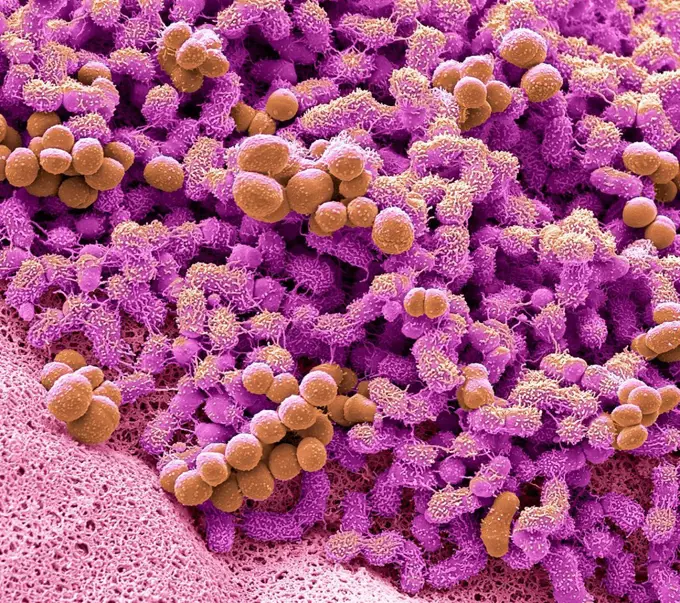
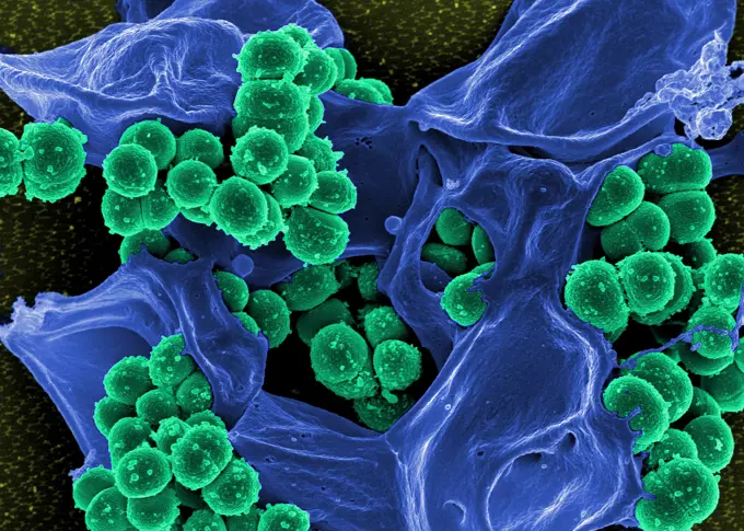
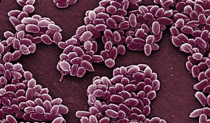
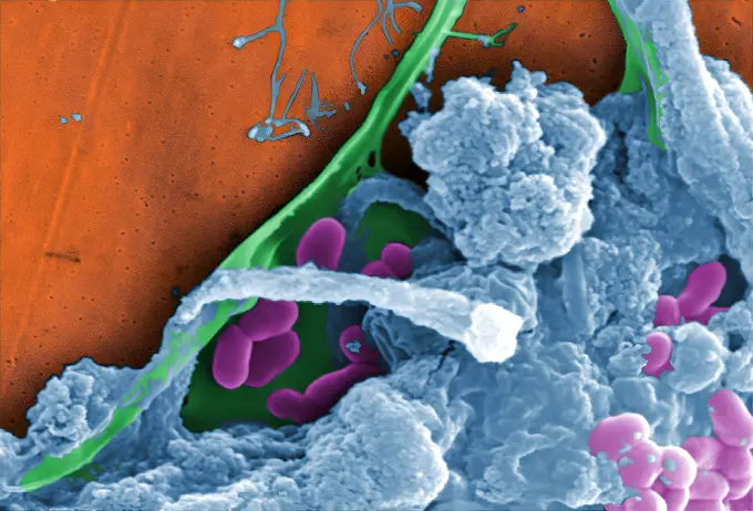
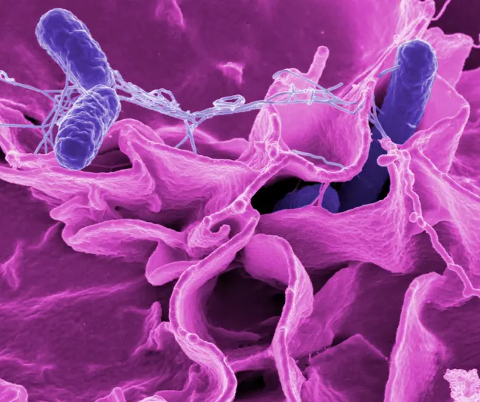
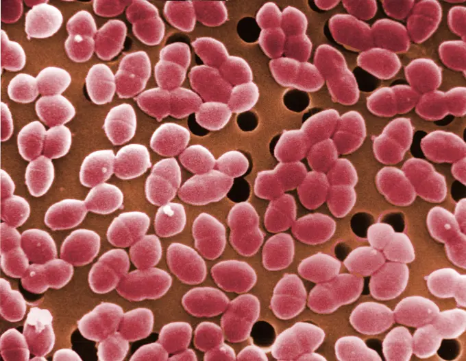
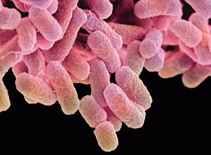
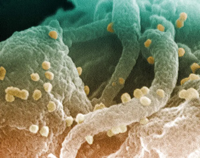
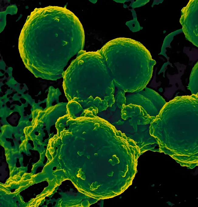
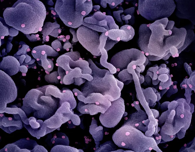
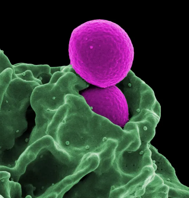
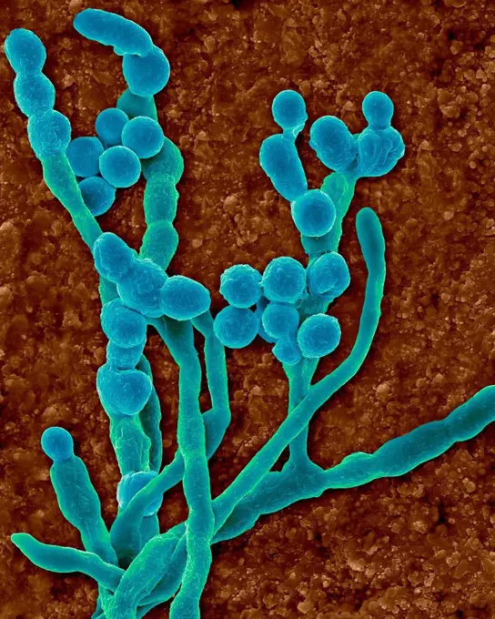
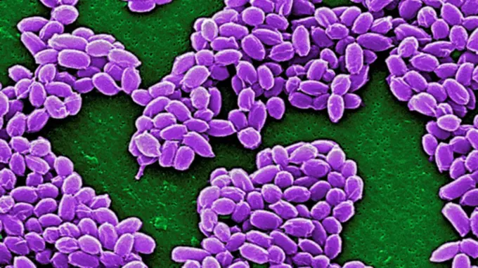
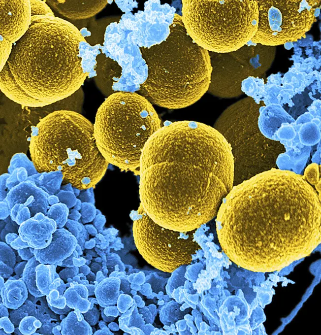
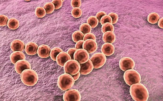
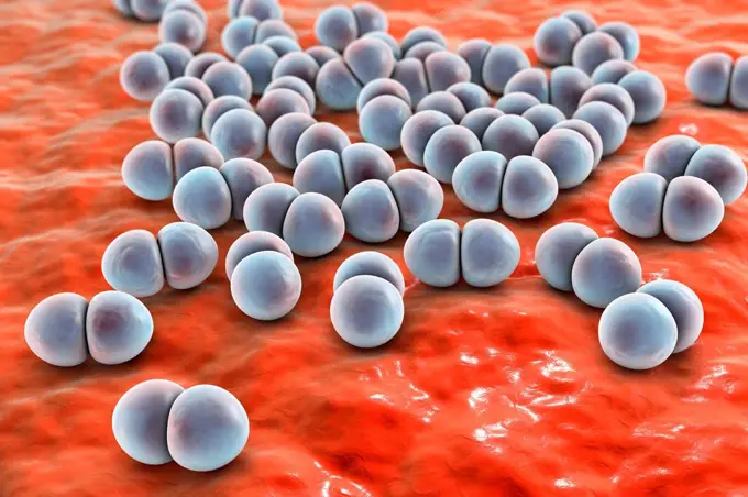
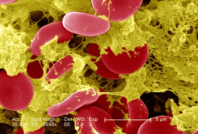
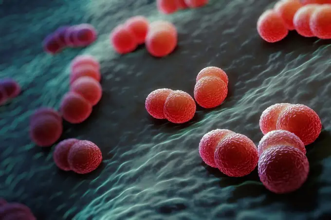
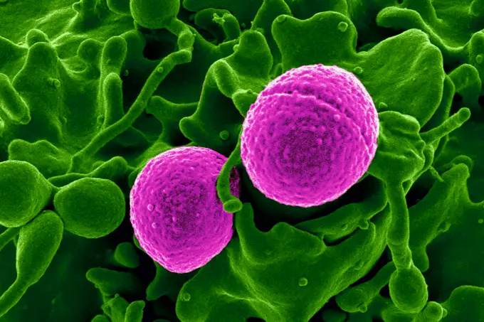
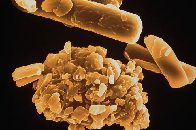
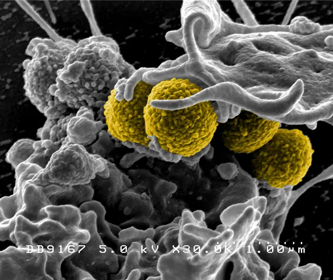
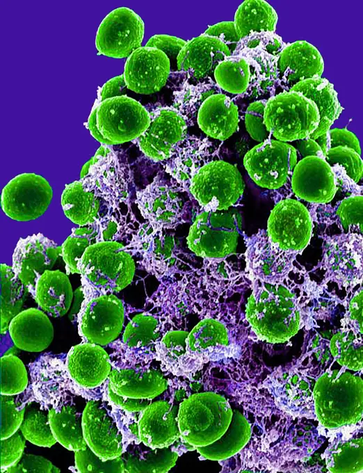
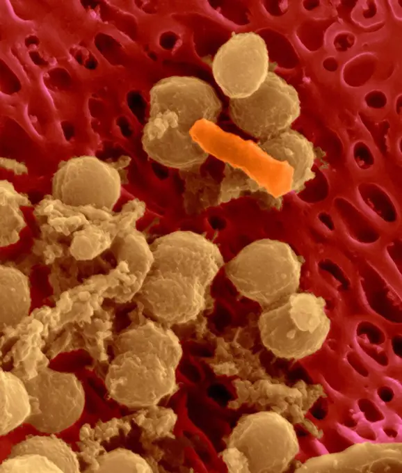
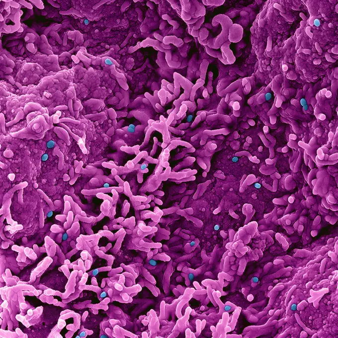
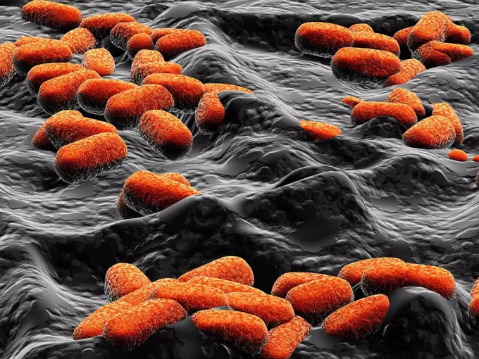
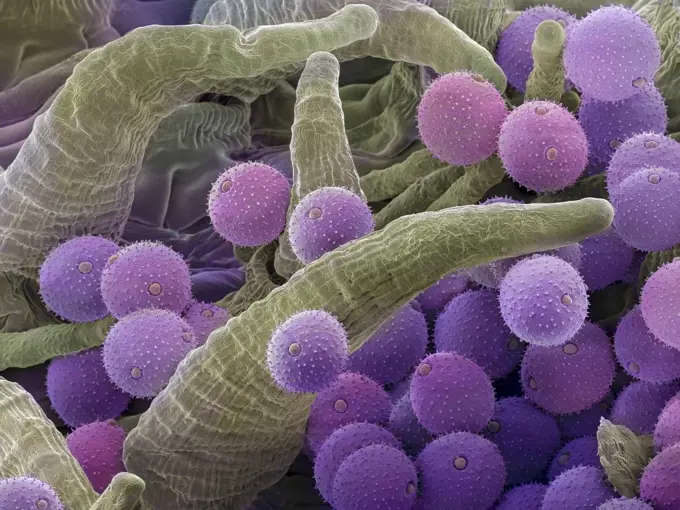
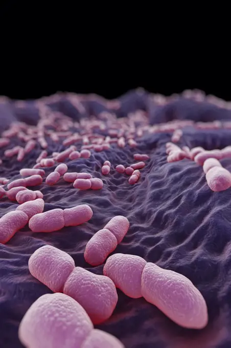
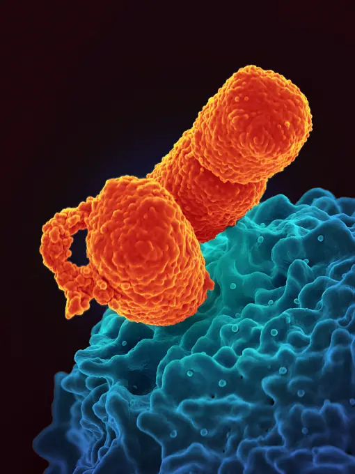
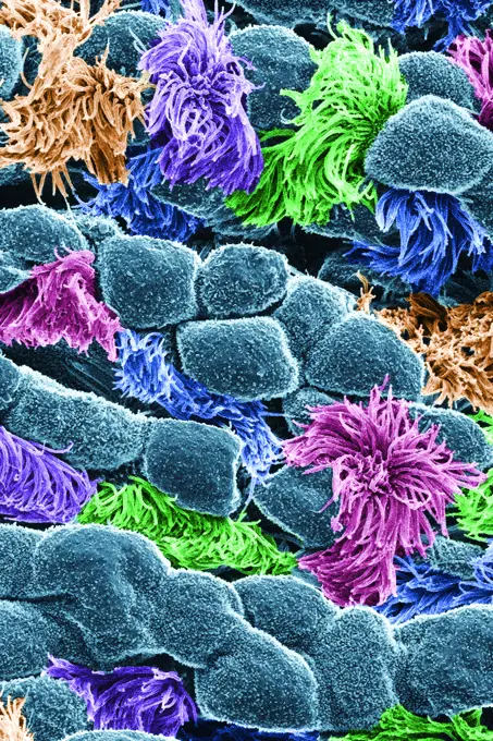
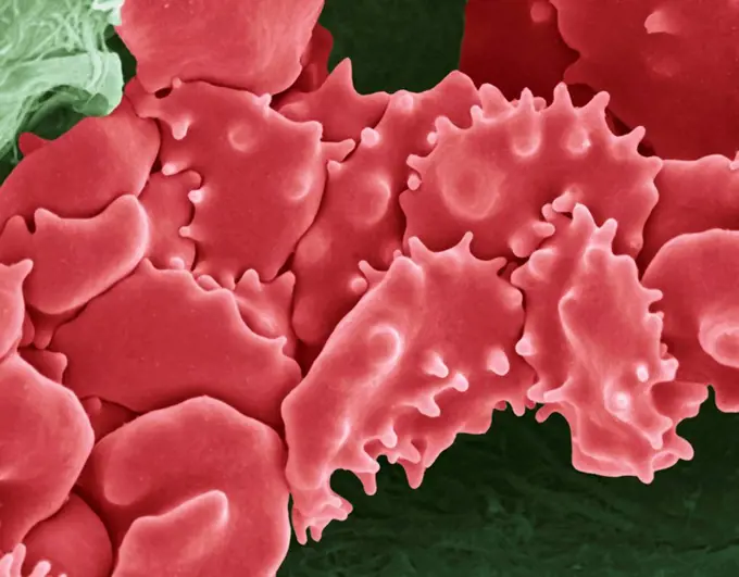
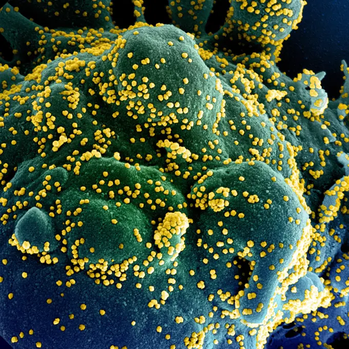
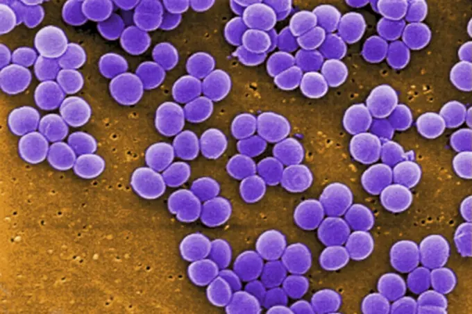
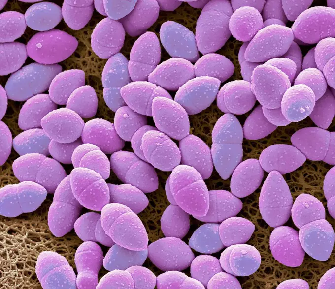
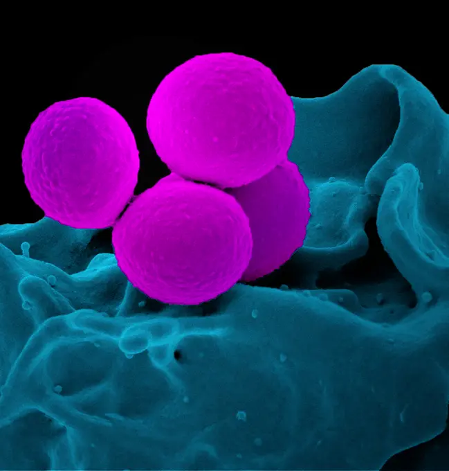
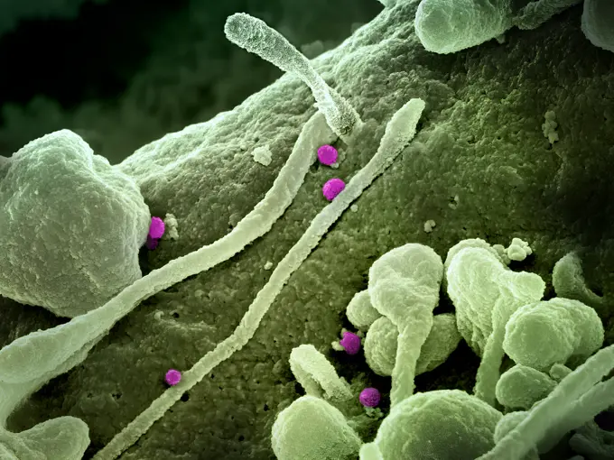
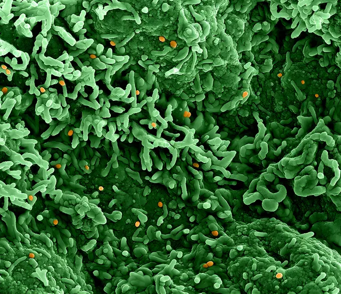
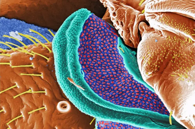
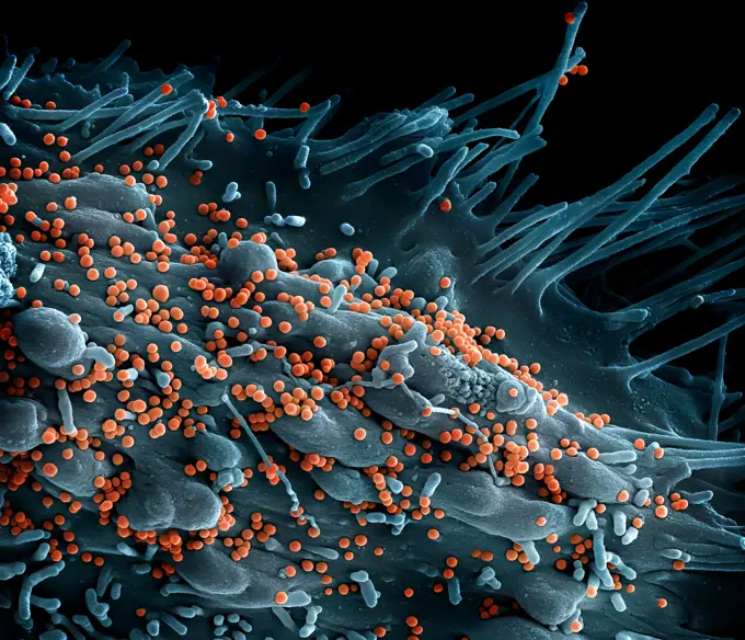
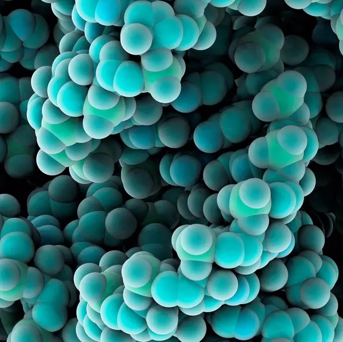
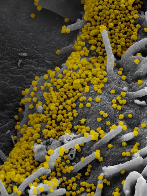
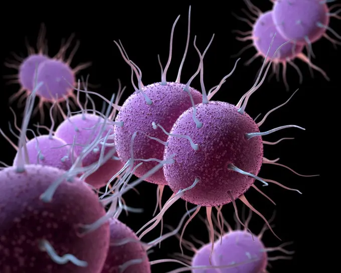
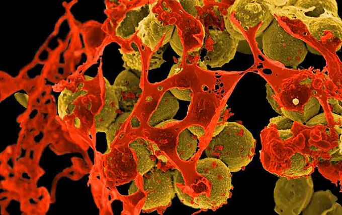
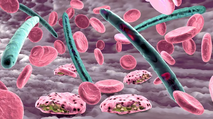
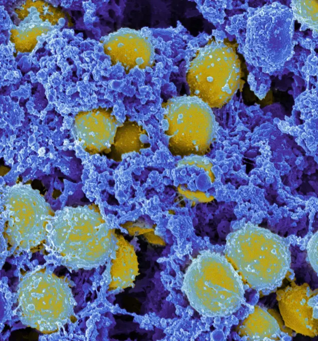
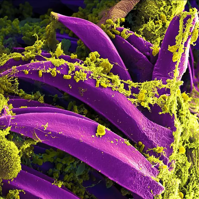
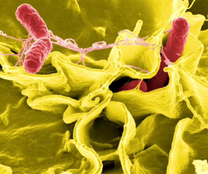
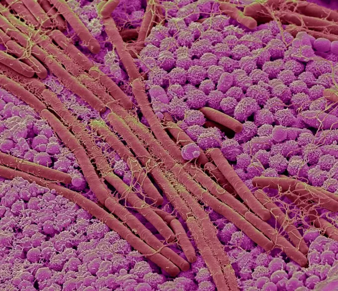
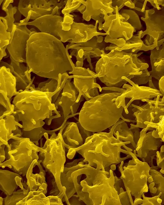
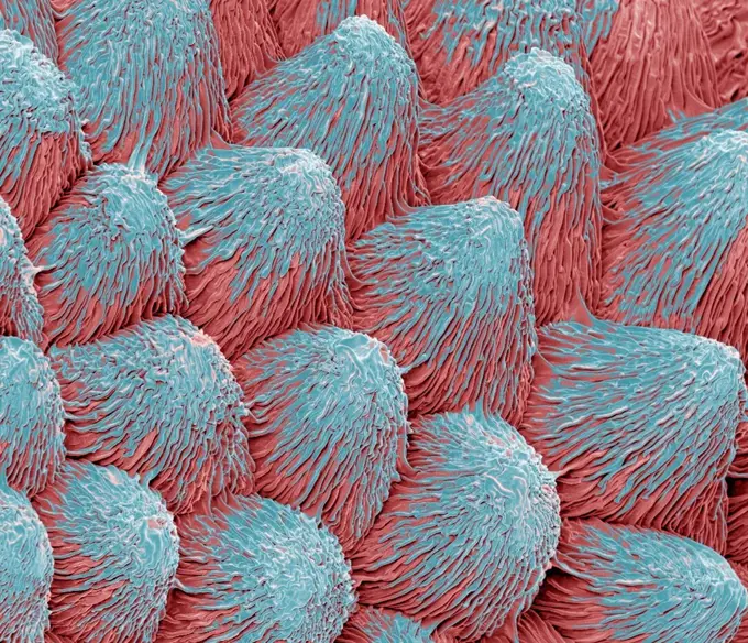
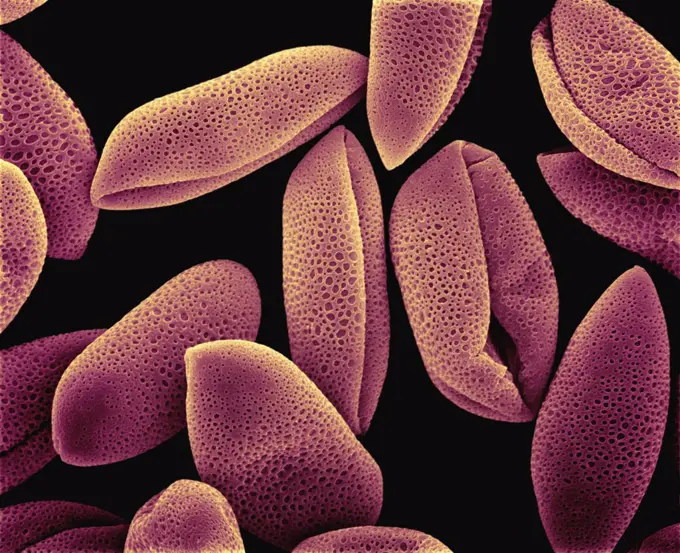
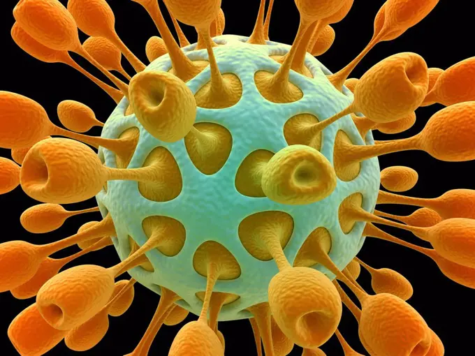
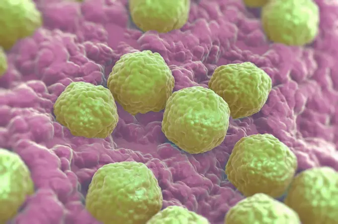
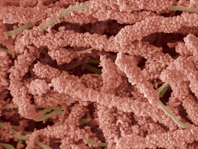
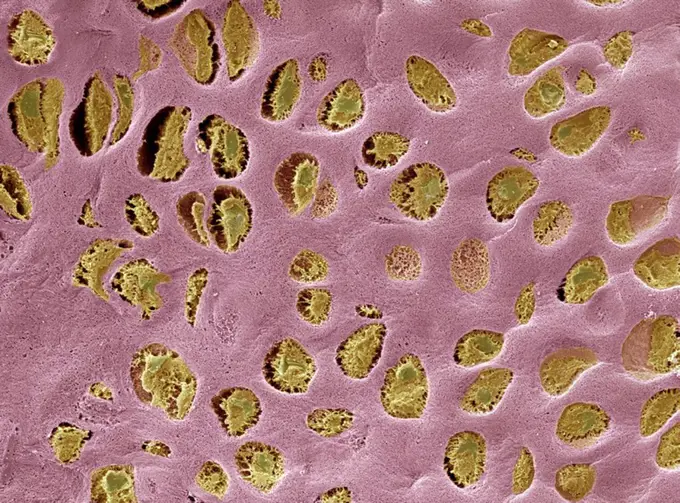
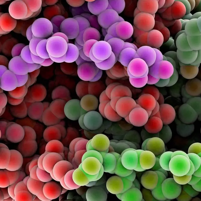
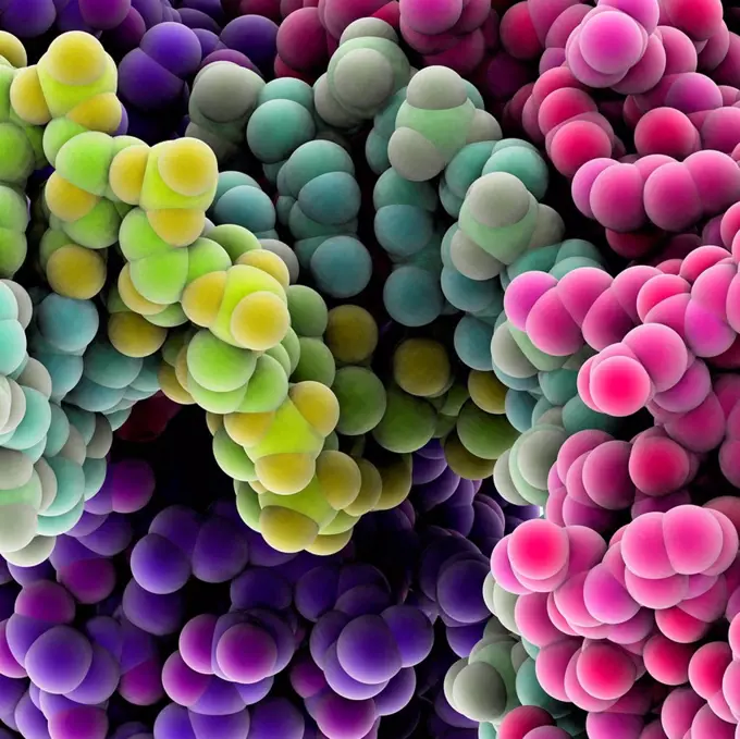
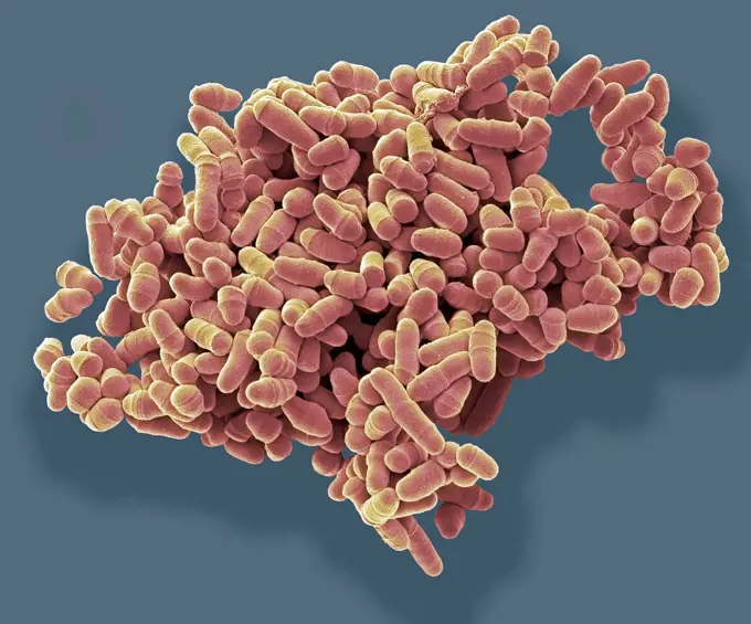
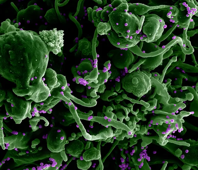
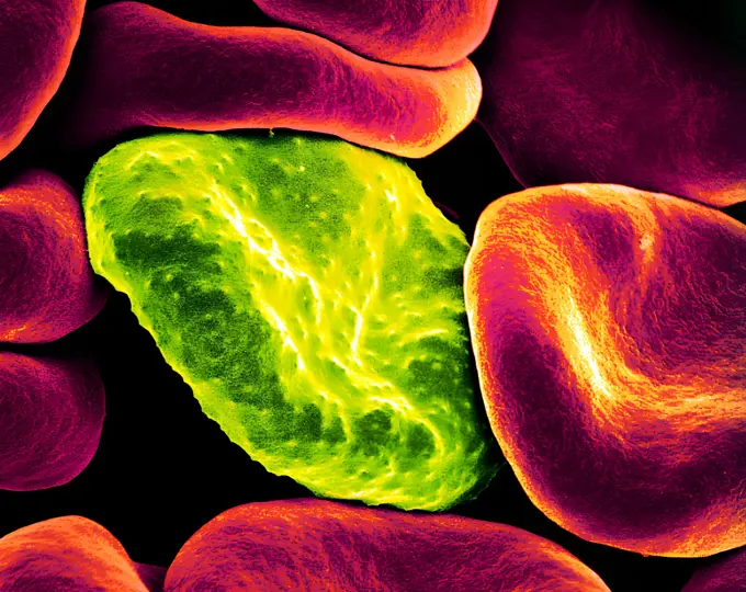
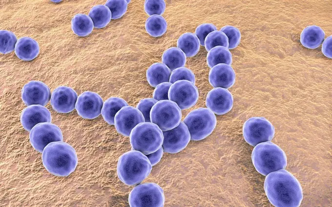
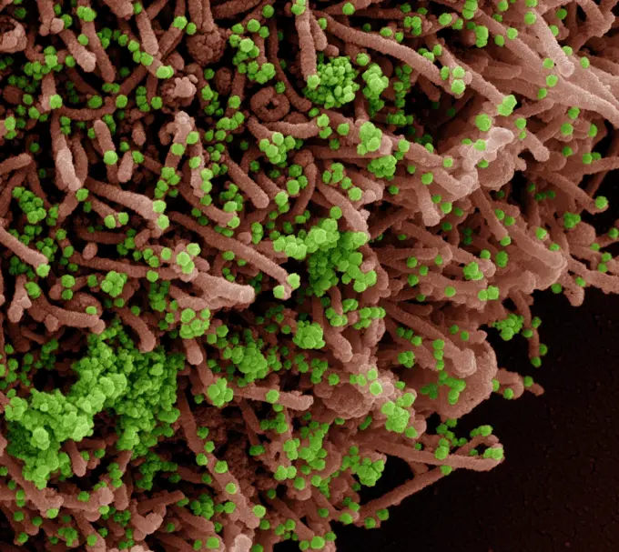
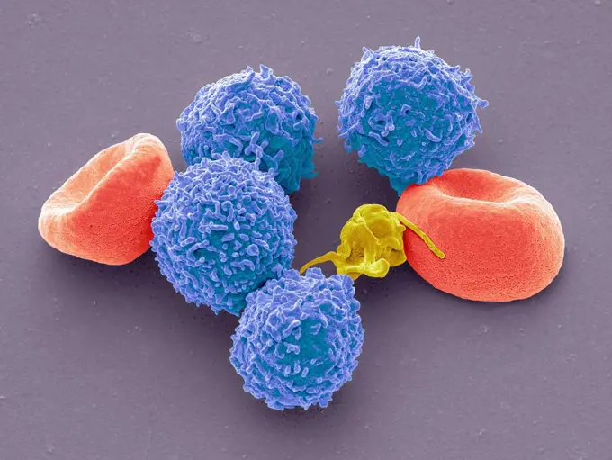
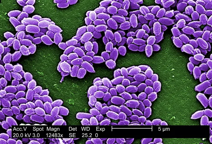
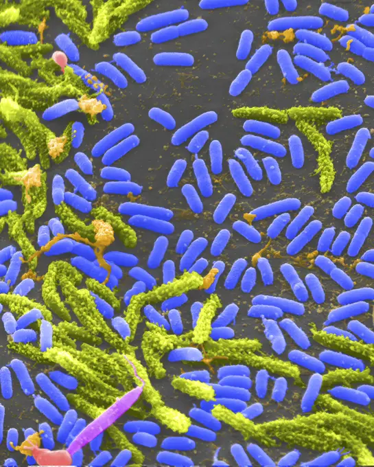
Fine ArtVintageFootageSubscriptionBoardsFree ResearchPricing
  • Fine Art
  • Vintage
  • Footage
  • Subscription
  • Boards
  • Free Research
  • Pricing

Microscopic Bacteria and Cells

Detailed scanning electron micrographs of bacteria, including Staphylococcus and SARS-CoV-2, showcasing vibrant colors and intricate structures.

Foot bacteria, SEM
Foot bacteria, SEM
173 assets in this story
824-63227277
Scanning electron micrograph of methicillin-resistant Staphylococcus aureus bacteria (yellow) and a dead human white blood cell (colored red.)
824-63225897
Colorized scanning electron micrograph of a cell (blue) infected with SARS-CoV-2 virus particles (pink), isolated from a patient sample. Image captured at the NIAID Integrated Research Facility (IRF) in Fort Detrick, Maryland.
1899-65662503
Colorized scanning electron micrograph of a cell (green) infected with the Omicron strain of SARS-CoV-2 virus particles (orange), isolated from a patient sample.
824-63191256
Scanning electron micrograph of methicillin-resistant Staphylococcus aureus and a dead human neutrophil.
4269-27651
Anthrax bacteria. Electron micrograph of spores of Bacillus anthracis, a Gram-positive species of bacteria which is highly pathogenic and causes the disease anthrax in man.
4269-20408823
Coloured scanning electron micrograph (SEM) of methicillin-resistant Staphylococcus aureus (MRSA) bacteria.
1899-61460840
This digitally colorized scanning electron microscopic (SEM) image of an untreated water specimen extracted from a wild stream mainly used to control flooding during inclement weather, revealed the presence of unidentified organisms, which included bacteria, protozoa, and algae. In this particular view, a mass of gelatinous biofilm had enmeshed numbers of microorganisms, including amoebae and bacteria. CDC/ Janice Haney Carr 2009
824-63194443
Salmonella bacteria, a common cause of food poisoning, invade an immune cell.
4128R-15545702
Foot bacteria, SEM
4269-27454
Enterococcus. Scanning electron micrograph (SEM) of Enterococcus faecalis (formerly known as Streptococcus faecalis). This Gram-positive bacterium is commonly found in human intestine. It can also cause urinary tract and  skin infections. E. faecalis is resistant to a number of antibiotics and is often involved in nosocomial diseases.
4128R-6552
E. coli bacteria, SEM
4269-27248
AIDS virus. Scanning electron micrograph (SEM) of human immunodeficiency virus (HIV-1) appearing as small spheres on the surface of a lymphocyte.
824-66066007
Scanning electron micrograph of neutrophil ingesting methicillin-resistant Staphylococcus aureus bacteria. Credit: National Institute of Allergy and Infectious Diseases, National Institutes of Health.
824-65831031
Colorized scanning electron micrograph of a cell (purple) infected with the Omicron strain of SARS-CoV-2 virus particles (pink), isolated from a patient sample.
824-63191247
MRSA being ingested by neutrophil.
824-63194666
Colorized scanning electron micrograph of Middle Eastern Respiratory Syndrome virus particles attached to the surface of an infected VERO E6 cell.
824-65830617
Colorized scanning electron micrograph of Marburg virus particles (purple) both budding and attached to the surface of an infected VERO E6 cell (green).
4128R-13622316
Coloured scanning electron micrograph (SEM) of the common environmental and allergenic mould (Cladosporium sp.); fungal hyphae producing spores. Also ...
4128-V58557894
4128-V58562055
4384-107
Under a high magnification of 12,483X, this scanning electron micrograph (SEM) detected spores from the Sterne strain of  Bacillus anthracis  bacteria. A key characteristic of the Sterne strain of  B. anthracis is the wrinkled surface of the protein coat of these bacterial spores. These spores can live for many years which, enables the bacteria to survive in a dormant state. Photo by Janice Carr.
4269-20408822
Coloured scanning electron micrograph (SEM) of methicillin-resistant Staphylococcus aureus (MRSA) bacteria.
4128R-14846670
Peptostreptococcus bacteria, illustration
4128R-13193038
Computer illustration of Pneumococci (Streptococcus pneumoniae) bacteria.
824-63123772
This scanning electron micrograph (SEM) depicted a closer view of number of red blood cells found enmeshed in a fibrinous matrix on the luminal surface of an indwelling vascular catheter; Magnified 5698x. In this instance, the indwelling catheter was a tu
4378-4616
Neisseria meningitidis
824-63191205
Colorized transmission electron micrograph showing USA 300 strain of Staphlococcus aureus, outside a white blood cell.
4269-24636
Cholesterol crystals. SEM (Electron Scanning Microscope) of crystals of cholesterol.
824-63191259
Interaction of MRSA with a human white cell. The bacteria shown is strain MRSA252, a leading cause of hospital-associated infections in the United States and United Kingdom.
4128R-15545724
Sputum culture, SEM
824-63194640
Scanning electron micrograph of a clump of Staphylococcus epidermidis bacteria in the extracellular matrix, which connects cells and tissue.
4128-V58562021
4128R-13619989
Bacteria on an epithelial cell from the human tongue filiform papilla, coloured scanning electron micrograph (SEM). Coccoid bacteria (likely Streptoco...
4128R-11476191
Protein molecules, computer artwork.
824-57657564
Colorized scanning electron micrograph of monkeypox virus (blue) on the surface of infected VERO E6 cells (pink).
4128R-14038
Escherichia coli bacteria, computer artwork. E. coli bacteria are a normal part of the intestinal flora in humans and other animals, where they aid di...
4070-48287209
Pollen grains on the style of a Harebell (Campanula rotundifolia), Hoe Grange Nature Reserve, Derbyshire, UK. August. 0.14mm across, x715 magnification when printed at 10cm wide.
1899-65662314
Colorized scanning electron micrograph of Marburg virus particles (green) both budding and attached to the surface of an infected VERO E6 cell (red).
824-65830621
Colorized scanning electron micrograph of Marburg virus particles (green) both budding and attached to the surface of an infected VERO E6 cell (red).
4378-4592
Listeria Bacteria
1899-65662364
A human neutrophil interacting with Klebsiella pneumoniae (orange), a multidrugresistant bacterium that causes severe hospital infections.
824-63227139
In this image, the community of cells lining the airways of a mouse is magnified more than 10,000 times. This collection of cells, known as the mucociliary escalator, is also found in humans. It's our first line of defense against inhaled bacteria, allergens, pollutants and debris. System malfunctions can cause or worsen lung infections and conditions such as asthma and chronic obstructive pulmonary disease. Cells shown in gray secrete mucus, which traps inhaled particles. The colored cells remove the mucus layer from the lungs.
4128R-2849
Crenated red blood cells, SEM
4269-20408844
Yersinia pestis bacteria that had gathered on the proventricular spines of a Xenopsylla cheopis flea. The Y. pestis bacterium is the pathogen that causes bubonic plague, scanning electron micrograph (SEM).
4128R-15545722
Sputum culture, SEM
824-57657675
Colorized scanning electron micrograph of monkeypox virus (blue) on the surface of infected VERO E6 cells (pink).
824-63223234
Colorized scanning electron micrograph of an apoptotic cell (blue/green) heavily infected with SARS-COV-2 virus particles (yellow), isolated from a patient sample. Image captured at the NIAID Integrated Research Facility (IRF) in Fort Detrick, Maryland. Credit: NIAID.
4384-130
Under a high magnification of 10,000x, this scanning electron micrograph (SEM) shows a strain of Staphylococcus aureus  bacteria taken from a vancomycin intermediate resistant culture (VISA). Under SEM, one can not tell the difference between bacteria that are susceptible, or multidrug resistant, but with TEM, at least with VISA isolates one can see a thickening in the cell wall that may attribute to their reduced susceptibility to vancomycin .VISA and VRSA are specific types of antimicrobial-re
824-63225851
Colorized scanning electron micrograph of a cell (pink) infected with SARS-CoV-2 virus particles (teal), isolated from a patient sample. Image captured at the NIAID Integrated Research Facility (IRF) in Fort Detrick, Maryland.
1899-61460371
Colorized scanning electron micrograph of monkeypox virus (blue) on the surface of infected VERO E6 cells (pink)
4128-20041776
Kefir bacteria, SEM
824-63194427
MRSA (yellow) being ingested by neutrophil (purplish blue).
824-65830671
Scanning electron micrograph of Neisseria gonorrhoeae bacteria, which causes gonorrhea.
824-63191222
Scanning electron micrograph of a human neutrophil ingesting MRSA.
4128-V58562023
1899-65662406
Colorized scanning electron micrograph of Crimean-Congo hemorrhagic fever (CCHF) virus particles (pink) budding from the surface of cultured epithelial cells from a patient. NIAID Research Team Develops 2nd Model of Crimean-Congo Fever.
824-65830727
Colorized scanning electron micrograph of Crimean-Congo hemorrhagic fever (CCHF) virus particles (pink) budding from the surface of cultured epithelial cells from a patient. NIAID Research Team Develops 2nd Model of Crimean-Congo Fever.
824-65830953
Colorized scanning electron micrograph of mpox virus (orange) on the surface of infected VERO E6 cells (green).
4384-162
A digitally-colorized scanning electron micrograph (SEM) of a  a beautiful diaphanous structure in the mesothoracic region of a bedbug (Cimex lectularius).  It is speculated that this wondrous ultrastructural organ is most probably a scent gland, or related to the dissemination of scent, which may be pheromonal in nature. Photo by Janice Carr.
824-63227045
Scanning electron micrograph of Lassa virus budding from a cell. Lassa virus (LASV) is an arenavirus that causes Lassa hemorrhagic fever, a type of viral hemorrhagic fever (VHF) in humans and other primates. Image purchased and colorized from the NIAID Integrated Research Facility (IRF) in Fort Detrick, Maryland. Credit: NIAID
824-63225894
Colorized scanning electron micrograph of a cell (orange) infected with SARS-CoV-2 virus particles (green), isolated from a patient sample. Image captured at the NIAID Integrated Research Facility (IRF) in Fort Detrick, Maryland.
4128R-11476211
Protein molecules, computer artwork.
824-63227068
This scanning electron microscope image shows SARS-CoV-2 (round gold particles) emerging from the surface of a cell cultured in the laboratory. SARS-CoV-2, also known as 2019-nCoV, is the virus that causes COVID-19. Image captured and colorized at Rocky Mountain Laboratories in Hamilton, Montana. Credit: NIAID
1525-56278064
Neisseria gonorrhoeae, the bacterium responsible for the sexually transmitted infection Gonorrhea. 3D illustration. Neisseria gonorrhoeae Bacteria
824-63194424
Scanning electron micrograph of methicillin-resistant Staphylococcus aureus (MRSA, brown) surrounded by cellular debris. MRSA resists treatment with many antibiotics.
824-63194421
This scanning electron micrograph revealed the presence of the human immunodeficiency virus (HIV-1), (spherical in appearance), which had been co-cultivated with human lymphocytes. Note the lymphocyte in the lower left, and some of its extended pseudopodia. HIV-1 virions can be seen on the surface of this lymphocyte.
6188-65540199
Malariaerreger, der die Malariaerkrankung verursacht, und Blutzellen in den Blutkreislauf Malaria pathogen causing malaria illness and blood cells into blood circulation - 3d illustration Copyright: xZoonar.com/ChristophxBurgstedtx 22577638
824-63227304
MRSA bacteria bound to a neutrophil, a type of white blood cell that fights infection.
4128R-4399
Dental plaque, SEM
824-63223261
Scanning electromicrograph of Staphylococcus aureus bacteria. Credit: NIAID.
824-63191239
Scanning electron micrograph of Yersinia pestis, which causes bubonic plague, on proventricular spines of a Xenopsylla cheopis flea.
4269-27694
Salmonella typhimurium. Scanning electron micrograph (SEM) of Salmonella typhimurium (red), a gram negative bacterium responsible for salmonellosis.
4128R-15448749
Tongue bacteria. Coloured scanning electron micrograph (SEM) of bacteria on the surface of a human tongue. Large numbers of bacteria can form a visibl...
4269-20408847
Coloured scanning electron micrograph (SEM) of methicillin-resistant Staphylococcus aureus (MRSA) bacteria.
4128R-13619964
Activated platelets with channels of the open canalicular system (OCS), coloured scanning electron micrograph (SEM). Platelets are blood cell fragment...
4128R-8940
Orchid petal, SEM
824-63123135
Scan electron micrography.
4201-66343
Common Hyacinth (Hyacinthus orientalis) SEM close-up view of pollen at 700x magnification
824-57657674
Colorized scanning electron micrograph of monkeypox virus (purple) on the surface of infected VERO E6 cells (tan).
4128R-32069
Computer artwork of a generic virus particle, depicting virus types like corona, bird flu, aids, influenza, swine flu and herpes.
4378-3705
Rubella virus particles on a cellular surface. Rubella disease, commonly known as German measles, is the cause of congenital rubella syndrome when infection occurs during the first weeks of pregnancy.
4128R-8072
Plaque_forming bacteria, SEM
1899-61460368
Colorized scanning electron micrograph of monkeypox virus (purple) on the surface of infected VERO E6 cells (tan)
4128R-8486
Hyaline cartilage, SEM
824-63223182
Colorized scanning electron micrograph of MERS virus particles (yellow) both budding and attached to the surface of infected VERO E6 cells (blue). Image captured and color-enhanced at the NIAID Integrated Research Facility in Fort Detrick, Maryland. Credit: NIAID.
4128-V58567178
4128R-11476225
Protein molecules, computer artwork.
4128R-11476141
Protein molecules, computer artwork.
4128R-15465096
Dividing yeast cells, SEM
824-63227049
Scanning electron micrograph of Lassa virus budding from a cell. Lassa virus (LASV) is an arenavirus that causes Lassa hemorrhagic fever, a type of viral hemorrhagic fever (VHF) in humans and other primates. Image purchased and colorized from the NIAID Integrated Research Facility (IRF) in Fort Detrick, Maryland. Credit: NIAID
824-65830624
Colorized scanning electron micrograph of red blood cell infected with malaria parasites (green). The small bumps on the infected cell show how the parasite remodels its host cell by forming protrusions called 'knobs' on the surface, enabling it to avoid destruction and cause inflammation. Uninfected cells (pink) have smoother surfaces.
4128R-14846668
Peptostreptococcus bacteria, illustration
4384-127
Under a magnification of 3841X, this scanning electron micrograph (SEM) revealed some of the ultrastructural morphologic details exhibited by a number of Gram-positive bacilli, or rod-shaped,  Mycobacterium fortuitum bacteria. M. fortuitum is classified as a rapidly-growing Mycobacterium, due to the fact that it can be grown on laboratory culture medium in less than 7 days.  As a human pathogen, this organism has been determined to be the cause of skin infections, including furunculosis, i.e
4128-111494385
Hair follicles. Coloured scanning electron micrograph (SEM). The outer layer of hair (the cuticle) has overlapping scales of keratin. These scales are...
824-65830619
Colorized scanning electron micrograph of Marburg virus particles (yellow) both budding and attached to the surface of an infected VERO E6 cell (blue).
824-63225920
Colorized scanning electron micrograph of a cell (red) infected with a variant strain of SARS-CoV-2 virus particles (green), isolated from a patient sample. Image captured at the NIAID Integrated Research Facility (IRF) in Fort Detrick, Maryland.
4128R-15545714
Blood cells, SEM
4048-6159
Spores from an anthrax bacteria. These spores live for many years enabling the bacteria to survive in a dormant state. Photo by Janice Haney Carr, 2002.
824-63227433
Vibrio, a type (genus) of rod-shaped bacteria. Some species of Vibrio cause cholera in humans.
4128R-11476181
Protein molecules, computer artwork.
PREVIOUS
of 2
NEXT
2401 S. Ervay, Suite 206
Dallas, Texas 75215
United States
Browse
CategoriesGalleriesFree Research
For Creators
License Your ContentSuperStock For StudentsFrame of Mind BlogContributor PortalNew Image SpotlightLicense AI Imagery
Resources
About UsLicensingTerms & ConditionsAPI AccessPrivacyPricing
Contact
+1 866 236 0087help@superstock.com Contact form
©2026 SuperStock. All Rights Reserved. -A
This site is protected by reCAPTCHA and Google Privacy Policy and Terms of Service apply.