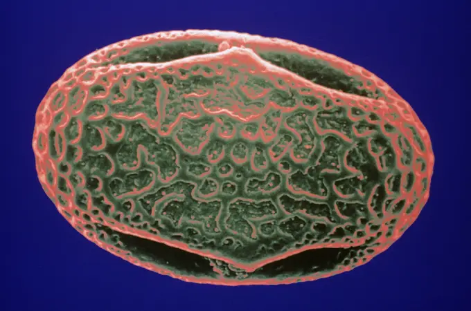
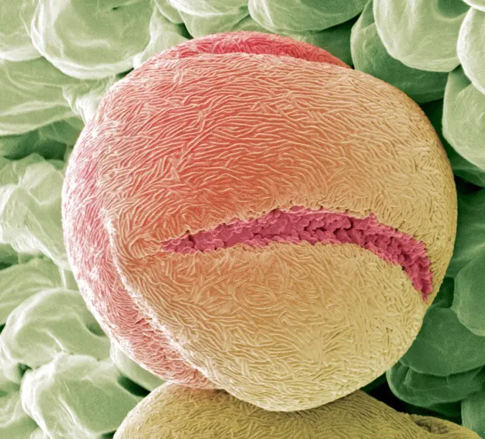
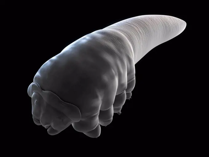
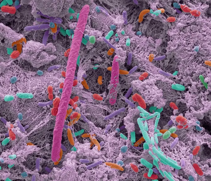
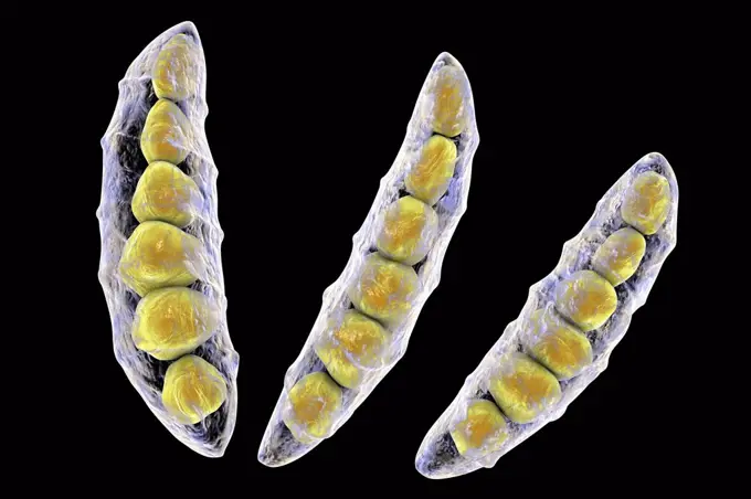
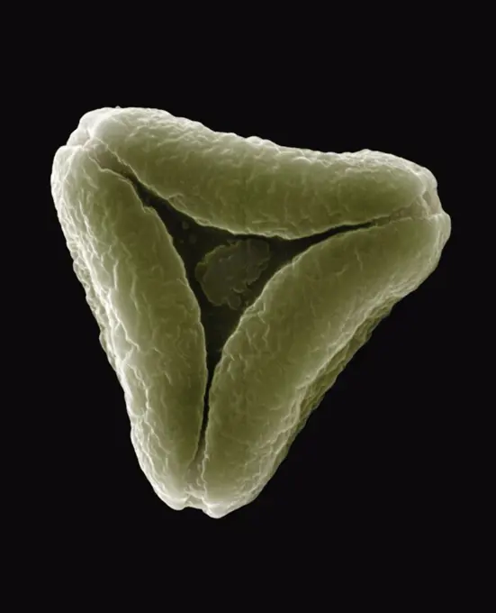
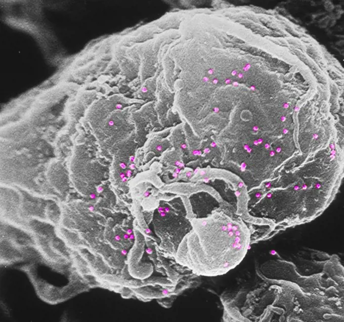
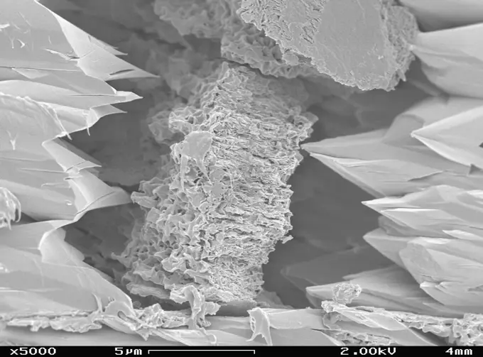
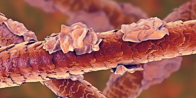
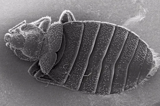
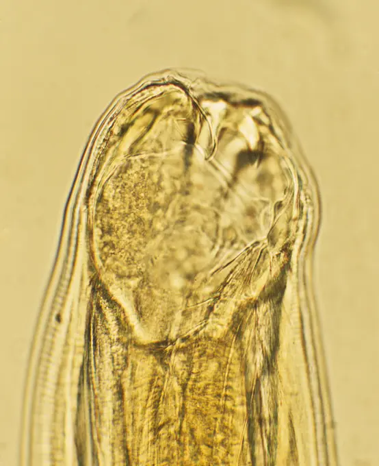
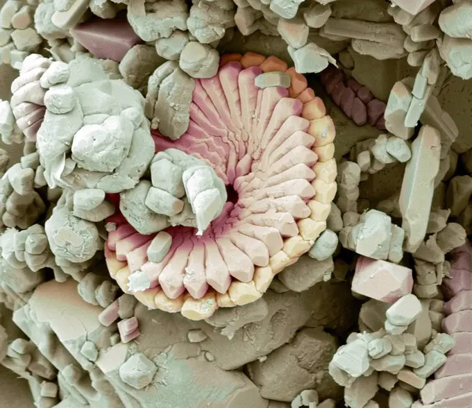
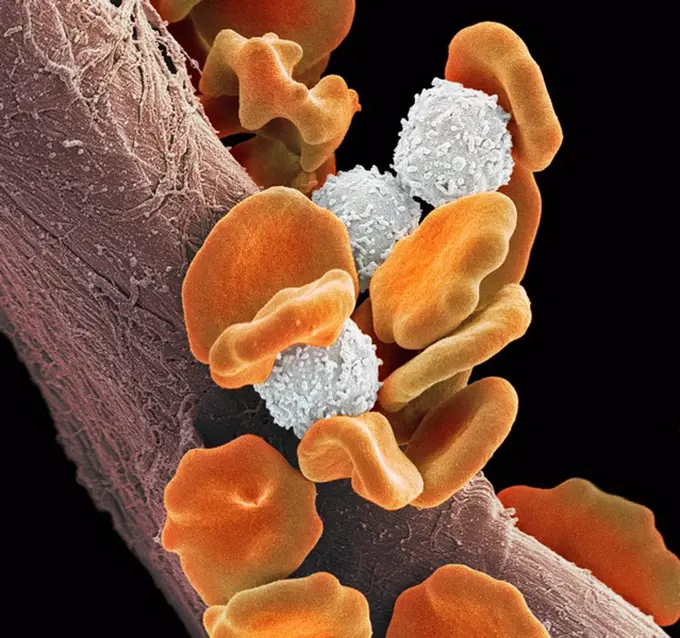
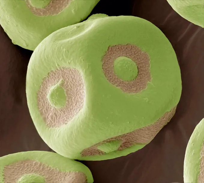
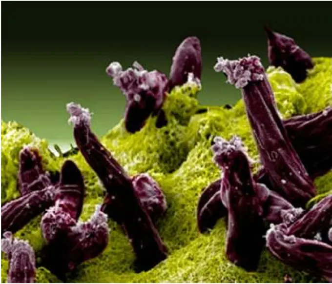
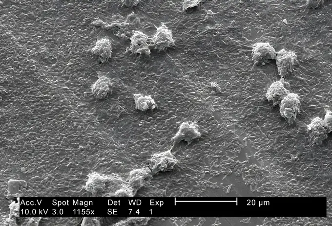
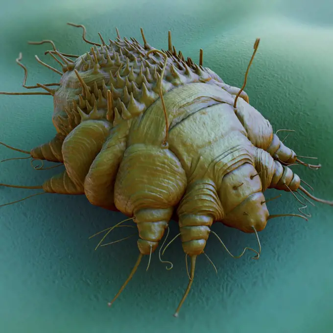
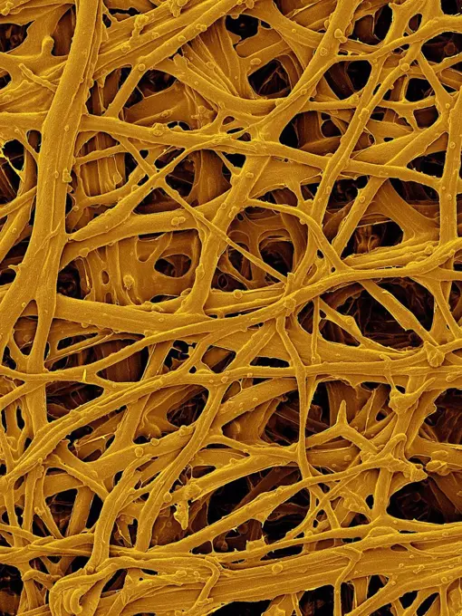
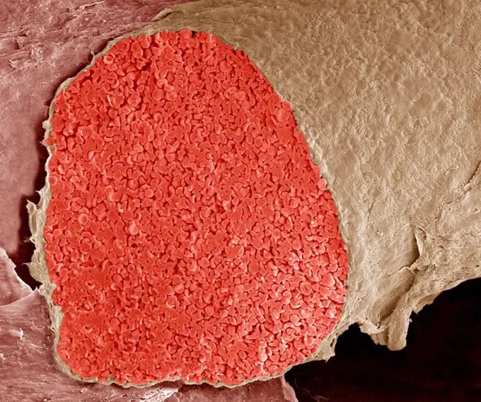
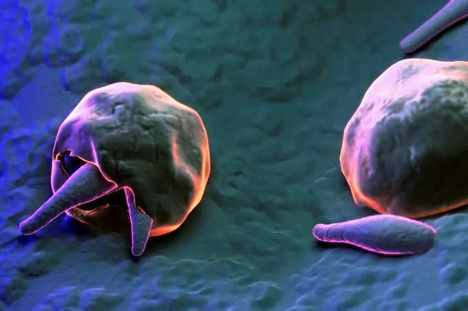
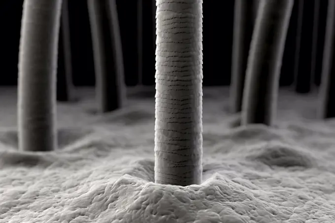
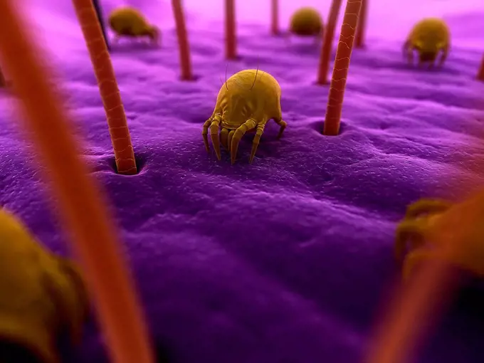
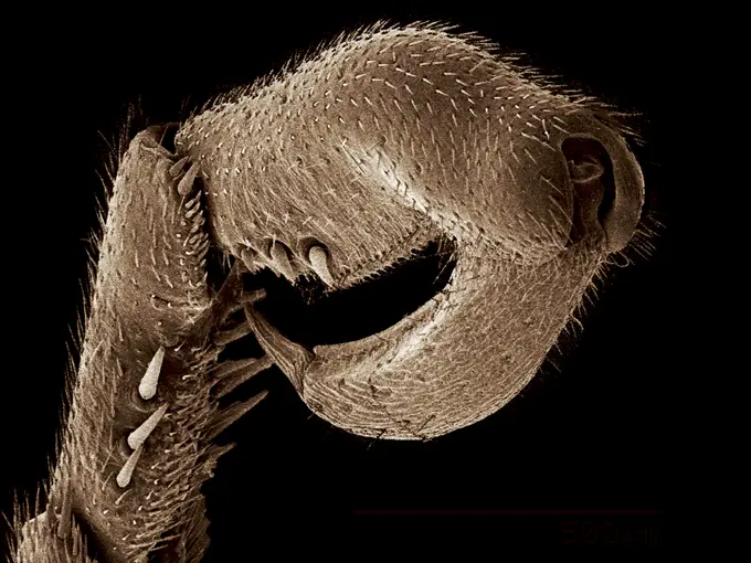
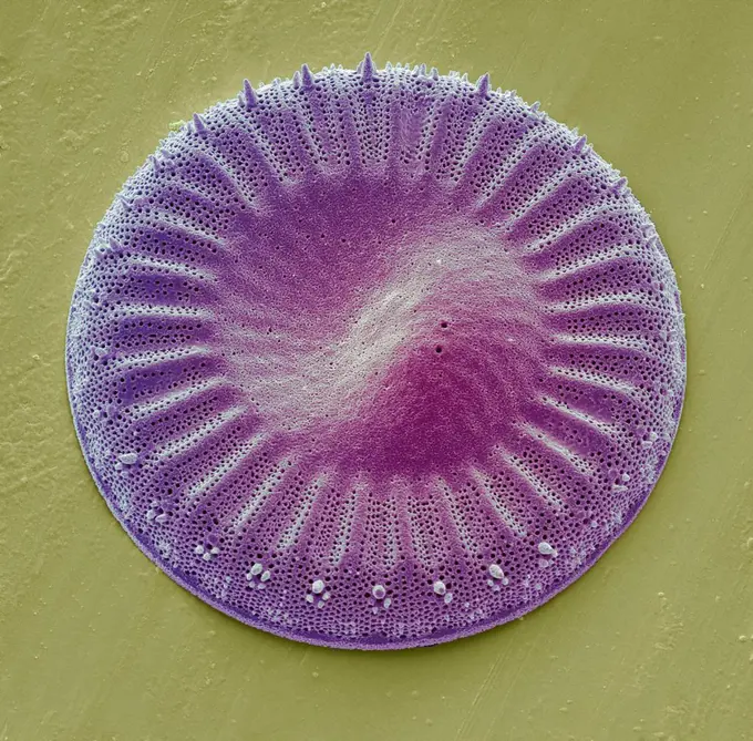
Fine ArtVintageFootageSubscriptionBoardsFree ResearchPricing
  • Fine Art
  • Vintage
  • Footage
  • Subscription
  • Boards
  • Free Research
  • Pricing

Microscopic Life Forms

Detailed close-ups of microscopic textures and insect features captured under an electron microscope, revealing the complexity of natural patterns.

Closeup view of a microscopic surface revealing intricate textures
190 assets in this story
4128R-34264
Eyelash mites. Coloured scanning electron micrograph (SEM) of human eyelash mites (Demodex sp.) around hair follicles. Magnification: x35 when printed...
4128R-14146
Cat flea. Coloured scanning electron micrograph SEM of the head of a cat flea Ctenocephalides felis. Magnification: x130 when printed at 10 centimetre...
4128R-6435
Collagen, SEM
4269-24739
Pollen. Pollen of clover SEM (Scanning Electron Microscope)
4378-2795
Diagram of a Water Bear (Tardigardes) which is a polyextremophile.
4128R-9675
Strawberry pollen, SEM
4384-417
Tiny Mite on Lizard's Skin
4128R-14829
Eyelash mite Demodex sp., computer artwork.
4128R-13573028
Oral bacteria. Coloured scanning electron micrograph (SEM) of mixed oral bacteria. The mouth contains a large number of bacteria, most of which are ha...
4128R-13620155
Papillae on the upper surface of a rose flower petal (Rosa sp), coloured scanning electron micrograph (SEM). Papillae are projections from epidermal c...
1439-57940051
Closeup view of a microscopic structure resembling a canyon or wave in grayscale tones.
1439-57940167
Extreme closeup of a textured surface under a microscope
4128-V58559201
4128R-8935
Bottlebrush pollen, SEM
255-31004
Close-up of a Hornet's nest
6145-29266766
Tectorial membrane from cochlea, SEM
4128R-13620204
Coloured scanning electron micrograph (SEM) Chicken eggshell cross section (Gallus gallus domesticus). Shown from left to right in the image. hard min...
824-63123768
Scanning electron micrograph of a laboratory-grown potable water biofilm, with the presence of Hartmanella vermiformis cysts. Aquatic bacteria were grown as biofilm on steel for one week. Hartmanella vermiformis protozoans were then added, which phagocytized the bacteria, that multiplied within vesicles and became cysts in which the bacteria will live until they rupture.
6145-44641469
NASA's Mars Exploration Rover Spirit used its microscopic imager to capture this spectacular, jagged mini-landscape on a rock called 'GongGong.'
6188-65540173
3d-Illustration von vielen verschiedenen Pollenkörpern in schwarz-weiß 3d illustration of many different pollen bodies in black and white Copyright: xZoonar.com/ChristophxBurgstedtx 22576133
1773-100039
SEM image of Taraxacum seed detail
1899-85571
Yersinia Pestis SEM
4128R-9186
Euglypha amoeba shell, SEM
4128R-6421
Bone tissue, SEM
1439-57940117
Extreme closeup of a textured surface under magnification
4128R-13587447
Computer illustration of conidia (asexual spores) from a Fusarium sp. fungus. Some Fusarium fungi are pathogens of plants and humans.
4269-24818
Skin. Section of human skin with with 3 hairs. ESM (Electron Scanning Microscope)
4201-66250
Bottlebrush (Callistemon sp) SEM close-up view of a grain of pollen at 2800x magnification
4297-1421
Scanning electron micrograph of human immunodeficiency virus (HIV), grown in cultured lymphocytes. Virions are seen as small spheres on the surface of the cells
6145-44727927
A scanning electron microscope captured this image of terresterial soil containing a phyllosilicate mineral from Koua Bocca, Ivory Coast, West Africa. This soil shares some similarities with Martian soil scooped by NASA's Phoenix Lander.
4384-340
Detail of Domesticated Cat's Claw
4384-377
Microscopic Detail of a Millipede
4128-V58562016
4220-21334731
Scanning Electron Micrograph: Woodboring Weevil.     Date:
1439-57949041
Upclose view of an ant's head under the microscope
4128R-13573030
Chinese hibiscus pollen. Coloured scanning electron micrograph (SEM) of pollen grains on the anther of a Chinese hibiscus (Hibiscus rosa-sinensis) flo...
4128R-15556
kiwi fruit Actinidia deliciosa pollen grain, coloured scanning electron micrograph SEM. Magnification: x3000 when printed at 10 centimetres wide.
4128R-869
Pyoderma skin disease, coloured scanning electron micrograph SEM. Large numbers of lymphocytes yellow are present on the surface of the fractured skin...
1439-57952643
Closeup of a dragonfly's compound eyes and head details
4384-371
Microscopic Detail of Dragonfly's Wing Attachment
4201-82340
Wolf Spider (Lycosa tarantula) SEM close-up view of face at 19x magnification, showing six eyes
4391-337
Macrophages are the string-like projectile-looking items interacting with the spherical lymphocyte.
4384-160
This scanning electron micrograph (SEM) revealed some of the ultrastructural morphology displayed on the ventral surface of a bedbug, Cimex lectularius.  From this view you can see the insects skin-piercing mouthparts used it uses to obtain its blood meal. Although bedbugs have been found naturally-infected with blood-borne pathogens, they are not effective vectors of disease.  The primary medical importance is inflammation associated with their bites (due to allergic reactions to components in
824-63227051
This image shows how the mouth design makes ticks generally difficult to remove once they have attached for a blood meal. Credit: NIAID
4378-3945
Sickle-cell disease, sickle-cell anaemia or drepanocytosis is a recessive genetic blood disorder characterized by red blood cells that assume an abnormal, rigid, sickle shape. Here healthy blood cells are seen along with diseased cells.
1439-57952401
Closeup of a microscopic specimen on a slide
4128R-15366451
Human hair with dandruff, illustration
4384-155
This scanning electron micrograph (SEM) revealed some of the ultrastructural morphology displayed on the dorsal exoskeletal surface of a bedbug, Cimex lectularius.  Note the numbers of what appear to be hairs, but are actually known as setae, which are sensorial in nature, and are composed of chitin, as is the entire exoskeleton.Chitin is a molecule made up of bound units of acetylglucosamine, joined in such a way as to allow for increased points at which hydrogen bonding can occur. In this wa
4220-21334549
Hookworm.     Date:
4128R-12701
Fossil debris in chalk. Coloured scanning electron micrograph SEM of a fossilised fragment of a coccolithotrope skeleton. Magnification: x8000 when pr...
4384-234
Scanning Electron Micrograph of Mosquito
4128R-507
Leukaemia blood cells, SEM
4128R-9539
Flowering currant pollen, SEM
824-63223171
Scanning electron micrograph of Plasmodium gallinaceum invading mosquito midgut. Credit: NIAID.
824-63194412
Under a magnification of 2500X, this colorized scanning electron micrograph (SEM) revealed the presence of a large number of Gram-negative Salmonella typhimurium bacteria that had been isolated from a pure culture.
824-57659291
Magnified 1155X, this 2002 scanning electron microscope (SEM) image revealed some of the ultrastructural morphology exhibited by a number of Hartmannella vermiformis amoeba trophozoites. As part of a study to determine if Legionella pneumophila bacteria can colonize and grow in biofilms with or without the presence of H. vermiformis, here these protozoa were located on top of a base biofilm composed of Pseudomonas aeruginosa , Klebsiella pneumoniae and a Flavobacterium sp. bacteria. Note that the amoebas were grazing on these bacteria. CDC/Dr Rodney Donlan 2002
4378-2439
A close up view of the cause of scabies - the mite Sarcoptes scabiei.
4128R-13620186
Coloured scanning electron micrograph (SEM) Chicken eggshell inner membrane (Gallus gallus domesticus). Shown here is the eggshell inner membrane that...
4128R-4503
Foetal vein, SEM
4378-1189
Scanning electromicrograph styled parasitic infection Cryptosporidium.
4201-66317
Mediterranean Fan Palm (Chamaerops humilis) SEM close-up view of pollen at 270x magnification
1439-57943345
Intricate closeup of a microchip circuit with vibrant color enhancement
4128R-13587449
Computer illustration of conidia (asexual spores) from a Fusarium sp. fungus. Some Fusarium fungi are pathogens of plants and humans.
4128-111494398
Coloured scanning electron micrograph (SEM) of Pediculus humanus capitis, human head lice. P. humanus is divided into two subspecies; the head louse P...
4128R-6422
Prorocentrum dinoflagellates, SEM
4128R-863
Copepod, SEM
4128R-22306
Chromosomes, computer artwork.
4220-21334511
Scanning Electron Micrograph (SEM): Early human embryo, one cell removed for genetic analysis.     Date:
1439-57953821
Closeup of an insect's head in striking detail
6145-45247426
Paul Ducheyne, a principal investigator in the microgravity materials science program and head of the University of Pernsylvania's Center for Bioactive Materials and Tissue Engineering, is leading the trio as they use simulated microgravity to determine the optimal characteristics of tiny glass particles for growing bone tissue. The result could make possible a much broader range of synthetic bone-grafting applications. Even in normal gravity, bioactive glass particles enhance bone growth in laboratory tests with flat tissue cultures. Ducheyne and his team believe that using the bioactive microcarriers in a rotating bioreactor in microgravity will produce improved, three-dimensional tissue cultures. The work is sponsored by NASA's Office of Biological and Physical Research. The bioreactor is managed by the Biotechnology Cell Science Program at NASA's Johnson Space Center (JSC). NASA-sponsored bioreactor research has been instrumental in helping scientists to better understand normal an
4128-V58568579
4297-1889
Scanning electron microscopic image of Vibrio cholerae bacteria of the serogroup 01
4128-V58557869
1439-57952645
Closeup of microscopic elements, resembling an abstract landscape.
4378-944
Microscope styled close up view of human skin and hair roots.
4128R-15050
Dust mites. Computer artwork of house dust mites Dermatophagoides pteronyssinus on human skin.
1439-57943315
Intricate details of an ant's head under the microscope
1439-57943281
Intricate geometric patterns of crystals under a microscope.
4128R-12571809
Diatom. Coloured scanning electron micrograph (SEM) of a single diatom (cylotella sp.). Diatoms may be extremely abundant in both freshwater and marin...
4128R-15545716
C elegans, SEM
824-63225812
Scanning electron micrograph of Plasmodium gallinaceum invading mosquito midgut.
4413-21778
Cut petiole of Raspberry cane France
4128-16224490
Sea urchin, X-ray
1439-57952398
Closeup of an insect head captured under a microscope.
1439-57940123
Closeup shot of an ant antenna under electron microscope
6145-45244852
An etched sample of the aluminum indium alloy (magnified). When the hypermonotectic mixture is cooled in the Advanced Gradient Hearing Facility (AGHF), aluminum transitions to a solid first, trapping the indium in cylindrical fibers within the solid. Principal Investigator Dr. Barry Andrews
4201-21255291
Pothos (Epipremnum aureum) stoma magnified over 2000x
1439-57940205
Extreme closeup of an insect's wing under a microscope
4128R-10572
Rabbit flea, SEM
1439-57943314
Intricate microstructure of a creature captured in stunning detail.
PREVIOUS
of 2
NEXT
2401 S. Ervay, Suite 206
Dallas, Texas 75215
United States
Browse
CategoriesGalleriesFree Research
For Creators
License Your ContentSuperStock For StudentsFrame of Mind BlogContributor PortalNew Image SpotlightLicense AI Imagery
Resources
About UsLicensingTerms & ConditionsAPI AccessPrivacyPricing
Contact
+1 866 236 0087help@superstock.com Contact form
©2026 SuperStock. All Rights Reserved. -A
This site is protected by reCAPTCHA and Google Privacy Policy and Terms of Service apply.