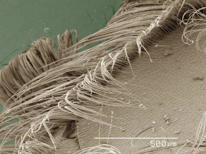
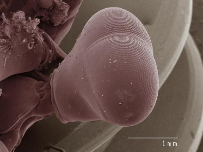
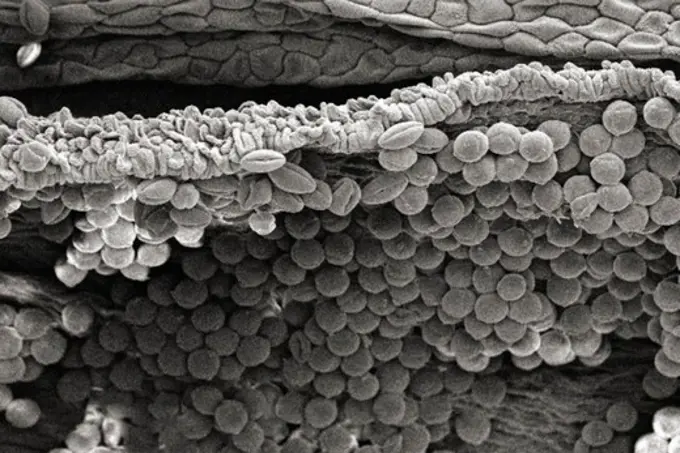
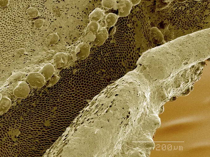
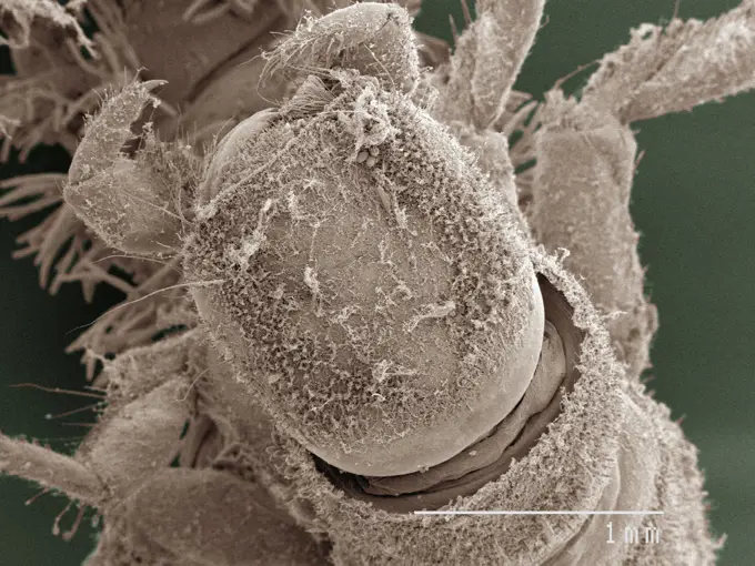
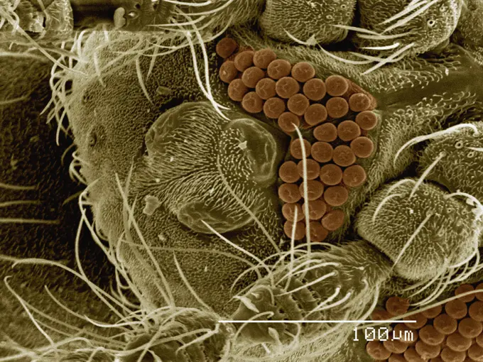
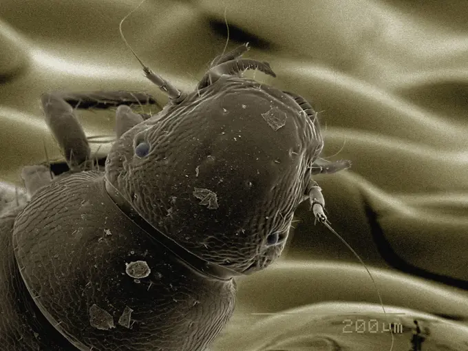
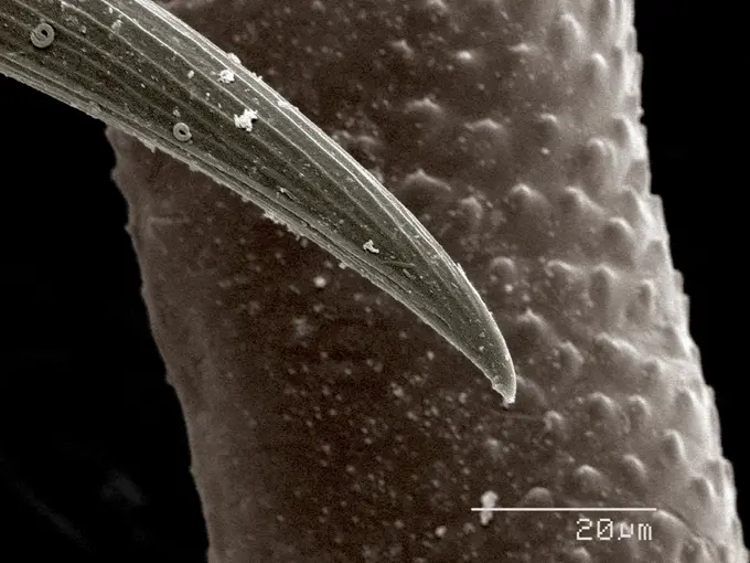
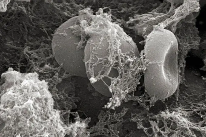
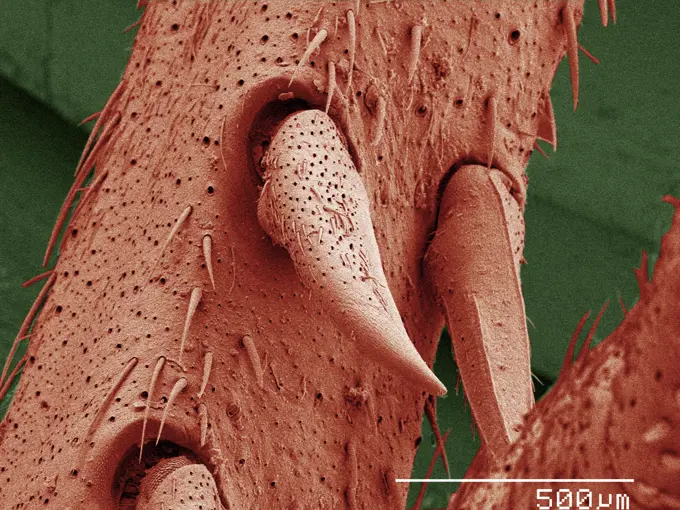
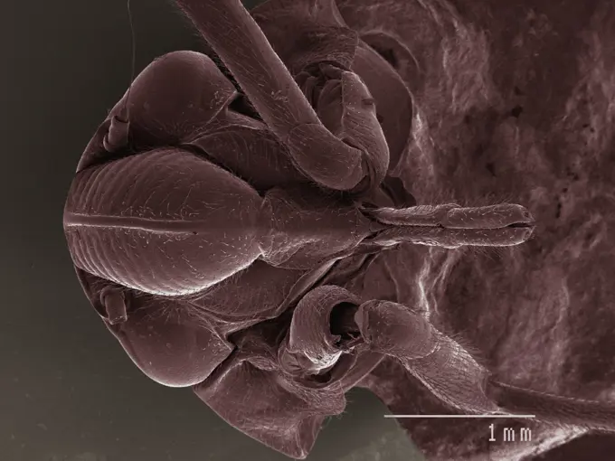
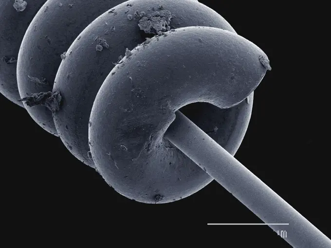
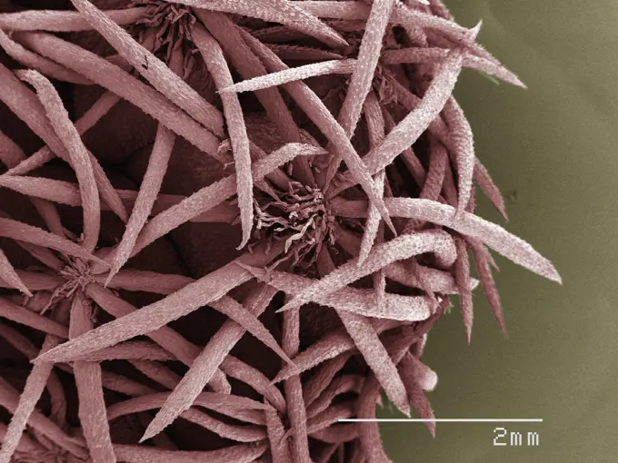
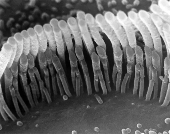
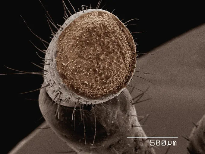
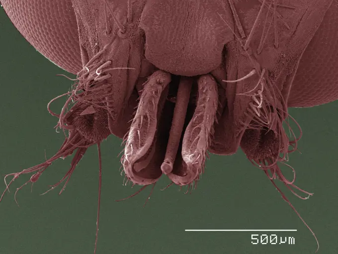
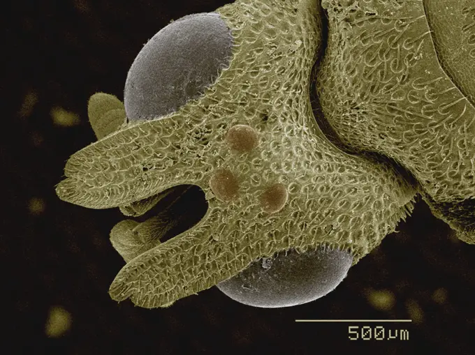
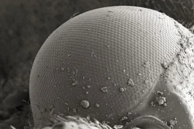
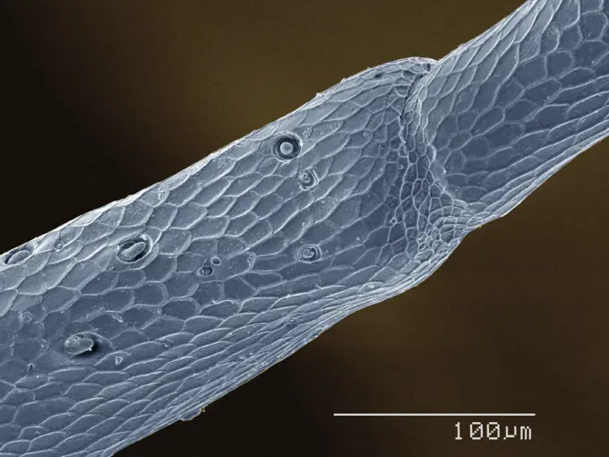
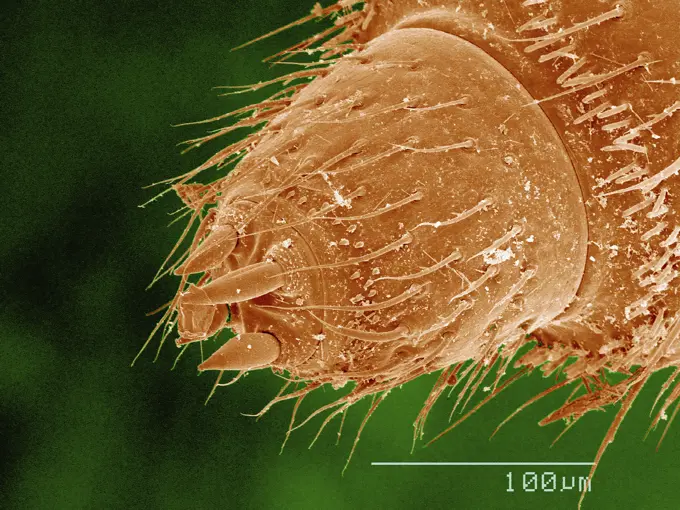
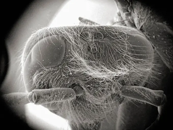
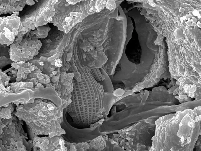
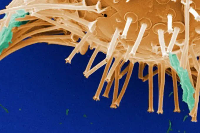
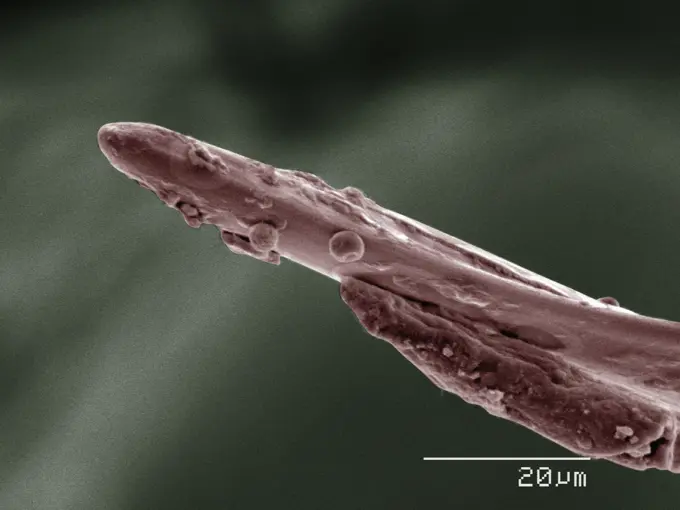
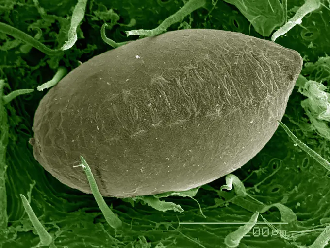
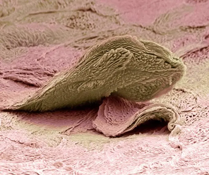
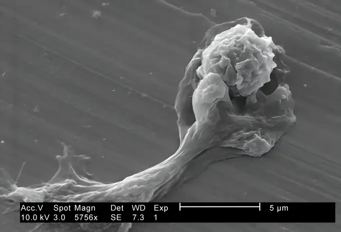
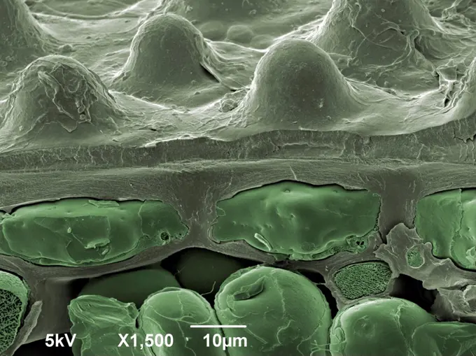
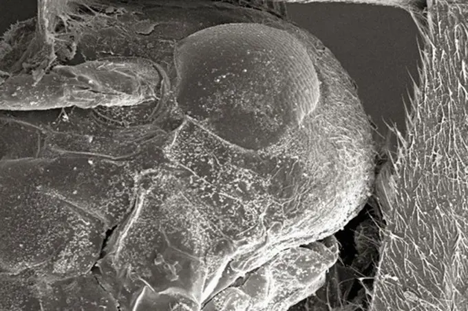
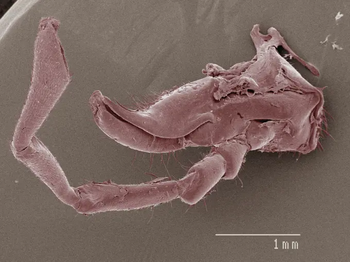
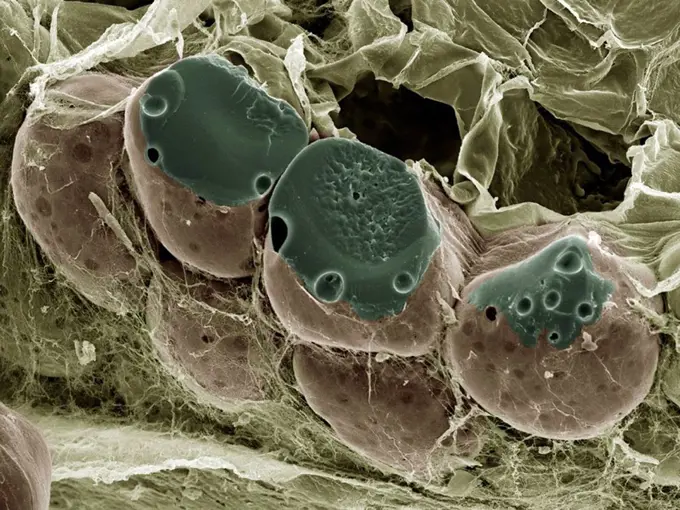
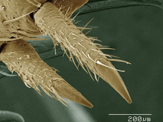
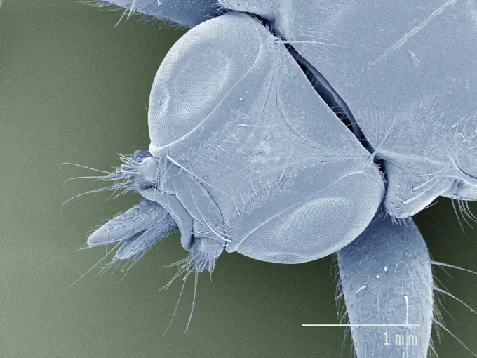
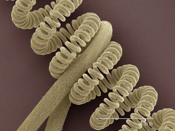
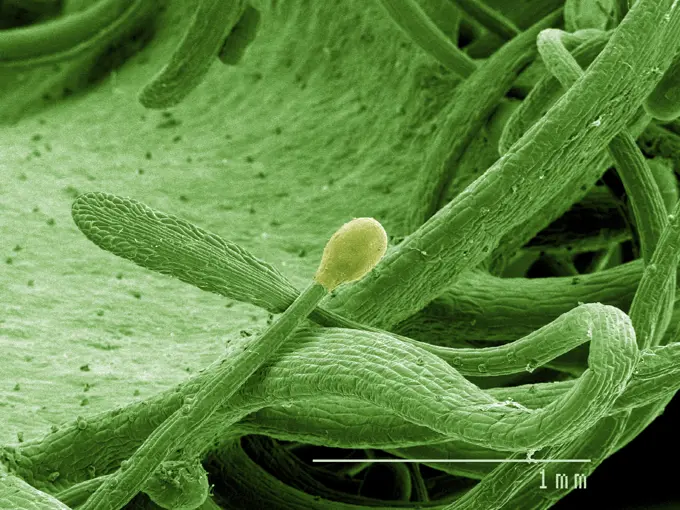
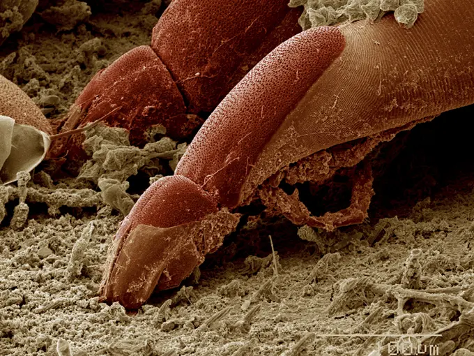
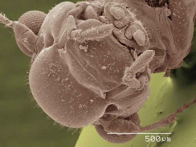
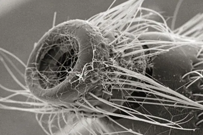
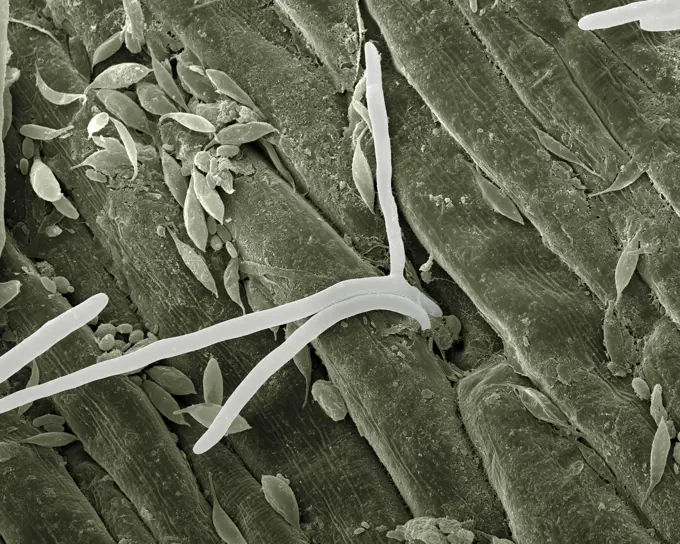
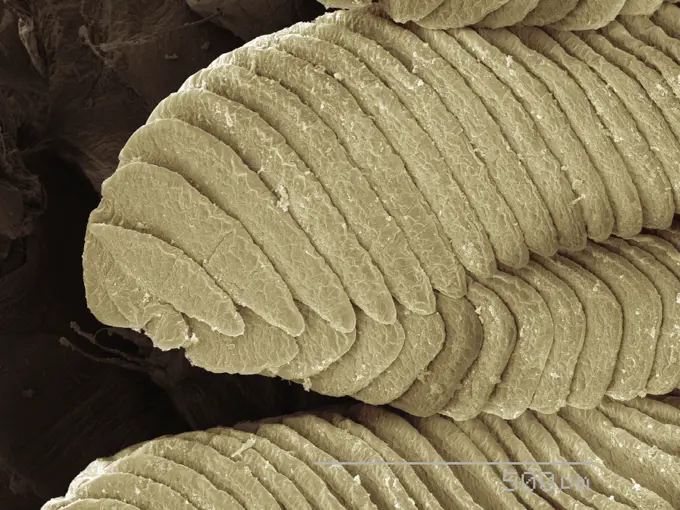
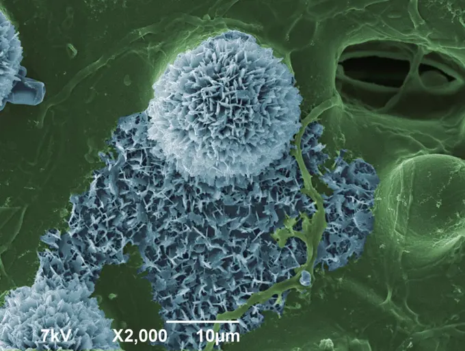
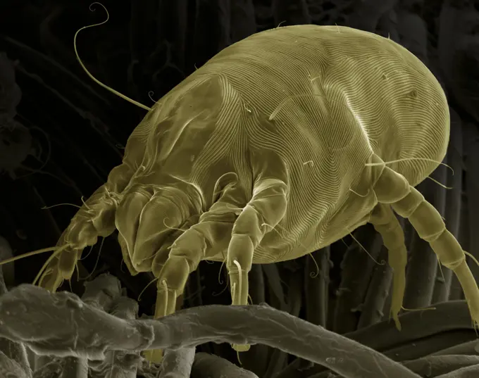
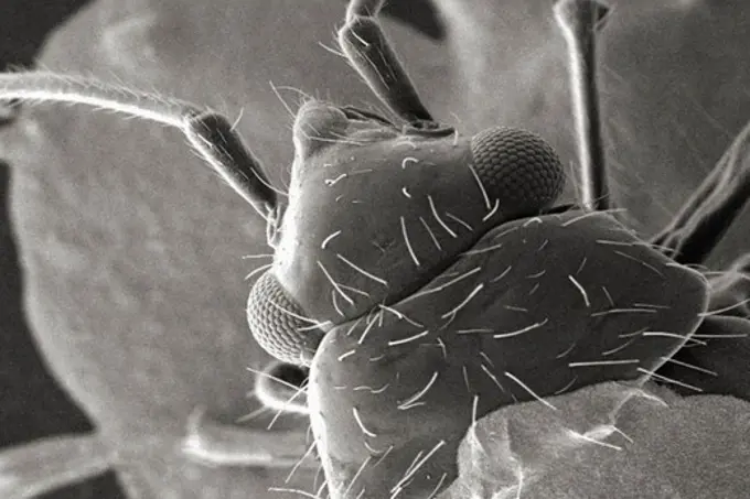
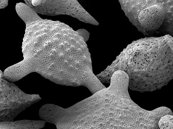
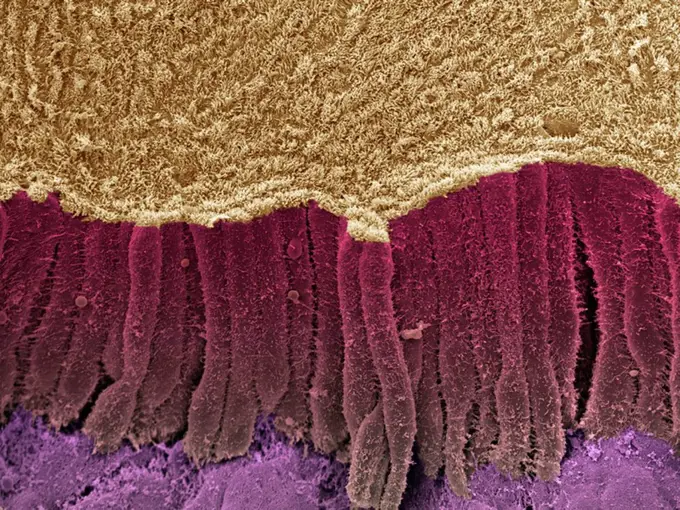
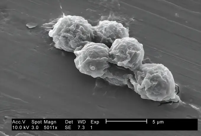
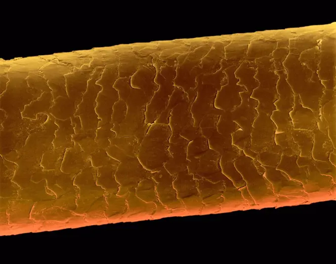
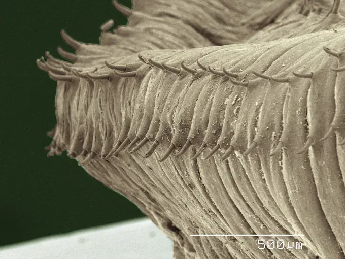
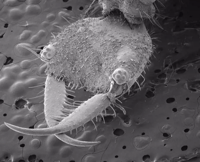
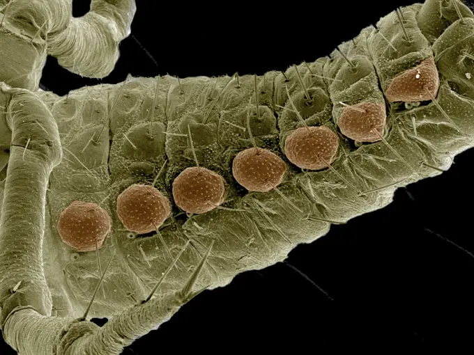
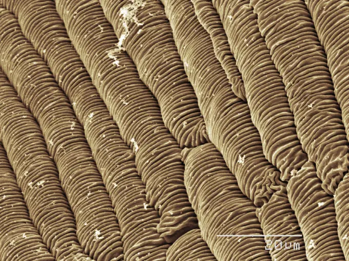
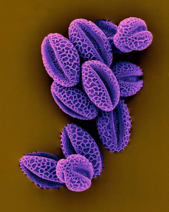
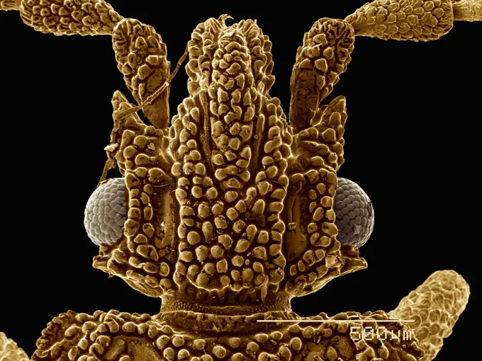
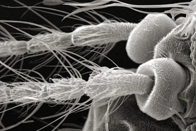
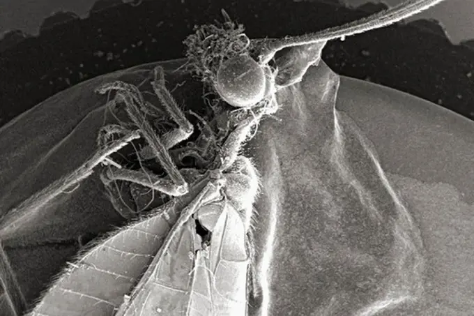
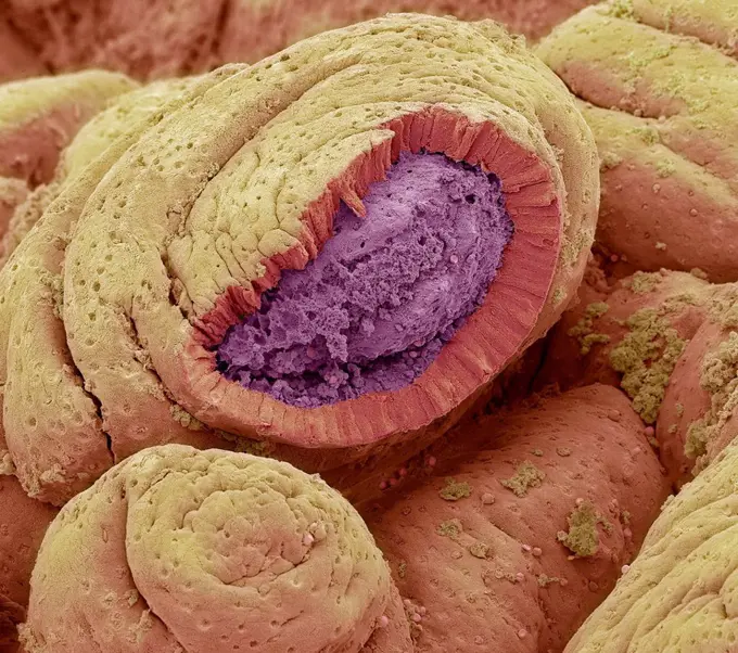
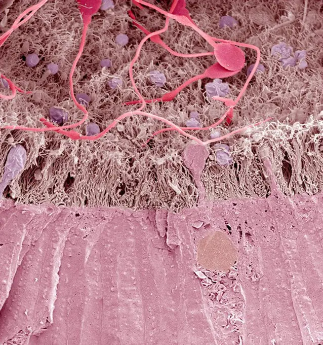
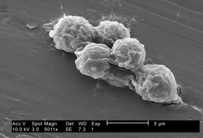
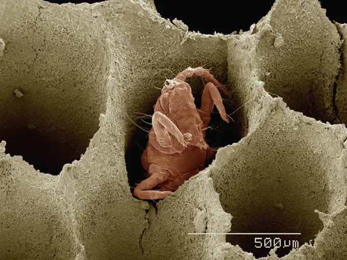
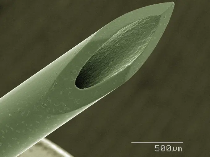
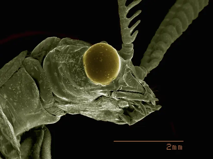
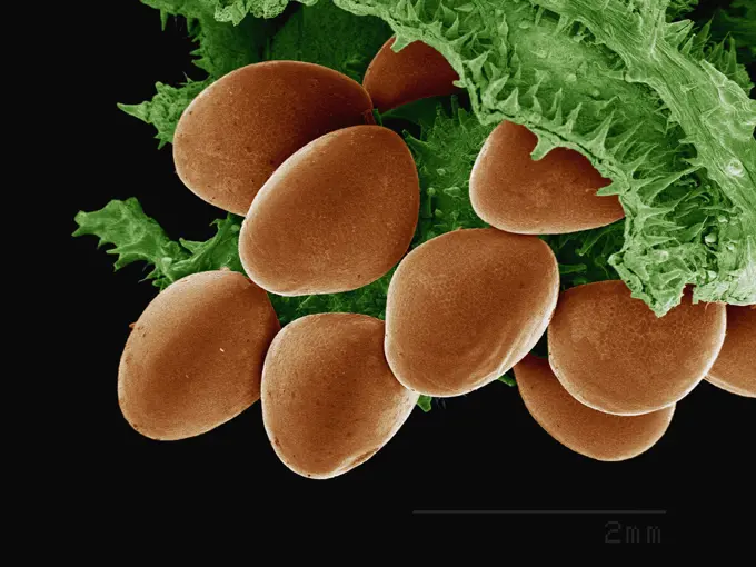
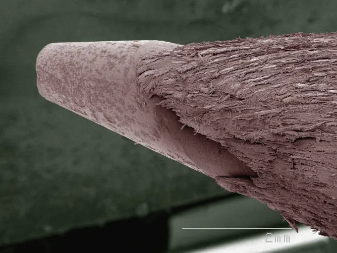
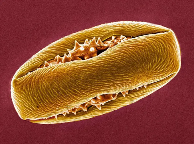
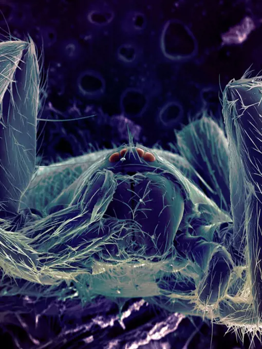
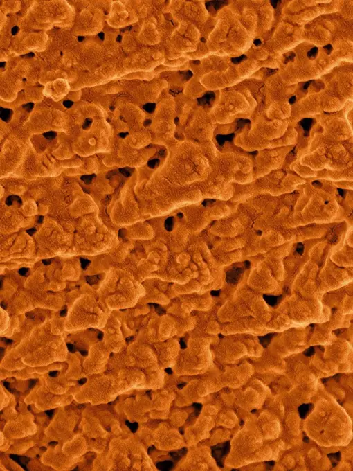
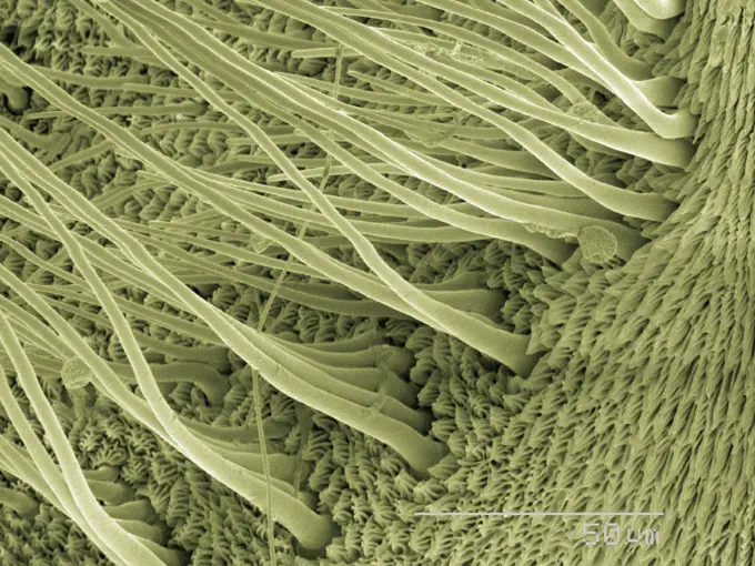
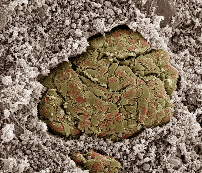
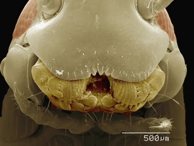
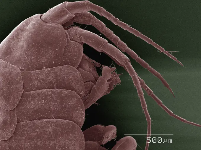
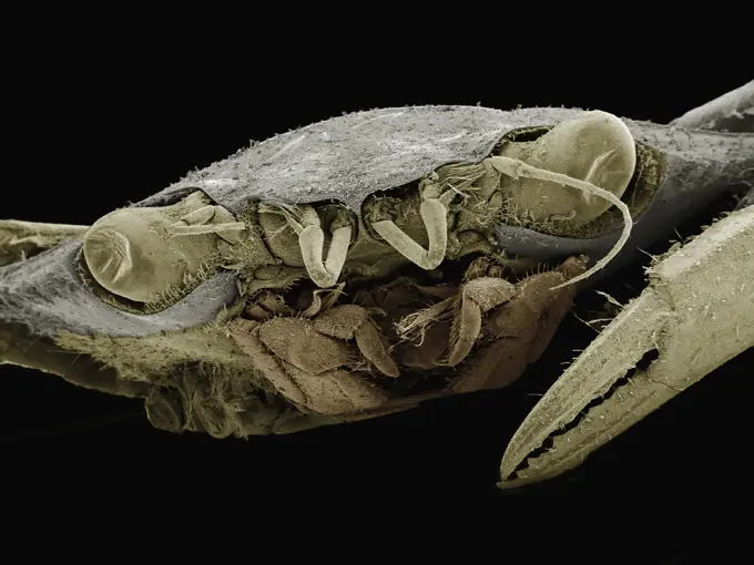
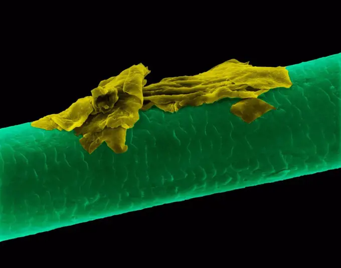
Fine ArtVintageFootageSubscriptionBoardsFree ResearchPricing
  • Fine Art
  • Vintage
  • Footage
  • Subscription
  • Boards
  • Free Research
  • Pricing

Microscopic Organisms

Close-up images capture various microscopic organisms and structures, showcasing intricate details and textures under high magnification.

Extreme closeup of fibrous material texture
175 assets in this story
1439-57940065
Closeup view of a microscopic organism under an electron microscope.
4384-396
Fireworks Flower With Pollen
1439-57940164
Extreme closeup of fibrous material texture
1439-57943329
Extreme close-up of an organic structure under a microscope
1439-57940094
Closeup of a microscopic creature revealing intricate details
1439-57949006
Closeup view of a textured surface under a microscope
1439-57949082
A microscopic view of intricate biological structures.
1439-57948993
Coloured SEM of Triungulin larva of Meloid beetle (Meloidae)
1439-57943317
Extreme closeup of a barbed plant hair under electron microscopy
4384-329
Red Blood Cells Enmeshed in Fibrinous Matrix
4201-66290
Wolf Spider (Lycosa tarantula) SEM close-up view of face at 21x magnification, showing six eyes
1439-57940220
Intricate closeup of a natural porous structure.
1439-57940159
Extreme closeup of a creature in sepia tones
1439-57949071
Closeup view of scientific marvel under the microscope.
1439-57940110
Closeup view of a textured natural object under magnification
1439-57940154
Magnified view of a microfiber under an electron microscope
6145-29256896
Inner hair bundle, Guinea pig cochlea
1439-57940155
Extreme closeup of a microscopic texture reminiscent of an alien landscape.
1439-57949010
Closeup of an insect antenna as seen through a scanning electron microscope.
4384-313
Electron Micrograph of Wasp's Head
1439-57940087
A closeup view of a spider under the microscope.
1439-57952386
Intricate patterns of microscopic structures revealed in stunning detail
1439-57949022
Extreme closeup of a bee's head under a scanning electron microscope
4384-299
Scanning Electron Micrograph of Insect's Compound Eye
1439-57940145
Closeup view of plant or animal cells under a microscope
1439-57952667
Closeup of a microstructure under a microscope
4384-374
Microscopic Detail of a Bee's Head
1439-57947641
Intricate Microscopic Landscape
4384-249
Scanning Electron Micrograph of Untreated Water Sample
4384-153
This digitally-colorized scanning electron micrograph (SEM) of an untreated water specimen extracted from a wild stream mainly used to control flooding during inclement weather, revealed the presence of unidentified organisms, which included bacteria, protozoa, and algae. In this particular image, unidentified sheets of algae were wrapped in a mass of what appeared to be a mucoid amorphous biofilm. Though many organisms found in untreated waters are harmless, there are many that are pathogenic t
1439-57940185
Extreme closeup of an organism under a microscope
1439-57952406
Close-up of a microorganism under a microscope.
4128R-1651
Skin cell. Coloured scanning electron micrograph SEM of a squamous cell on the surface of the skin. This is a flat, keratinised, dead cell. Squamous c...
1899-61460831
Magnified 5756X, this 2002 scanning electron microscope (SEM) image revealed some of the ultrastructural morphology exhibited by two Hartmannella vermiformis amoeba trophozoites. Note that one of the amoebas took on a very distorted shape, having extruded a number of pseudopodia, as it moved across the surface of the coupon. The trophozoite stage of an amoeba's life cycle is its vegetative phase, devoted to feeding, movement, and reproduction. This free-living protozoan moves in response to chemical cues in its environment by extending pseudopodia, or false feet, a number of which are visible in this image. CDC/Dr Rodney Donlan 2002
1773-209591
Leaf,  x1500 magnification
4384-225
Scanning Electron Micrograph of a Spider's Prey
1439-57940208
An upclose and personal view of a tiny critter's world under the microscope.
4128R-6264
Fat tissue, SEM
1439-57940214
Closeup of an ant's leg under the microscope
4128-V58557936
1439-57940120
Closeup of a coffee bean under the microscope
1439-57949040
This is a microscopic image showing a close-up of textured surfaces and spherical structures, likely taken with a scanning electron microscope (SEM).
4384-105
Seen emerging from behind its left compound eye, under a low magnification of 36X, this scanning electron micrograph (SEM) depicted the mid-region of this unidentified hornets left antenna. Found in the suburbs of Decatur, Georgia, this antenna was one of a pair, which along with other sensorial structures such as the hair-like setae, seen here coating the antennas surface, and the surface of insects head, provided the hornet with data indicative of changes encountered in its environment su
1439-57940111
Closeup of microscopic textures resembling alien landscapes.
1439-57940092
Upclose and personal with an insect's head under the microscope
1439-57940025
Intricate closeup of a microstructure resembling a zipper
4389-2061
Micrograph of Aerogel
1439-57943355
Closeup of microscopic structures with a green tint illustrating intricate biological details.
1439-57943290
An extreme closeup of a mite under high magnification
1439-57940095
A detailed look at the alien landscape of an insect's head under the microscope.
4384-211
Scanning Electron Micrograph of a Beetle's Antenna
4220-21334682
Scanning Electron Micrograph (SEM): Fusarium.     Date:
1439-57940016
A closeup view of textured layers resembling a natural formation or microscopic detail
4201-21262911
House Pseudoscorpion (Chelifer cancroides), magnified 64 times, Barcelona, Spain
1773-209594
Leaf micrograph, x2000 magnification
4220-21334697
Scanning Electron Micrograph (SEM) Dust Mite.     Date:
1439-57943369
Intricate details of a seed captured in a closeup shot against a black background.
4384-428
Microscopic Detail of Insect's Head
1439-57947636
Magnified beauty of microscopic structures
4128R-2609
Bile duct, SEM
824-57659300
Magnified 5011X, this 2002 scanning electron microscope (SEM) image revealed some of the ultrastructural morphology exhibited by a small group of Hartmannella vermiformis amoeba trophozoites. The trophozoite stage of an amoeba's life cycle is its vegetative phase, devoted to feeding, movement, and reproduction. This free-living protozoan moves in response to chemical cues in its environment by extending pseudopodia, or false feet, a number of which are visible in this image. CDC/Dr Rodney Donlan 2002
4128R-13620380
Human hair (Caucasian, brunette), coloured scanning electron micrograph (SEM). The outer layer of hair (the cuticle) has overlapping scales of keratin...
1439-57940049
Closeup texture of a natural specimen under electron microscopy.
4201-66327
Antlion (Myrmeleontidae) SEM close-up view of larva at 21x magnification
1439-57943311
A closeup electron microscope view of pollen grains clinging to a surface.
1439-57940112
Closeup view of textured surface under magnification
4128R-13622321
Weeping willow pollen (Salix babylonica), coloured scanning electron micrograph (SEM). The willow (and poplar) family, Saliaceae, is characterized by ...
1439-57953826
Intricate patterns of a plant under the microscope
4384-229
Scanning Electron Micrograph of Mosquito
4384-412
Microscopic Detail of Mosquito's Head
4384-331
Magnified Lateral View of Unidentified Insect
824-63227113
Yeast makes bread, beer and wine. And like us, yeast can reproduce sexually. A mother and father cell merges and a large cell which contains four offspring. When environmental conditions are favorable, the offspring are released, as shown here. Yeasts are also a popular subject of study for scientists. Yeast research has yielded vast knowledge of basic cellular and molecular biology as well as a myriad of human diseases, including colon cancer and various metabolic disorders.
4128-16171847
Intestinal lining. Coloured scanning electron micrograph (SEM) of a freeze-fractured of the small intestine. The surface consists of deep folds, calle...
4128R-10558
Epididymis, SEM
1439-57952654
Up close and personal with a microworld critter.
1899-61460834
Magnified 5011X, this 2002 scanning electron microscope (SEM) image revealed some of the ultrastructural morphology exhibited by a small group of Hartmannella vermiformis amoeba trophozoites. The trophozoite stage of an amoeba's life cycle is its vegetative phase, devoted to feeding, movement, and reproduction. This free-living protozoan moves in response to chemical cues in its environment by extending pseudopodia, or false feet, a number of which are visible in this image. CDC/Dr Rodney Donlan 2002
1439-57952392
A thrips (Thysanoptera) in bracket fungus
1439-57940093
Extreme closeup of a needle under a microscope showing intricate details
1439-57953827
Closeup view of an insect under a microscope
1439-57949079
Magnified view of pollen grains nestled among leafy textures
1439-57940039
Upclose texture of a sharp pencil point
4128R-2779
Horse chestnut pollen, SEM
824-57659293
Magnified 3850X, this 2002 scanning electron microscope (SEM) image revealed some of the ultrastructural morphology exhibited by what is thought to be a single Hartmannella vermiformis amoeba cyst and debris artifact. The amoeba assumes the cystic stage of its life cycle under harsh conditions such as drought, or when there is an accumulation of environmental toxins, thus reducing its metabolic needs. Under these circumstances, the protozoan produces a protective coat and goes into a dormant state. CDC/Dr Rodney Donlan 2002
1439-57940211
Closeup of an insect's hooked appendage under the microscope
4128R-1006
Nasturtium stem, SEM
1439-57940066
Closeup of microscopic structures with intricate details
4128-V58557876
1773-100037
SEM image of spider blue
4128R-13620179
Coloured scanning electron micrograph (SEM) Emu eggshell surface with tiny pores (Dromaius novaehollandiae). Calcium carbonate crystals make up the Em...
1439-57940004
Closeup view of microscopic structures showcasing intricate detail.
4220-21334754
Scanning electron micrograph (SEM) of Kidney glomerulus.     Date:
1439-57952666
Closeup view of an insect head under the microscope
1439-57940099
Closeup view of an insect under a scanning electron microscope
1439-57940132
Upclose view of the intricate texture under an electron microscope.
1439-57952390
Upclose encounter with a crab louse
4128R-13619952
Human hair with dandruff, coloured scanning electron micrograph (SEM). The outer layer of hair (the cuticle) has overlapping scales of keratin. These ...
1439-57940096
Intricate micro landscape captured in a highdetail microscopic image.
4128R-13619937
Used wax dental floss with dental plaque (green), bacteria (purple) and cheek cells (red) on the dental floss fibres (yellow), coloured scanning elect...
4297-1747
SEM image of the Death of a Cancer Cell (step 6 of a 6 step sequence)
1439-57943302
Intricate Patterns of Nature's Weave
PREVIOUS
of 2
NEXT
2401 S. Ervay, Suite 206
Dallas, Texas 75215
United States
Browse
CategoriesGalleriesFree Research
For Creators
License Your ContentSuperStock For StudentsFrame of Mind BlogContributor PortalNew Image SpotlightLicense AI Imagery
Resources
About UsLicensingTerms & ConditionsAPI AccessPrivacyPricing
Contact
+1 866 236 0087help@superstock.com Contact form
©2026 SuperStock. All Rights Reserved. -A
This site is protected by reCAPTCHA and Google Privacy Policy and Terms of Service apply.