Microscopic Organisms and Cells
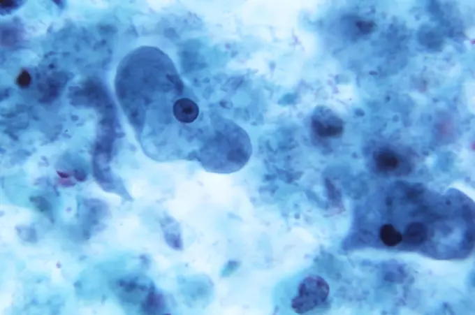
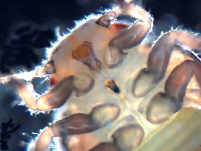
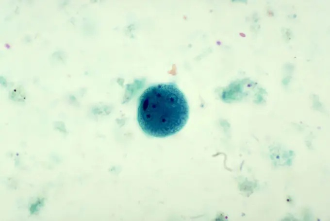
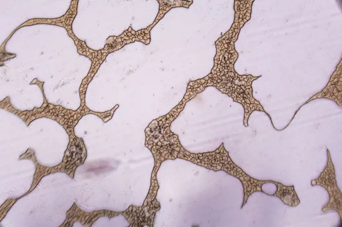
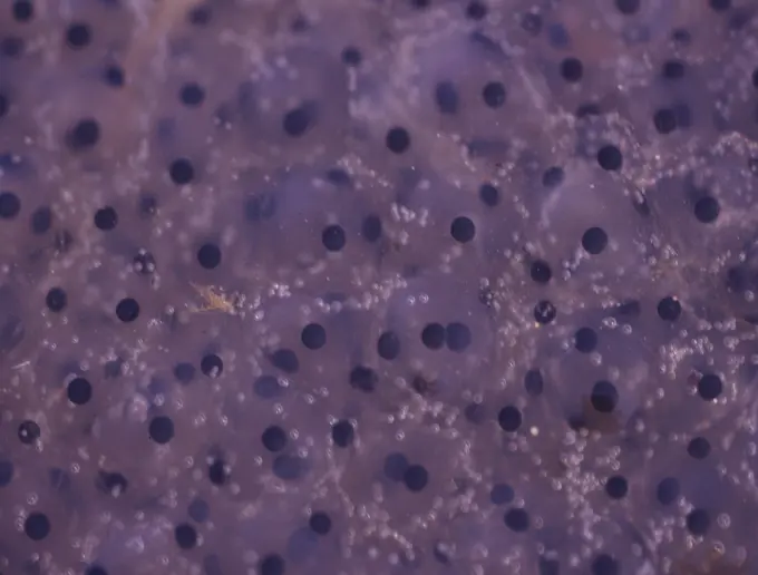
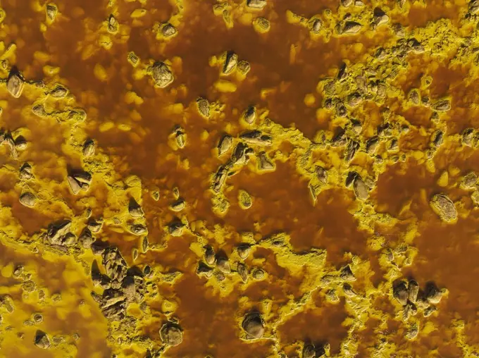
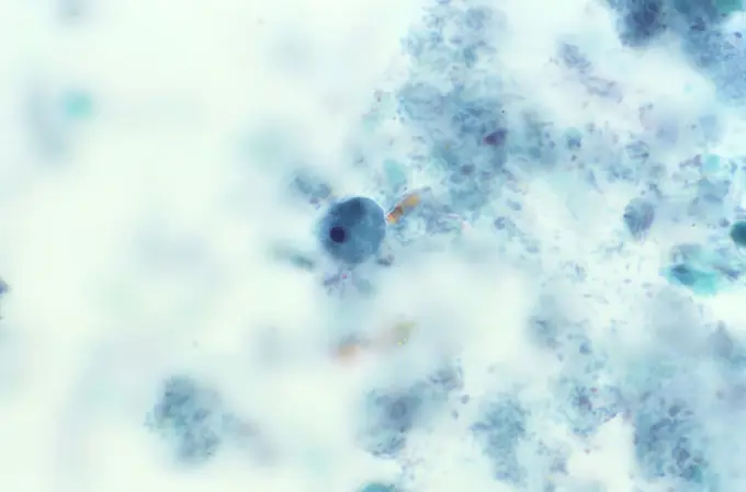
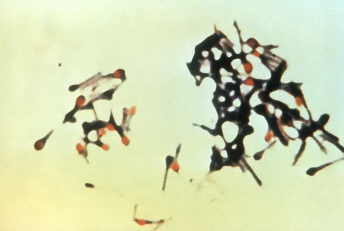
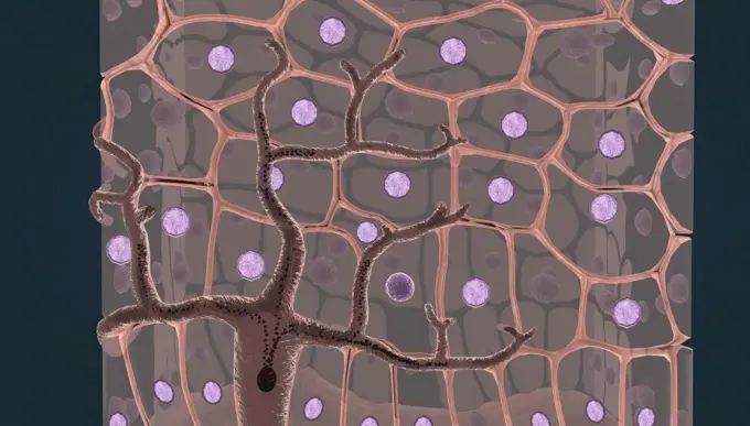
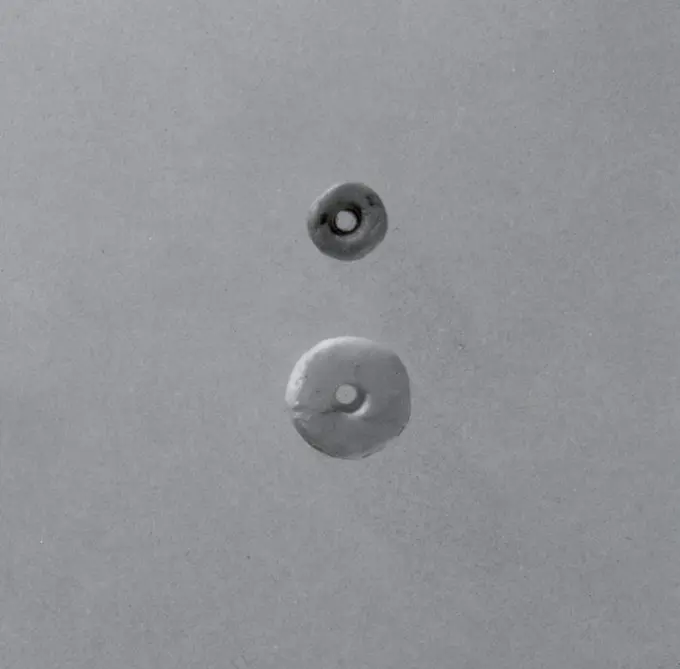
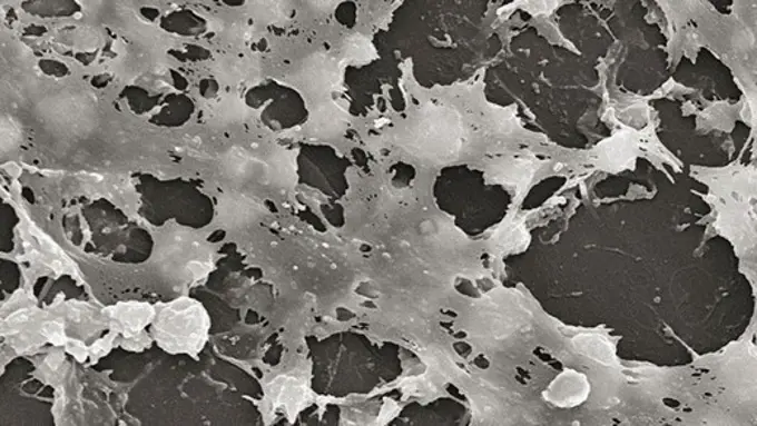
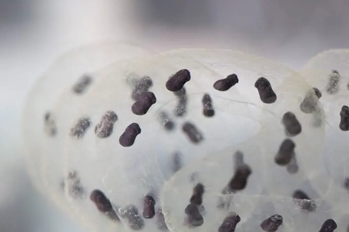
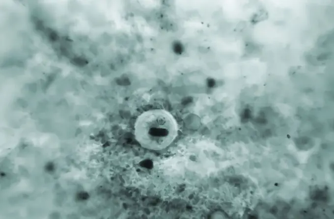
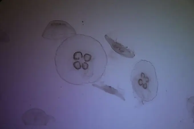
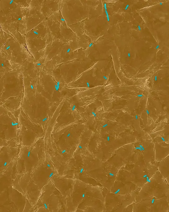
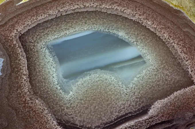
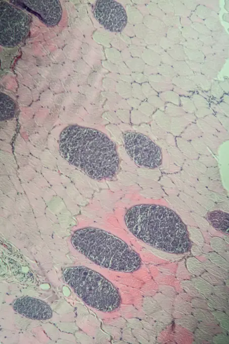
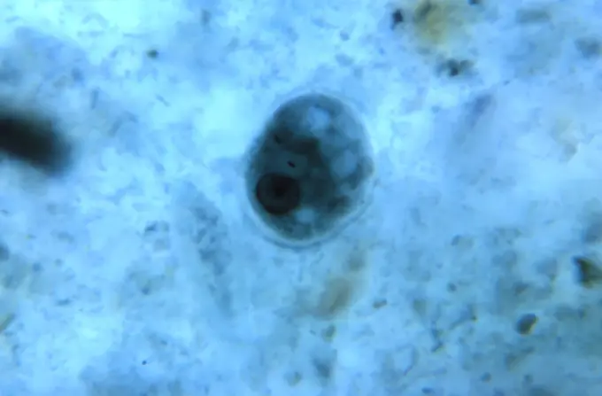
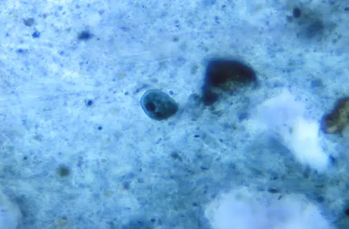
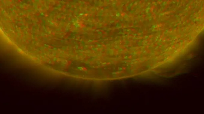
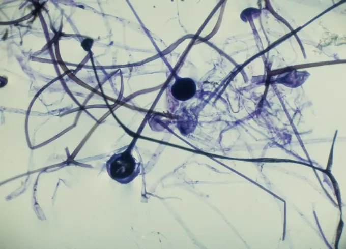
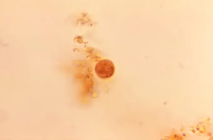
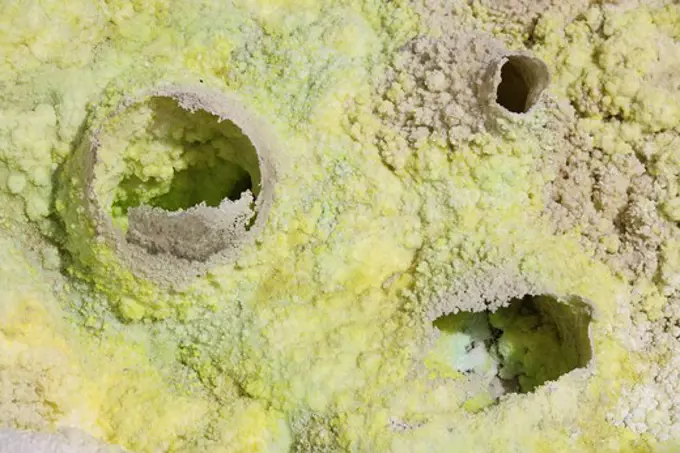
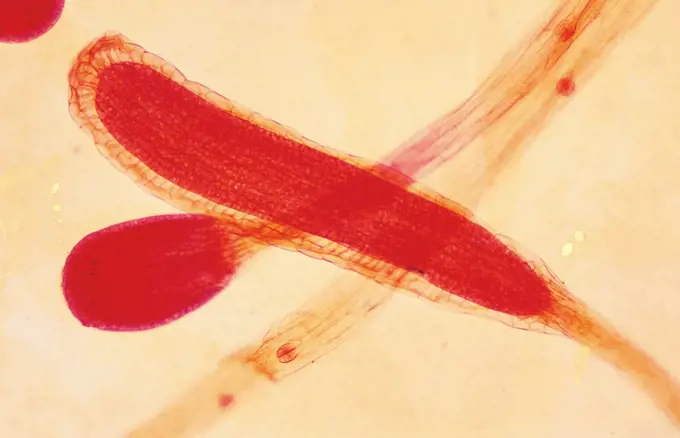
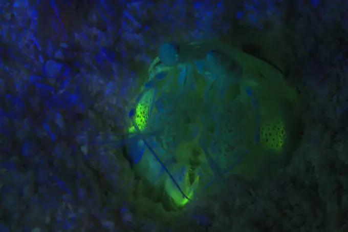
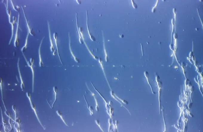
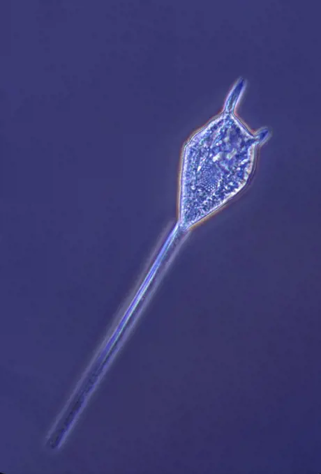
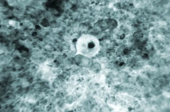
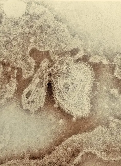
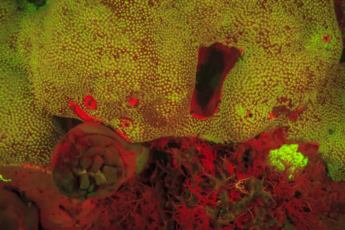
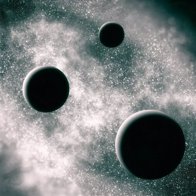
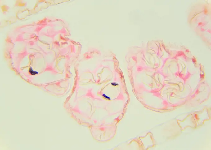
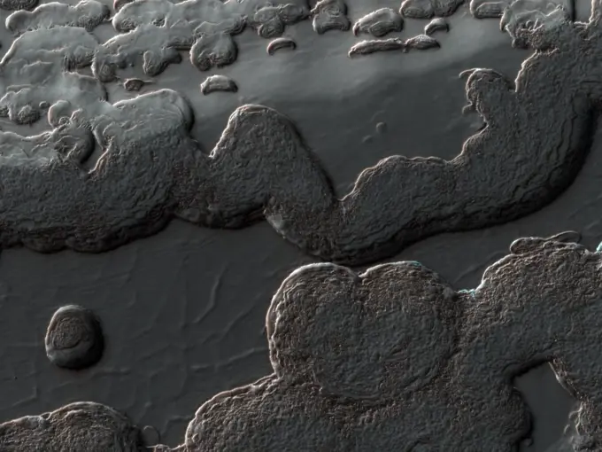
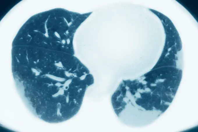
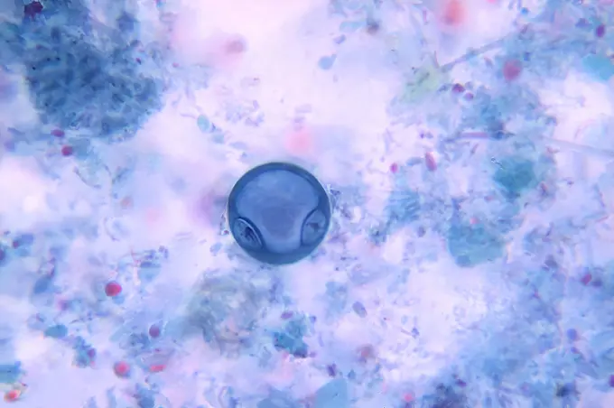
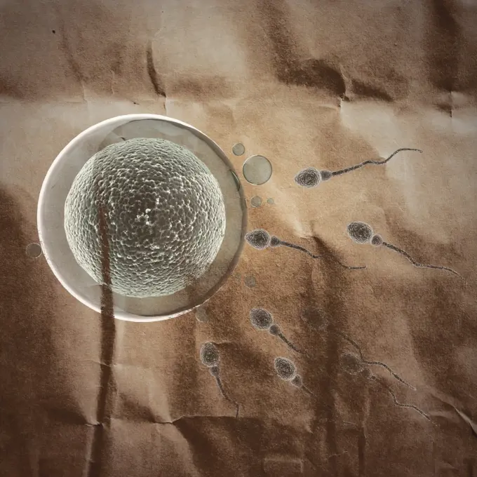
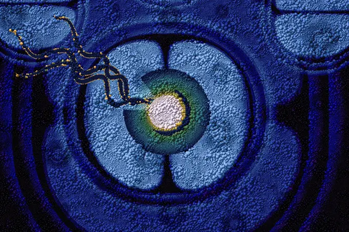
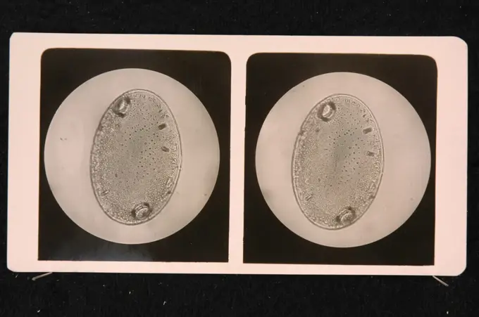
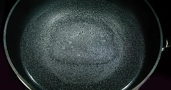
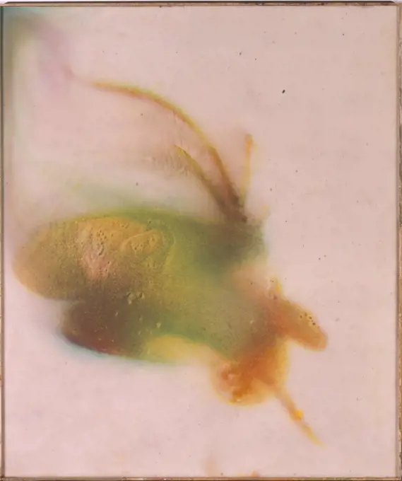
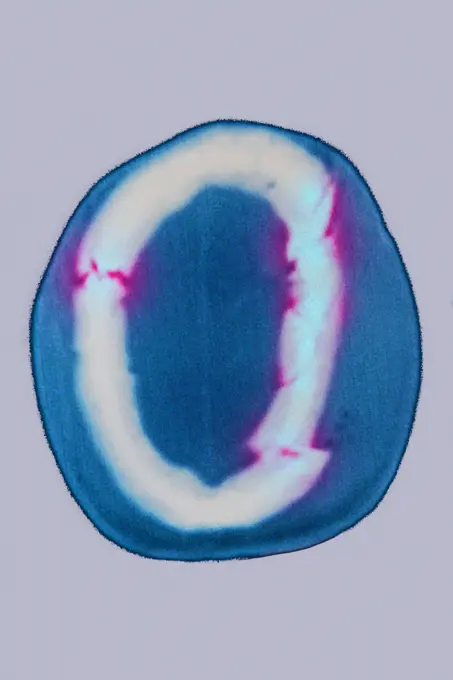
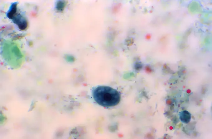
Microscopic images revealing various parasitic organisms and cellular structures with intricate details, combining shades of green, blue, and subtle hues.
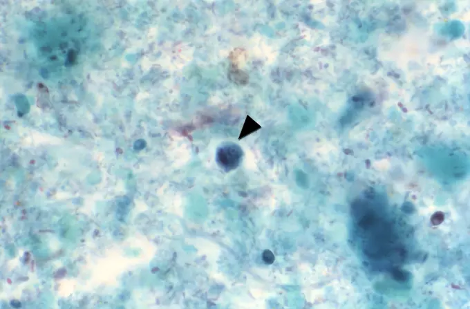
Microscopic images revealing various parasitic organisms and cellular structures with intricate details, combining shades of green, blue, and subtle hues.