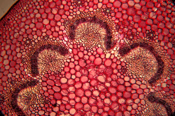
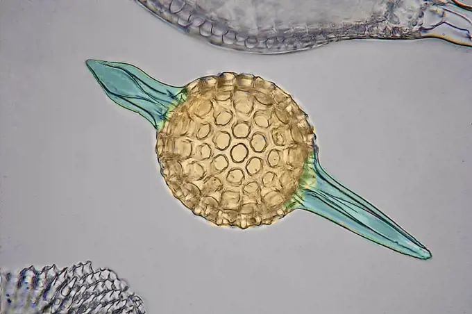
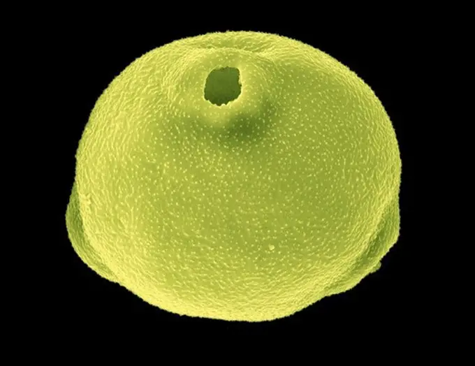
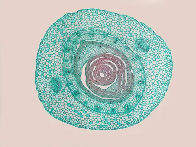
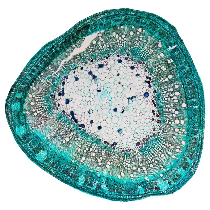
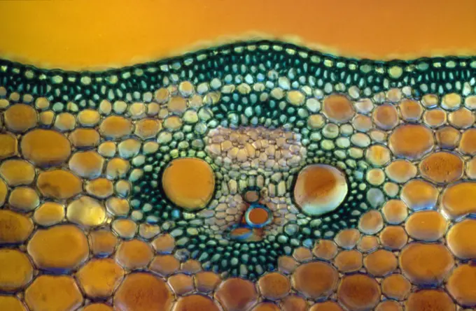
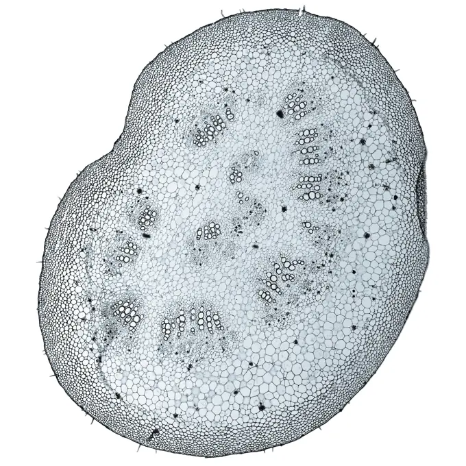
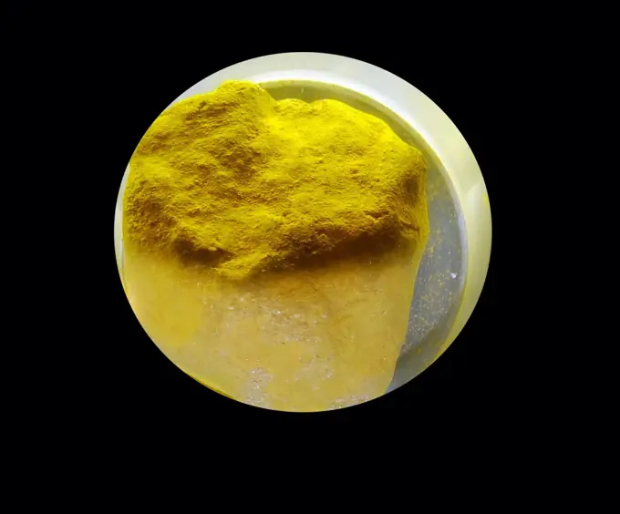
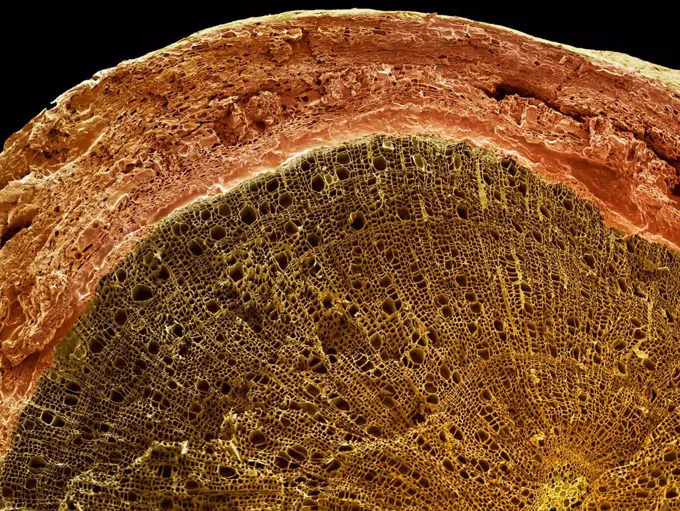
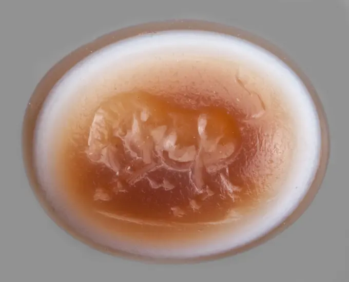
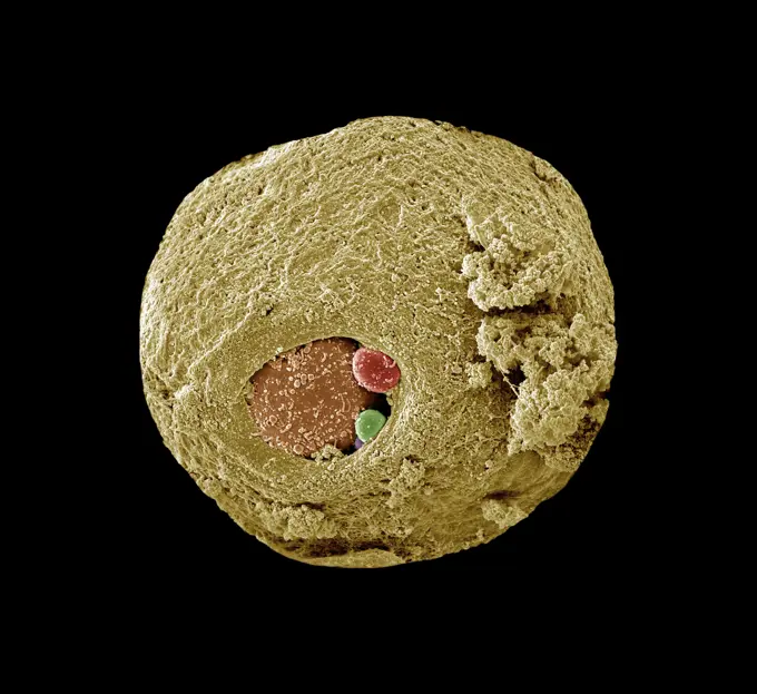
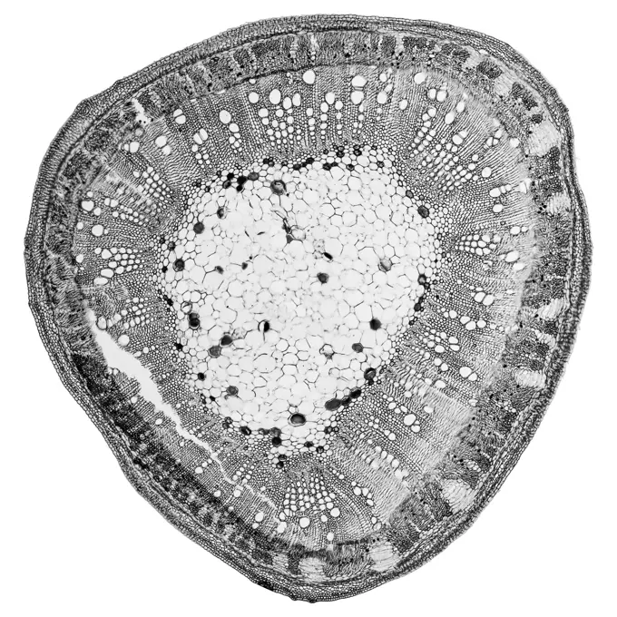
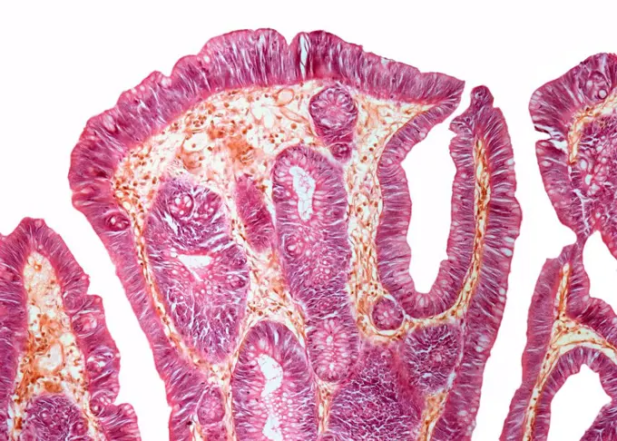
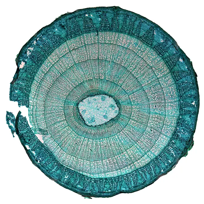
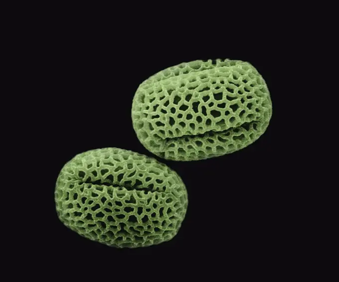
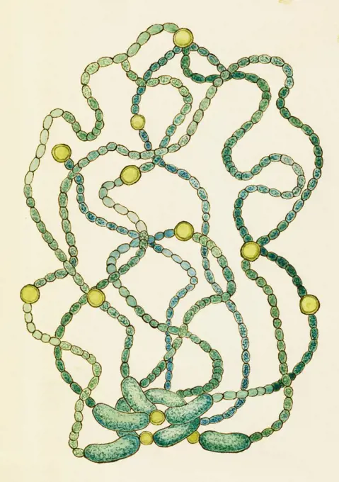
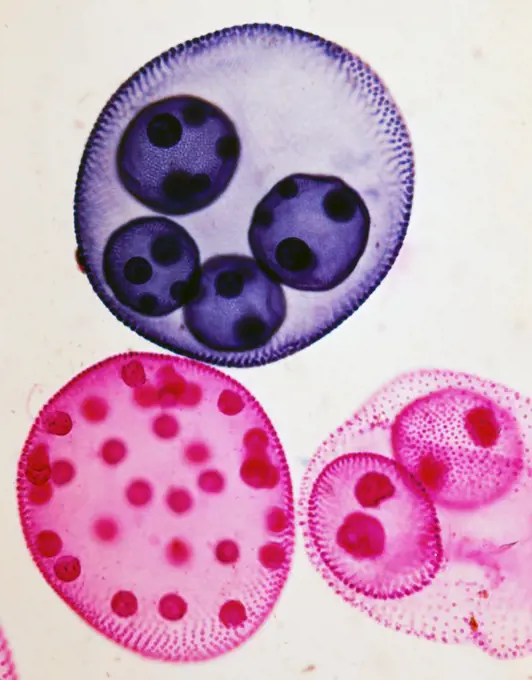
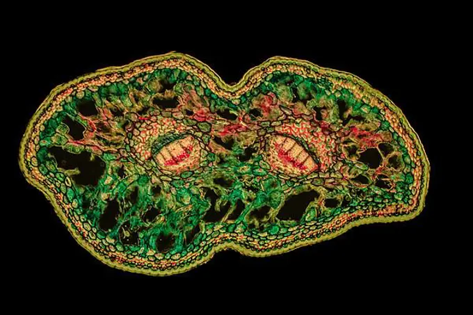
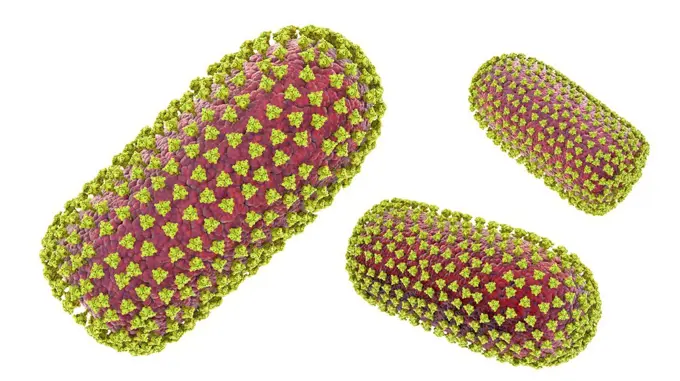
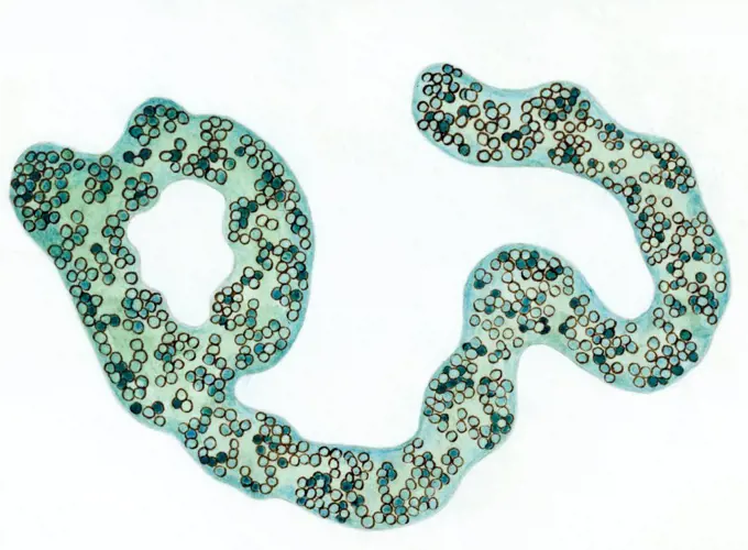
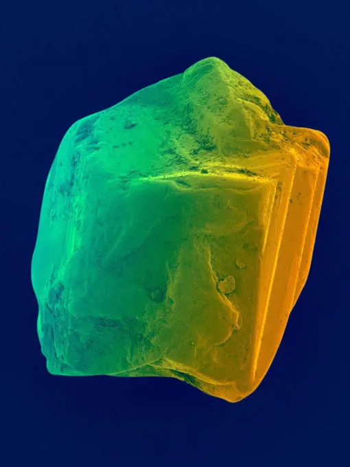
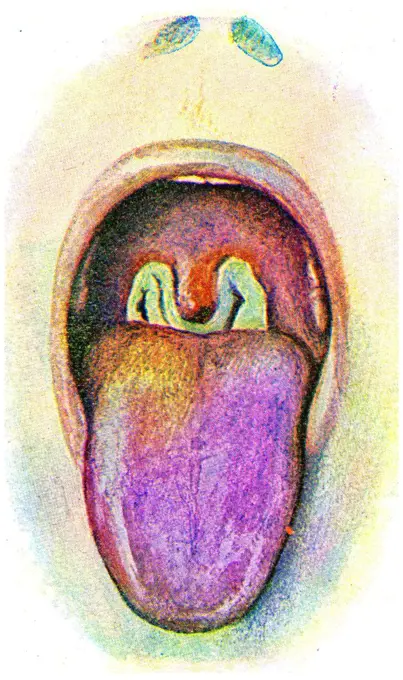
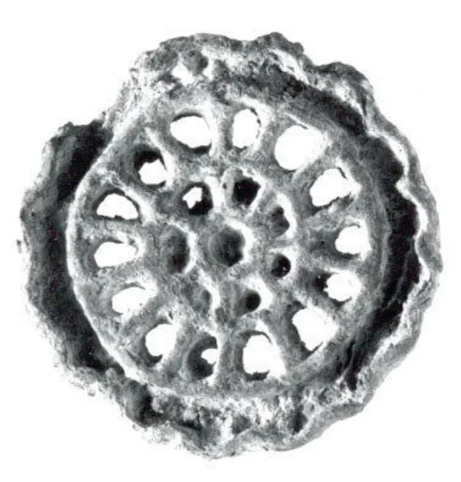
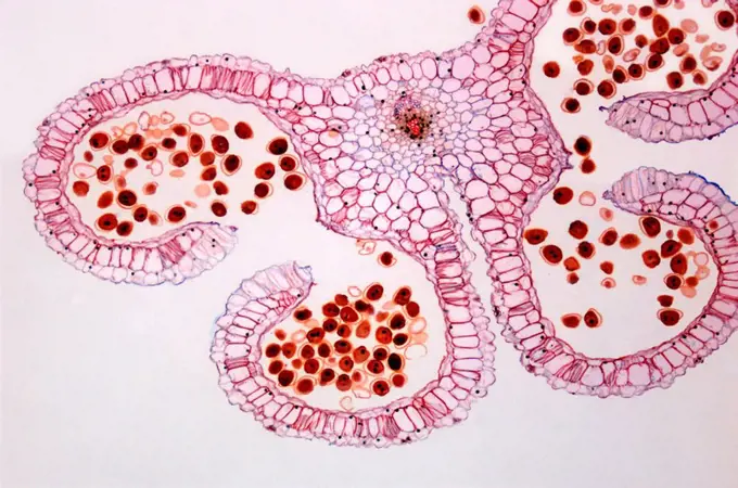
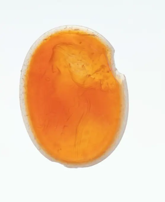
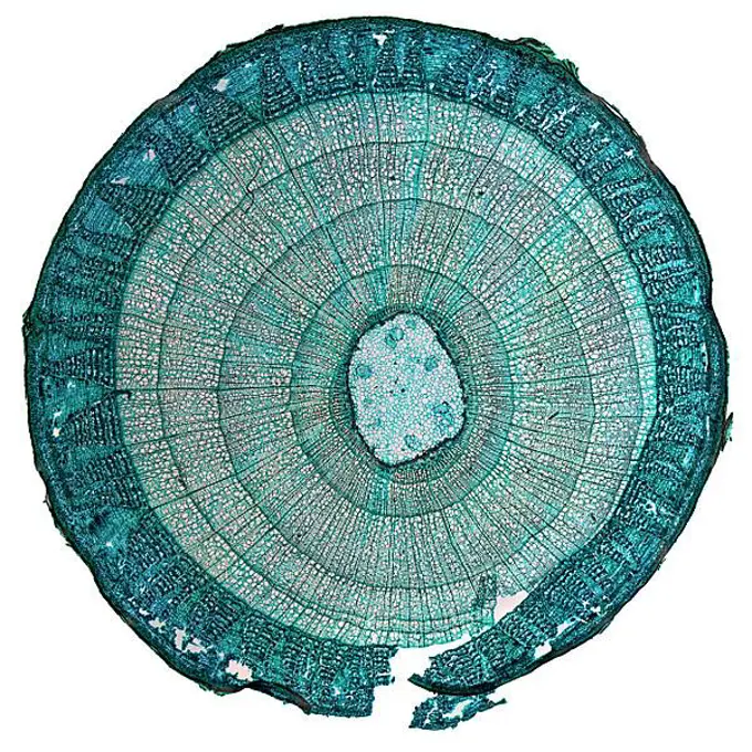
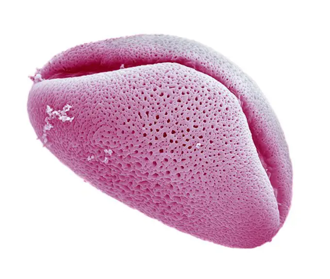
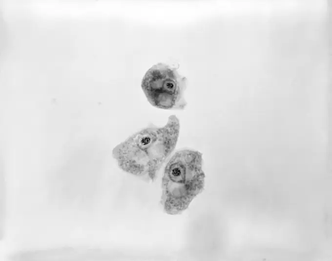
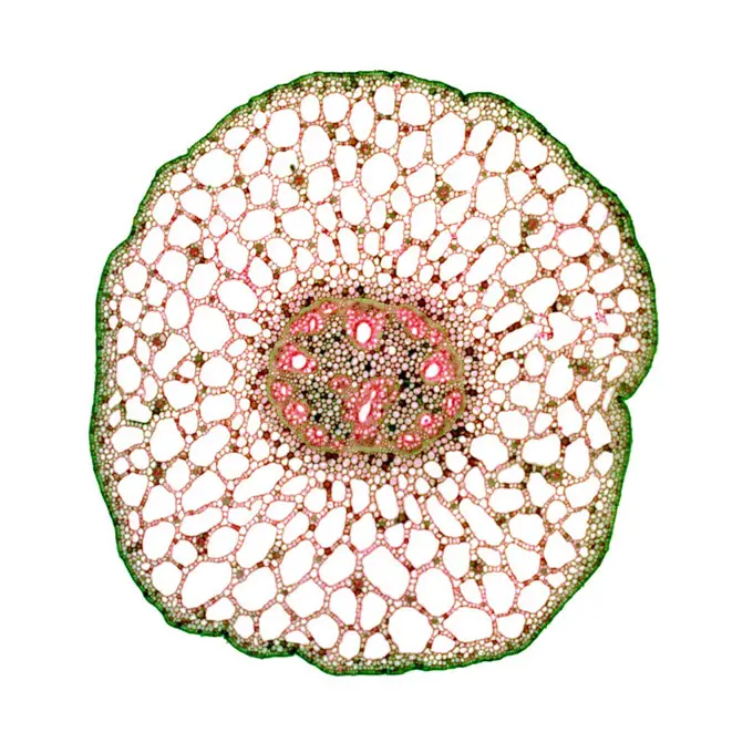
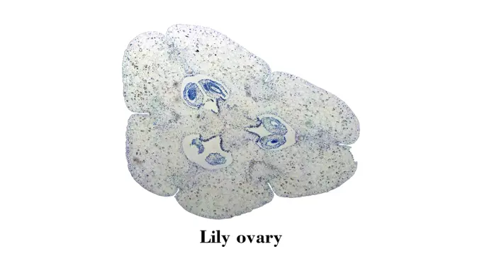
Fine ArtVintageFootageSubscriptionBoardsFree ResearchPricing
  • Fine Art
  • Vintage
  • Footage
  • Subscription
  • Boards
  • Free Research
  • Pricing

Microscopic Plant Structures

Detailed cross-sections of various plant tissues viewed under a microscope, highlighting cellular structures and colors, showcasing the complexity of plant anatomy.

Cutting of Ivy petiole
183 assets in this story
1540-111858042
Dance , Pictorial art of transverse section of ovary , It resembles a peacock feather in all it's glory and the dance , energy radiating outwards
4413-135027
Microscopic view of Radiolaire
4297-1264
Scanning electron microscopic image of a pollen grain of Birch
4128-17928335
Liver fluke, light micrograph
4298-1049
Cross section of an emerging corn (Zea) coleoptile, magnification 40x
1525-25939328
Structure of dried fruit of Echinocystis lobata, prickly and weird
6188-55585679
Cotton stem micrograph Light photomicrograph of Cotton stem cross section seen through microscope Copyright: xZoonar.com/ClaudioxDiviziax 8020463
4069-150
Platano or Plantain, watercolour, c.1585
4128-V58573794
6145-29806315
Sealing ca. 2030-1640 B.C. Middle Kingdom. Sealing 565405
7203-70646271
Natural occurring fluorescence in underwater red fluorescing sponges (Leucetta) and green hard coral (Acropora), captured by using special UV blocking barrier filters, Night dive at Wetar Island, Banda Sea, Indonesia
1890-107082
Light Micrograph LM of a transverse section of a Maize stem Zea sp. showing vascular bundle, cortex and epidermis, magnification x600
6188-55598793
Mulberry micrograph Light photomicrograph of Mulberry seen through microscope Copyright: xZoonar.com/ClaudioxDiviziax 11986382
4443-21932517
Blue tile of pilaster with 39 tiles, two logs, tile pilaster footage fragment ceramic pottery glaze, Three blue tiles with bunches of grapes leaves dragonfly basket of spiderweb and spider of pilaster with 39 tiles tree of bunches of grapes between which birds insect foot unknown, in black paint D 26.
4128-28515235
Bacteria and fungi cultured on Petri dish
4413-109790
Microscopic view of twig and moss Bryum sporangia
1746-21118099
Chrome yellow - crocoite. When the red mineral was crushed it would produce a yellow pigment.
6145-29606805
Collection Dutch Heidematschappij. Micro recording / cross section. undated
4128-18183824
Woody stem section, SEM
1848-18023535
Japanese umbrella fir (Sciadopitys verticillata), needle t.s., staining Wacker 3A, fluorescence and darkfield transmitted light
1525-28047255
Hydatid cyst, showing daughter cysts, vintage engraved illustration.
6145-45399670
Byk
4220-21334665
Scanning Electron Micrograph (SEM): Early human embryo, one cell removed for genetic analysis.     Date:
6188-55584504
Black and white Cotton stem micrograph Light photomicrograph of Cotton stem cross section seen through microscope in black and white Copyright: xZoonar.com/ClaudioxDiviziax 7744856
4128R-34258
Rectal papilloma, light micrograph. Magnification: x100 when printed at 10 centimetres wide.
6145-29606303
Collection Dutch Heidematschappij. Freshwater fishing, diatomee. undated
6188-55585871
Tilia stem micrograph High resolution light photomicrograph of tilia stem cross section seen through a microscope Copyright: xZoonar.com/ClaudioxDiviziax 8091797
4201-66356
Olive (Olea europaea) SEM close-up view of pollen at 1400x magnification
1788-111172661
Anabaena lemmermannii, Cyanobacteria or blue-green algae seen under a microscope. Drawing.
1788-111172794
Volvox colonies, genus chlorophytes, seen under a microscope.
1848-50837220
Japanese umbrella fir (Sciadopitys verticillata), needle t.s., staining Wacker 3A, fluorescence and darkfield transmitted light
4128-20239416
Rabies viruses, illustration
1848-51501677
Plastinate, cross section of human abdomen with skin cancer metastases, Body Worlds, Menschen Museum, Berlin, Germany, Europe
6188-55598495
Cells mitosis micrograph Light photomicrograph of Mitosis of onion root tip cells seen through microscope Copyright: xZoonar.com/ClaudioxDiviziax 11565715
4179-43458
Red Boring Sponge (Cliona delitrix) secretes Acid into Coral, killing it, Crb
1788-111172657
Microcystis aeruginosa, blue-green algae or cyanobacteria, seen under a microscope. Drawing.
4128R-13620018
Radiolarian tests (salt water), coloured scanning electron micrograph (SEM). The hard skeletons are composed of silica or strontium sulphate. The Radi...
1899-61460985
This photomicrograph revealed the presence of an Entamoeba histolytica trophozoite, which was photographed here while alive. This organism exhibited an explosive extrusion of a pseudopodia, pointing it to the left. CDC/Armed Forces Pathology Institute (AFIP) 1964
6145-44617844
Through the Haze
4297-1269
Scanning electron microscopic image of a pollen grain of corn
1746-21121910
Cake Sea Urchin (Clypeaster Reticulatus) oral view
4128R-13620337
Coloured scanning electron micrograph (SEM) of an uncut sapphire. Sapphire is a typically blue gemstone variety of the mineral corundum (aluminium oxi...
1525-27799743
Diphtheria, vintage engraved illustration.
6145-52771397
Microscopic photograph of scars on wheat and rye grains by Frana Jesenko on collodion paper mounted on a box box.
6145-29821092
Compartmented stamp seal ca. late 3rd-early 2nd millennium B.C. Bactria-Margiana Archaeological Complex. Compartmented stamp seal 326700
824-63208115
Osteoporosis with compression of the dorsal and lumbar vertebrae, seen on a sagittal plane MRI scan.
4128R-10352
Lily anther, light micrograph
6145-45502304
Bliżej Kultury
1848-50261391
(Tilia) stem micrograph
4297-1272
Scanning electron microscopic image of a pollen grain of an Alium
1899-61461029
This 1973 photomicrograph depicts three Naegleria gruberi, free-living amoebae. CDC/Dr George Healy 1973
1525-26226405
Abstract unique circle iridescent film from oil spill isolated on black background
824-63218216
Fracture of the skull, seen on a radial section scanner.
4449-28869912
Cucumber (macro photo)
1788-57305
Jellyfish Reticulomedusa greenei fossil, Scyphozoa, Carboniferous Period.
4128-30420964
Autosomal dominant vitreoretinochoroidopathy, illustration
1525-28047030
Kidney, chronic parenchymatous nephritis, vintage engraved illustration.
6145-29731032
Compartmented stamp seal ca. late 3rd-early 2nd millennium B.C. Bactria-Margiana Archaeological Complex. Compartmented stamp seal 326869
1746-21113128
Spatangus purpureus, a genus of heart urchins in the Spatangidae family. British Waters. Dated 1776
1899-61460428
Variola virus (poxviruses are large viruses of 300/200/100 nm. They are pathogenic for humans such as smallpox and vaccinia viruses as well as animal viruses that infect humans such as viruses monkeypox, cowpox, milker's nodule, pustular dermatitis in sheep Transmission electron microscopy
7008-69757139
Laguna agate from Mexico
6145-46765636
Painted image on the glass for SkiopTikon / Laterna Magica. Image of the starry sky.
1848-49597284
Rhodochrosite, Catamarca Province, Argentina Rhodochrosite is a manganese carbonate mineral that ranges in color from light pink to bright red
4115-5997
Deepsea Glass sponge (Hexactinellida) from 2500m, mid Atlantic ridge, June 2010
4297-1267
Scanning electron microscopic image of a pollen grain of a Petunia
4409-20923688
DRACMA DE ROSAS - ULTIMO TERCIO DEL S IV AC.
1788-20759729
Agate nodule, variety of Chalcedony, oxide. Section.
1439-57940019
A closeup view of human fingerprints
6145-52890670
Stereo photograph by Romanus Schmehlik depicting the diet of a lattle at a scale of 30:1, printed on silver gelatin paper in 1912. The image captures detailed microscopic observation of dietary structures.
6145-29606248
Collection Dutch Heidematschappij. egg deposit. undated
1848-51751781
Prehistorical insect trapped in Amber stone
6145-29724985
Compartmented stamp seal ca. late 3rd-early 2nd millennium B.C. Bactria-Margiana Archaeological Complex. Compartmented stamp seal 326828
4201-61054
Amu-Darya Trout (Salmo trutta trutta) alevin hatching from egg, Europe
1788-111172766
Cross section of a leaf affected by tobacco mosaic virus seen under a microscope.
1848-55286036
Historical book illustration, Table 9, Title Hexacoralla Six Radial Star Coral, Name Macandrina filograna, Family Astraeidae, Star Corals, from "Kunstformen der Natur" by Ernst Haeckel
4413-88881
Cross section of the reproductive system of a fern
4128R-8806
Pondweed stem, light micrograph
1899-65211
E-coli (Escherichia coli O157:H7) sample taken from swab
6145-45335895
Magic gemma with the performance of Jupiter Seapis
4413-193408
Olive fruit Fly larva dead infested
5514-55413222
Amazing ants carry fruit heavier than their bodies
4400R-1921
A colour chart, close-up.
1525-19790155
Lily ovary micrograph. Light photomicrograph of Lily ovary cross section seen through microscope
PREVIOUS
of 2
NEXT
2401 S. Ervay, Suite 206
Dallas, Texas 75215
United States
Browse
CategoriesGalleriesFree Research
For Creators
License Your ContentSuperStock For StudentsFrame of Mind BlogContributor PortalNew Image SpotlightLicense AI Imagery
Resources
About UsLicensingTerms & ConditionsAPI AccessPrivacyPricing
Contact
+1 866 236 0087help@superstock.com Contact form
©2026 SuperStock. All Rights Reserved. -A
This site is protected by reCAPTCHA and Google Privacy Policy and Terms of Service apply.