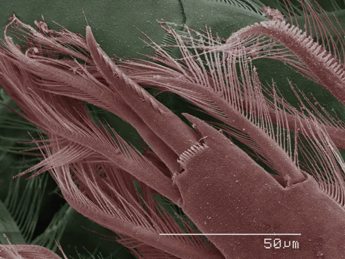
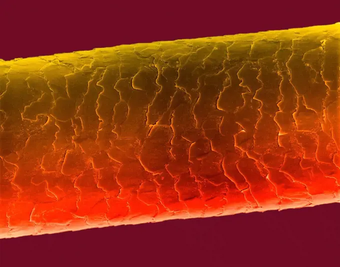
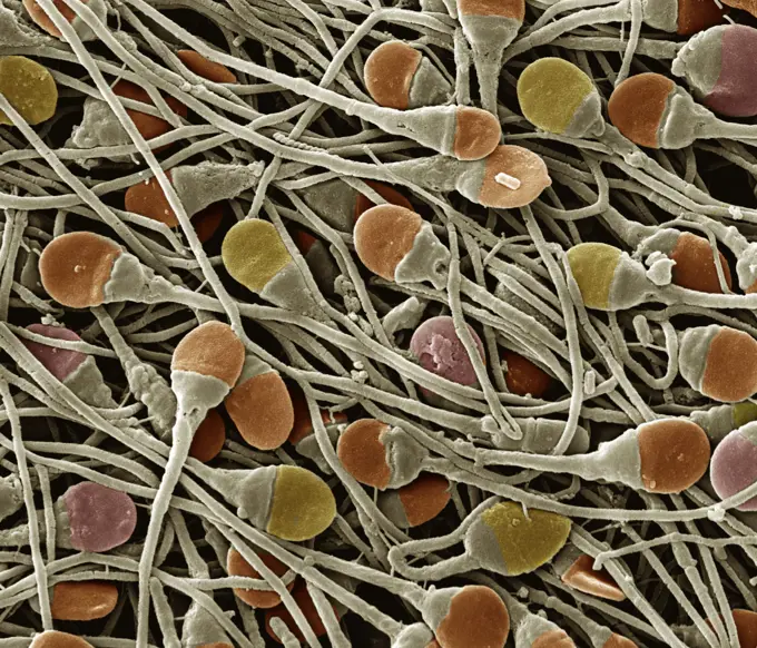
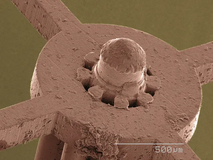
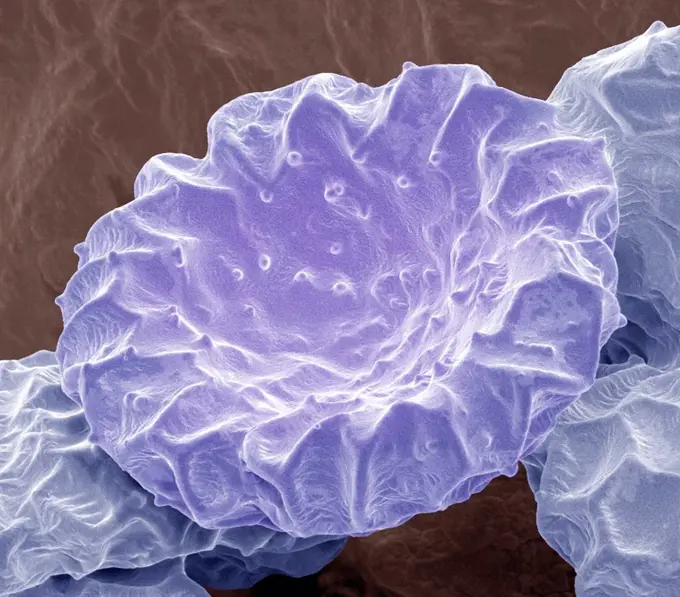
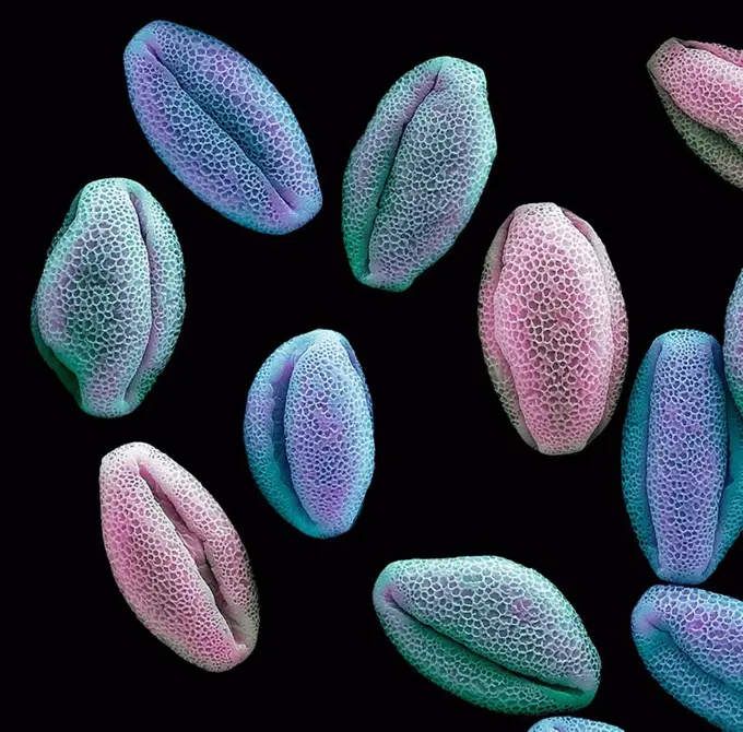
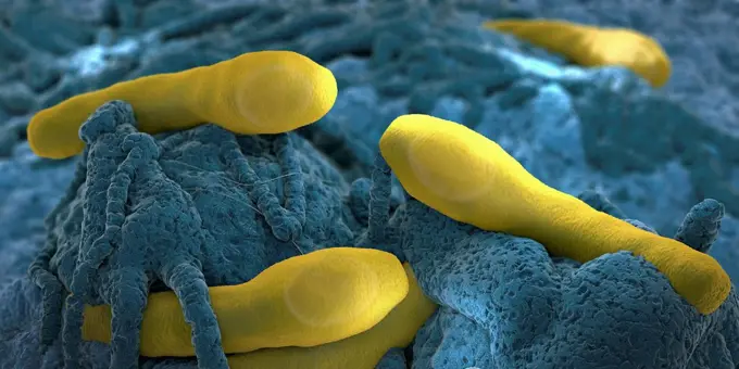
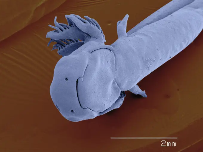
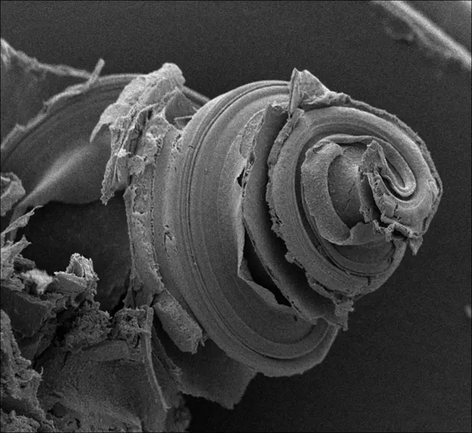
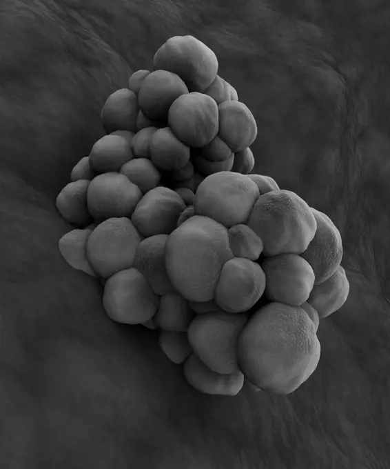
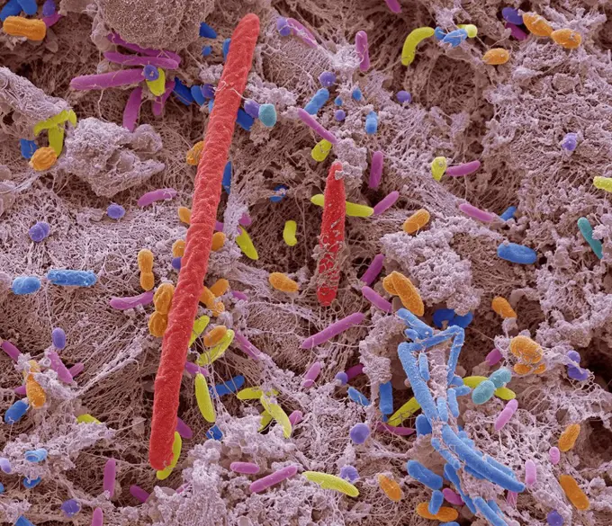
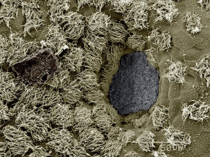
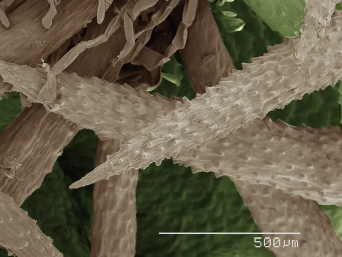
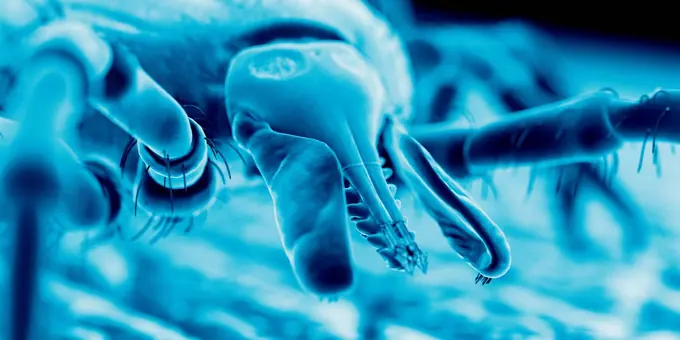
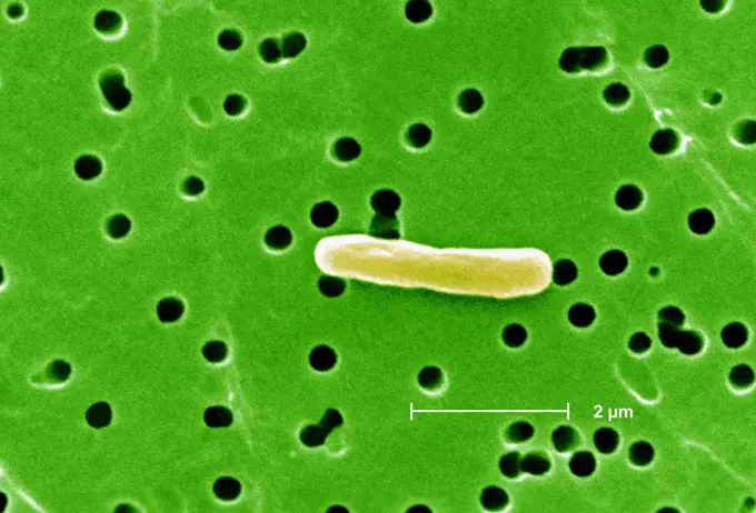
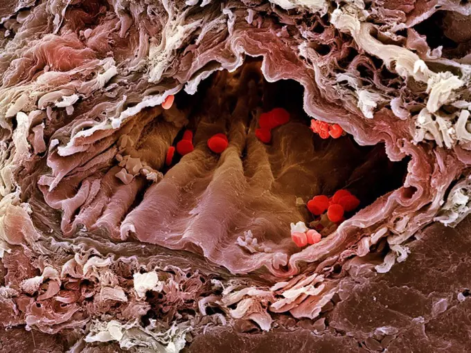
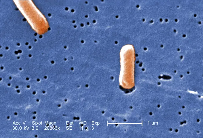
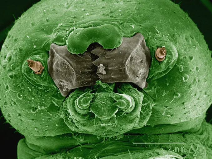
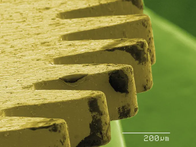
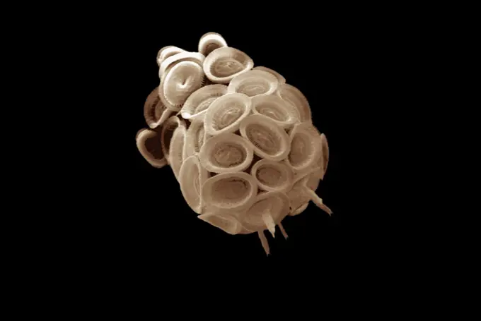
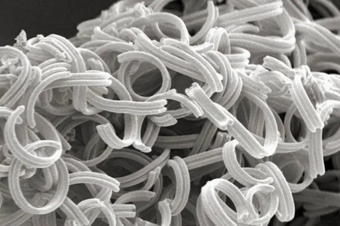
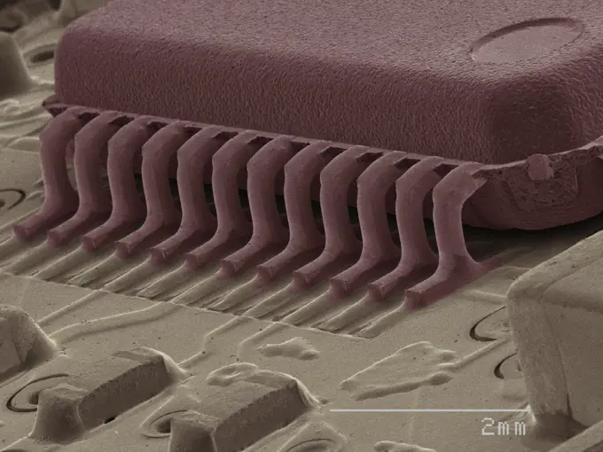
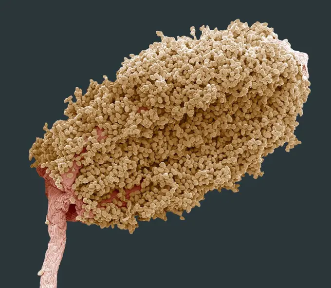
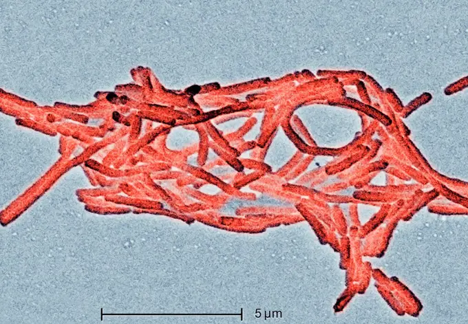
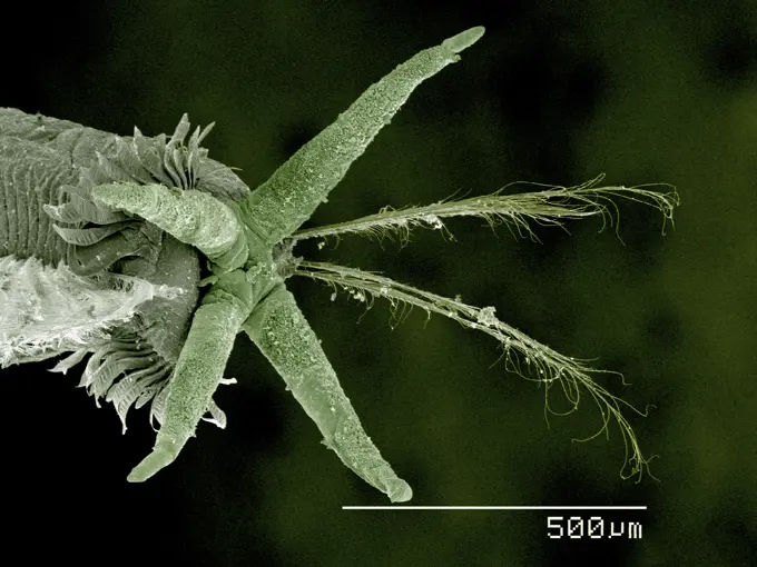
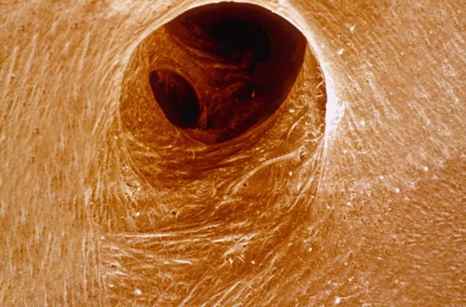
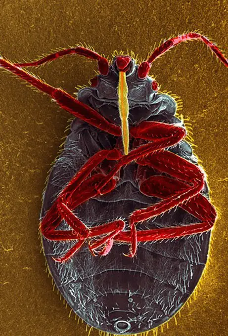
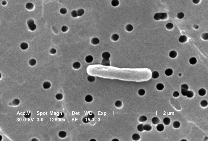
Fine ArtVintageFootageSubscriptionBoardsFree ResearchPricing
  • Fine Art
  • Vintage
  • Footage
  • Subscription
  • Boards
  • Free Research
  • Pricing

Microscopic Textures and Structures

Close-up views of various microscopic structures, including textile fibers and insect exoskeletons, showcasing intricate textures and organic forms.

A closeup view of a microscopic structure under high magnification
159 assets in this story
4128R-13620383
Human hair (Caucasian, brunette), coloured scanning electron micrograph (SEM). The outer layer of hair (the cuticle) has overlapping scales of keratin...
4220-21334744
Scanning Electron Micrograph (SEM): human sperm.     Date:
4128R-28137
Human coronavirus. Computer artwork of a Human coronavirus particle. Coronaviruses primarily infect the upper respiratory and gastrointestinal tract a...
1439-57939977
Microscopic view of a mechanical component.
4128R-14143
Heart strings, coloured scanning electron micrograph SEM. The heart strings, chordae tendineae, are cord_like tendons that connect the papillary muscl...
1439-57940131
A microscopic view of a bloodsucking tick
4269-6804
Scanning electron micrograph (SEM) of a glomerulus. On the surface, the ramifications of the podocytes which filter the blood and produce urine. Magnification x1000.
1899-61460830
Magnified 3000X, this 2002 scanning electron microscope (SEM) image revealed some of the ultrastructural morphology exhibited by four Hartmannella vermiformis amoeba cysts. The amoeba assumes the cystic stage of its life cycle under harsh conditions such as drought, or when there is an accumulation of toxins in its environment, which can reduce its metabolic needs. Under these circumstances, the protozoan produces a protective coat and goes dormant to wait for better conditions. CDC/Dr Rodney Donlan 2002
4128R-7664
Inner ear hair cells, SEM
1439-57949042
A closeup monochromatic view of a tiny helical structure and beads.
1899-61460823
Magnified 1155X, this 2002 scanning electron microscope (SEM) image revealed some of the ultrastructural morphology exhibited by a number of Hartmannella vermiformis amoeba trophozoites
4128R-8936
Bougainvillea pollen, SEM
4128-111584778
Water lily pollen grains. Coloured scanning electron micrograph (SEM) of pollen grains from a water lily flower. Nymphaeaceae is a family of flowering...
4128-20041032
Clostridium botulinum growing on tissue, illustration
4128R-9311
Wood anemone anther, SEM
1439-57940201
Extreme closeup of a tardigrade under the microscope
6145-29256365
The cochlea is a vital part of our ear, allowing us to detect a wide range of frequencies of sound. This is a picture showing the characteristic snail shell structure of the cochlea in the inner ear, captured on a high-resolution electron microscope. The bone usually covering the cochlea has been removed to open up a window into the hidden world of hearing.
4239R-20483596
Microscopic view of tumor.
4128R-13620194
Coloured scanning electron micrograph (SEM) Chicken eggshell cross section (Gallus gallus domesticus). Shown from left to right in the image. hard min...
4128R-13573038
Oral bacteria. Coloured scanning electron micrograph (SEM) of mixed oral bacteria. The mouth contains a large number of bacteria, most of which are ha...
4384-178
Under a moderate magnification of 340X, this digitally-colorized scanning electron micrograph (SEM) depicted the exoskeletal surface of a larval staged antlion, sometimes referred to as a doodlebug, because of the trails it leaves in the soft sand as it hunts for prey.  This arthropod undergoes dramatic morphologic changes when it metamorphoses into a beautiful flying antlion lacewing. In this particular view, the distal end of one of the larvas extremities is highlighted, revealing the claw
1439-57943337
Intricate textures of a microscopic landscape
1439-57940109
An upclose view of organic textures under magnification
4128-30418011
Tick on human skin, illustration
4128R-13620206
Coloured scanning electron micrograph (SEM) Cat hair (Felis silvestris catus). Cats have four types of hair. There are short fluffy hairs called secon...
824-63123756
This scanning electron micrograph (SEM) depicts a highly magnified view of a rod-shaped Escherichia coli bacterium; Magnification 12800x. Escherichia coli is a Gram-negative bacterium that normally colonizes the digestive tract of most warm-blooded anima
4128-111494391
Coloured scanning electron micrograph (SEM) of a freeze fracture of the vagina showing the stratified squamous epithelium and the underlying lamina pr...
6162-76256891
Aerial view of earth's veins flowing with pink hues and beige tones as geometric shapes meet, creating a stunning contrast., Eilat, South District, Israel.
4128R-6414
Artery SEM
4269-27448
Escherichia coli. This scanning electron micrograph (SEM) of a Escherichia coli bacterium ; Magnification 12800x. E. coli are rod-shaped Gram-negative bacteria that normally colonize the digestive tract in humans and other animals. However, some strains can produce a toxin that leads to severe illness or even death.
1439-57949075
Closeup view of a pointed object tip with microscopic details.
1439-57947381
Monochrome microverse a grayscale scientific exploration.
4269-6803
Scanning electron micrograph (SEM) of a glomerulus. On the surface, the ramifications of the podocytes which filter the blood and produce urine. Magnification x1000.
1773-97832
Electron micrograph of Legionella bacteria, 6500x
824-63194911
This digitally-colorized scanning electron micrograph (SEM) depicted the dorsal (upper) surface of a Giardia protozoan that had been isolated from a rat's intestine. Some of the identifying morphologic characteristics include pairs of thread-like flagella that facilitate motility, and a ventolateral flange that appears as a ruffle around the anterior portion of the organism. Pairs of flagella seen here include an anterior, posterior-lateral, and caudal pairs. The protozoan Giardia causes the diarrheal disease called giardiasis.
4384-380
Microscopic Detail of a Wasp's Leg
1439-57947633
Intricate closeup of coral interaction
4384-345
Head and Leg Joint of Unidentified Insect
824-63194410
At a magnification of 20863X, this colorized scanning electron micrograph (SEM) revealed two Gram-negative bacilli, or rod-shaped Salmonella sp. bacteria. The genus Salmonella is a member of the taxonomic family, Enterobacteriaceae, and approximately 2000 serotypes of this genus are known to cause disease in human beings.
1439-57949000
Intricate details of an insect under the microscope
1439-57949039
Closeup view of an insect under the microscope
1439-57939979
Closeup of a microchip edge under magnification
4220-21334515
Scanning Electron Micrograph (SEM): Coccolithophore (Calcareous Nannoplankton), Syracosphaera pulchra.     Date:
1439-57952630
Extreme closeup of an insect head under a scanning electron microscope
4269-27366
Vibrio parahaemolyticus. Scanning electron micrograph (SEM) of Vibrio parahaemolyticus. bacteria. This Gram-negative bacterium causes gastointestinal infection to man. It is closely related to Vibrio cholerae, the  agent of cholera. Magnified 19058x.
4384-350
Microscopic Debris
1439-57940137
A microscopic view of a microchip resembling a scifi landscape.
4128R-9310
Wood anemone anther, SEM
1773-97828
Electron micrograph of Legionella bacteria, 6500x
1439-57943351
Microscopic closeup of an insect with detailed appendages
1439-57943373
Closeup of a seed under the microscope in sepia tones
824-63123121
The human skeleton is made of bone composed of calcium, an important element in body functions. The surface of bones are covered with a connective membrane called the periosteum. Below the periosteum the bone is dense like ivory (cortical bone) and very r
4297-1135
Scanning electron micrograph revealing the ultrastructural morphology displayed on the ventral surface of a Bedbug (Cimex lectularius)
4269-27449
Escherichia coli. This scanning electron micrograph (SEM) of a Escherichia coli bacterium ; Magnification 12800x. E. coli are rod-shaped Gram-negative bacteria that normally colonize the digestive tract in humans and other animals. However, some strains can produce a toxin that leads to severe illness or even death.
4128-V58569338
255-58492129
Vintage Photograph. Tomato horn worm with cocoons of the Braconid wasp attached to the body of the worm.
824-57659297
Magnified 1796X, this 2002 scanning electron microscope (SEM) image revealed some of the ultrastructural morphology exhibited by three Hartmannella vermiformis amoeba cysts. The amoeba assumes the cystic stage of its life cycle under harsh conditions such as drought, or when there is an accumulation of toxins in its environment, which can reduce its metabolic needs. Under these circumstances, the protozoan produces a protective coat and goes dormant to wait for better conditions. CDC/Dr Rodney Donlan 2002
824-63194915
This digitally-colorized scanning electron micrograph (SEM) revealed some of the ultrastructural morphology of a sickle cell red blood cell (RBC) found in a blood specimen of an 18 year old female patient with sickle cell anemia, (HbSS), People who have this form of sickle cell disease inherit two sickle cell genes (S), one from each parent. This is commonly called sickle cell anemia, and is usually the most severe form of the disease.
4384-233
Scanning Electron Micrograph of Mosquito
PREVIOUS
of 2
NEXT
2401 S. Ervay, Suite 206
Dallas, Texas 75215
United States
Browse
CategoriesGalleriesFree Research
For Creators
License Your ContentSuperStock For StudentsFrame of Mind BlogContributor PortalNew Image SpotlightLicense AI Imagery
Resources
About UsLicensingTerms & ConditionsAPI AccessPrivacyPricing
Contact
+1 866 236 0087help@superstock.com Contact form
©2026 SuperStock. All Rights Reserved. -A
This site is protected by reCAPTCHA and Google Privacy Policy and Terms of Service apply.