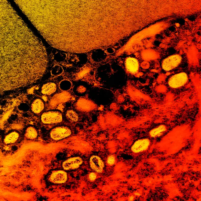
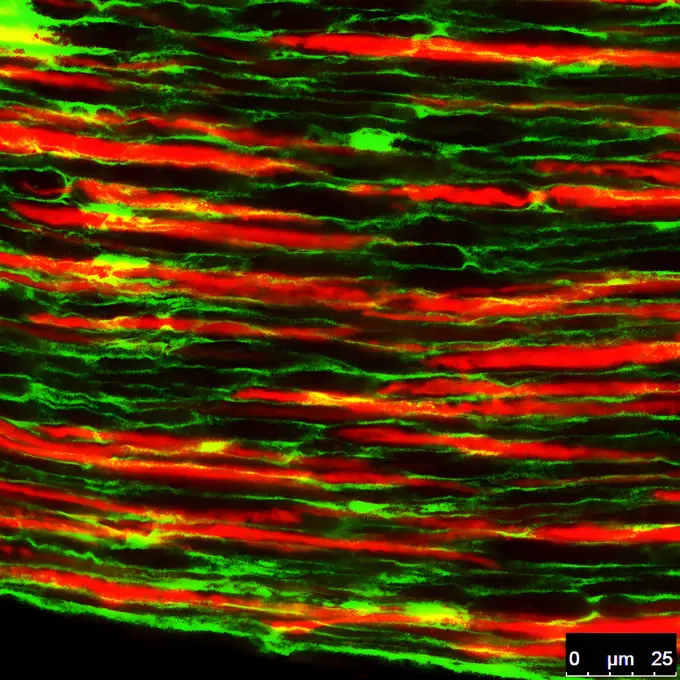
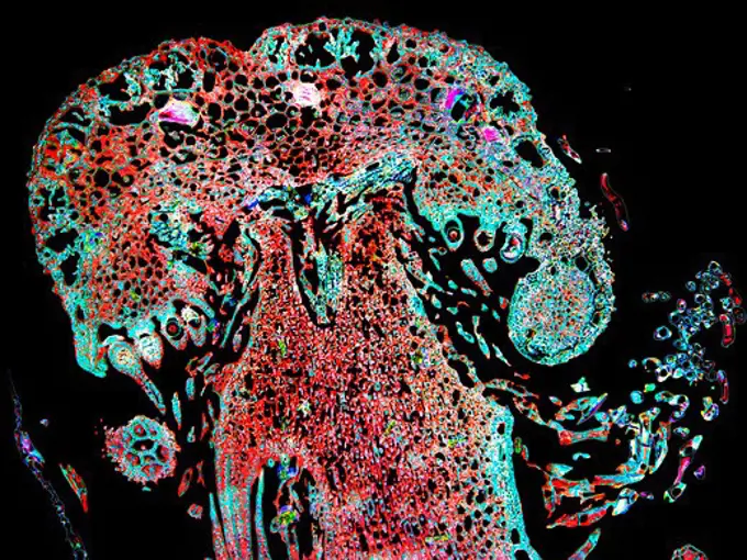
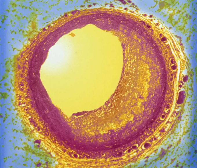
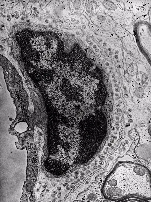
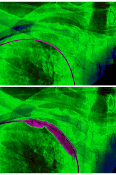
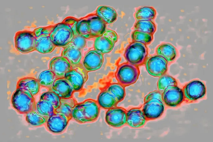
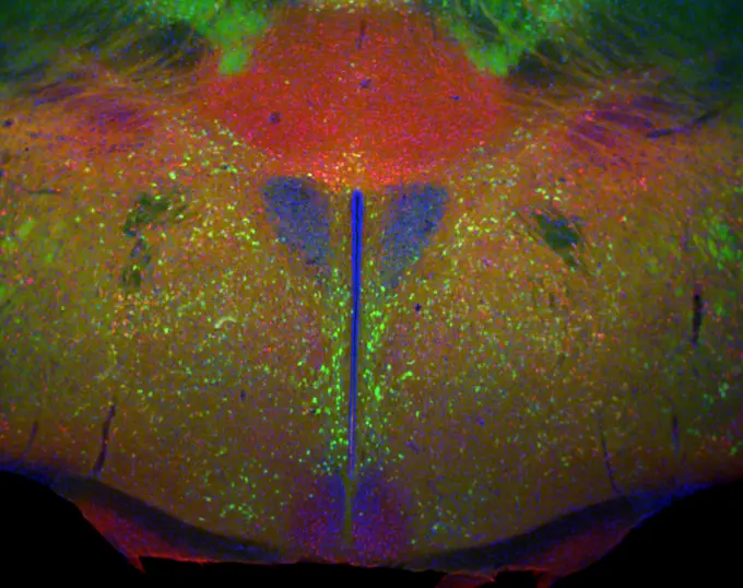
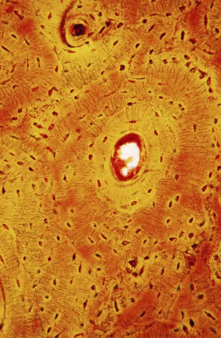
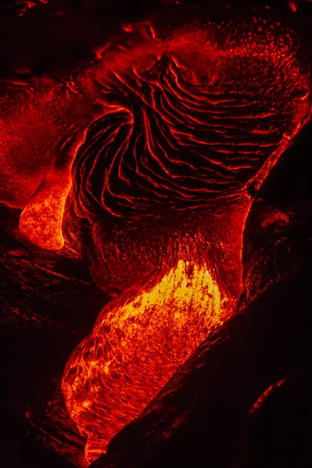
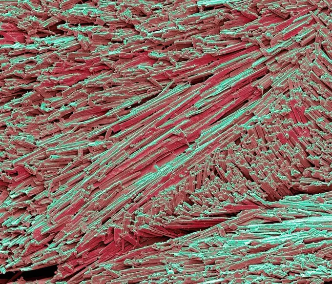
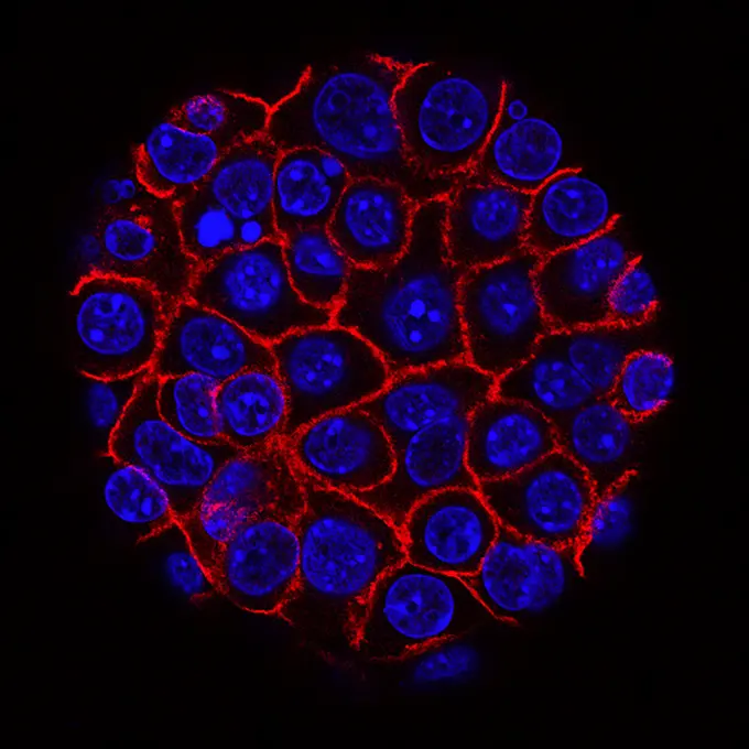
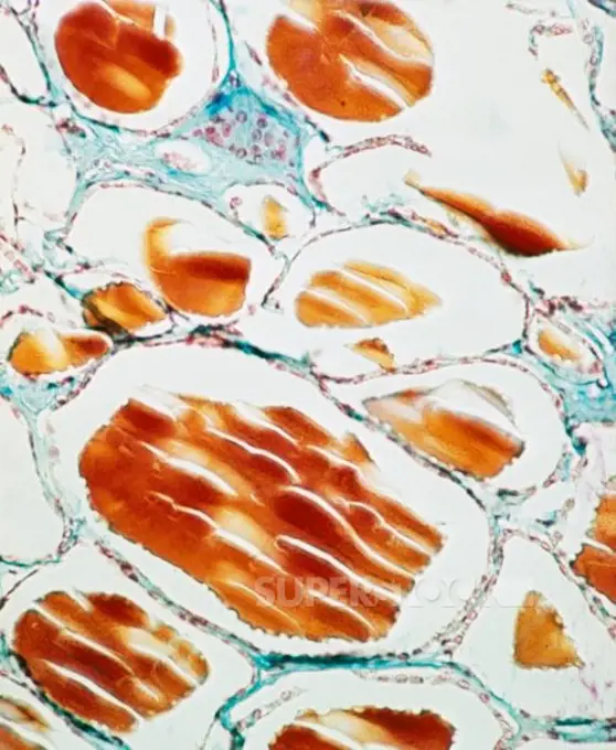
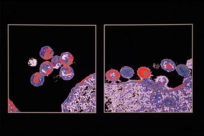
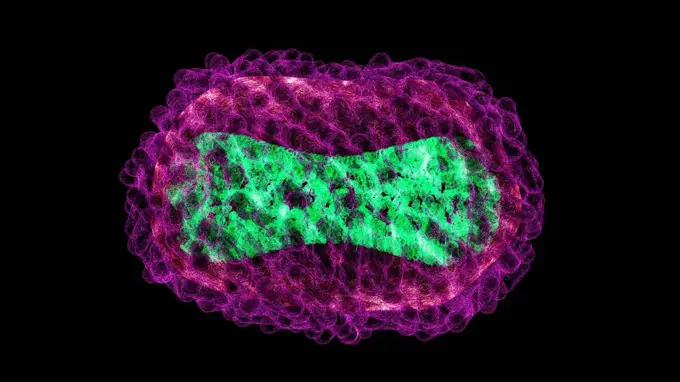
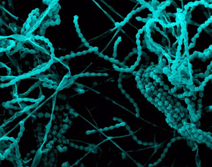
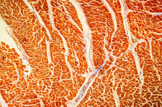
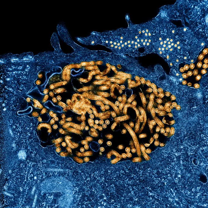
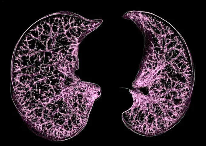
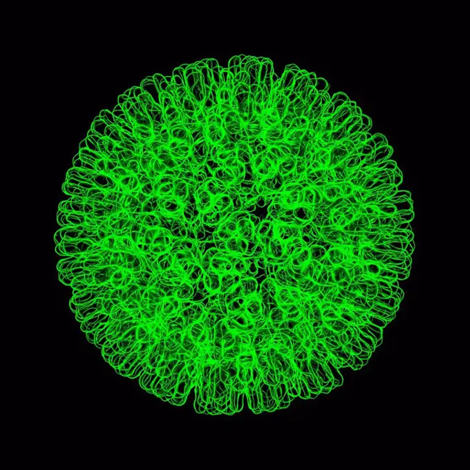
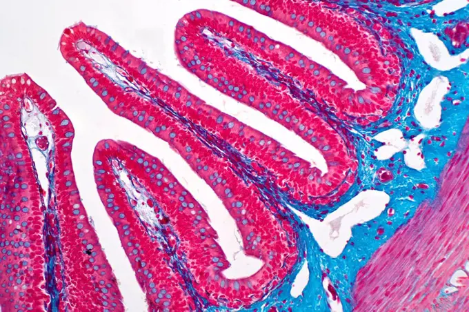
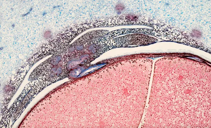
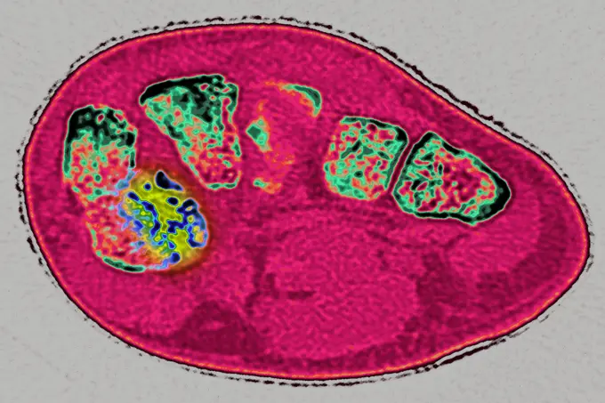
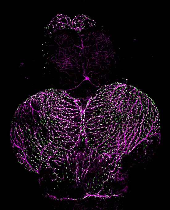
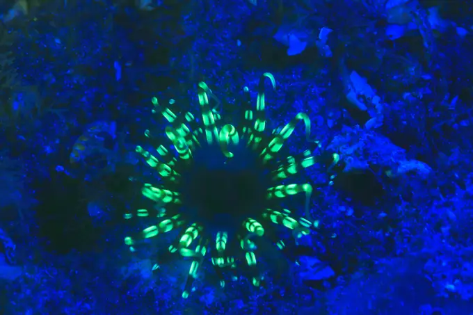
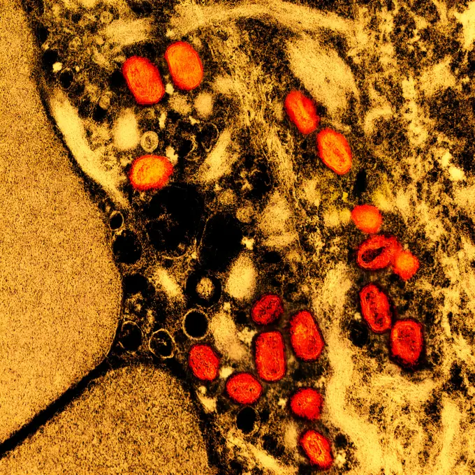
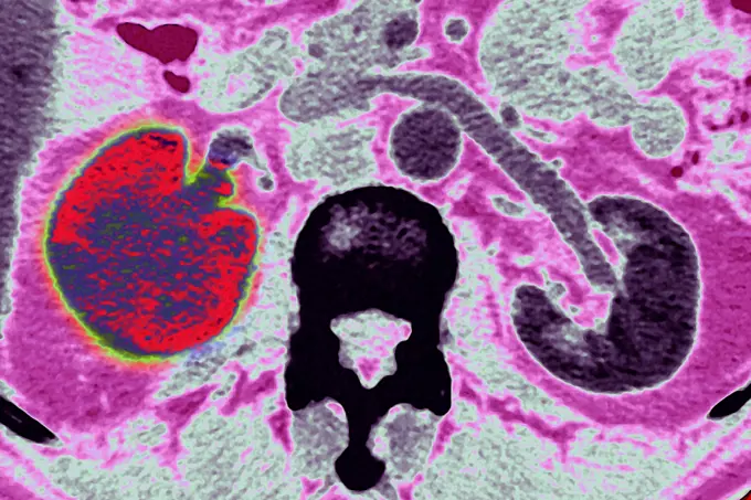
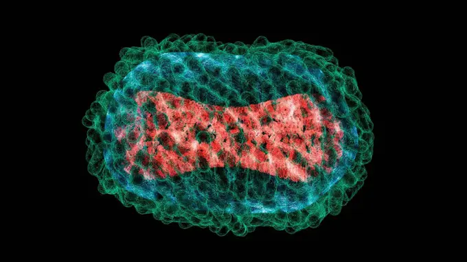
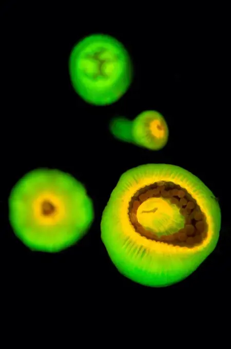
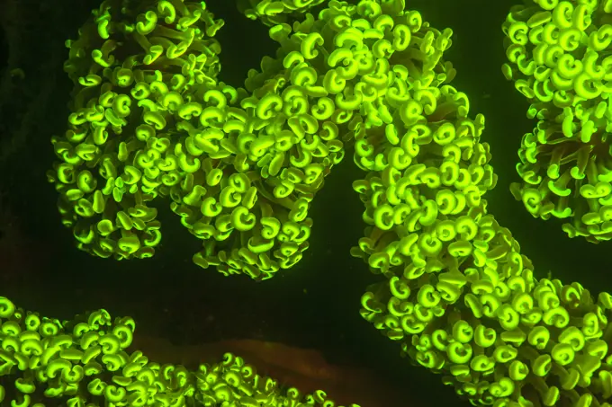
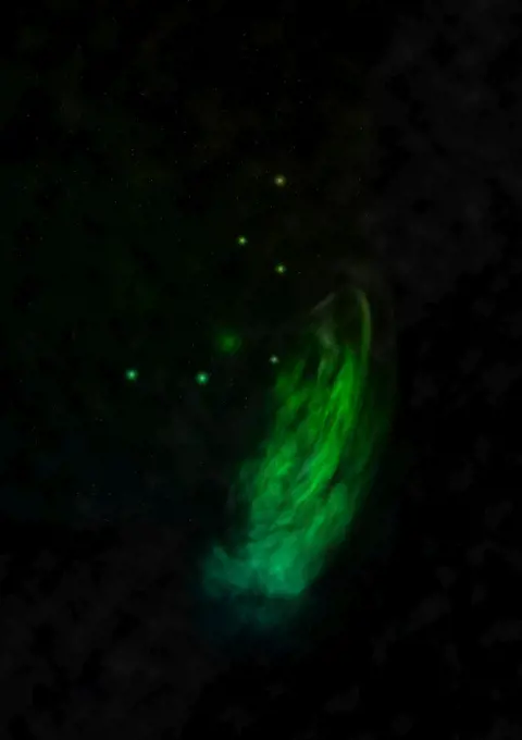
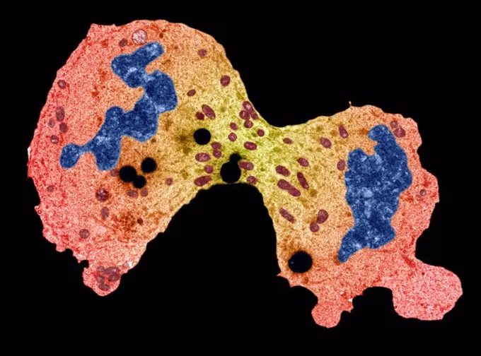
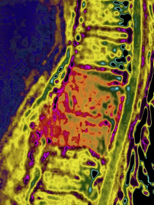
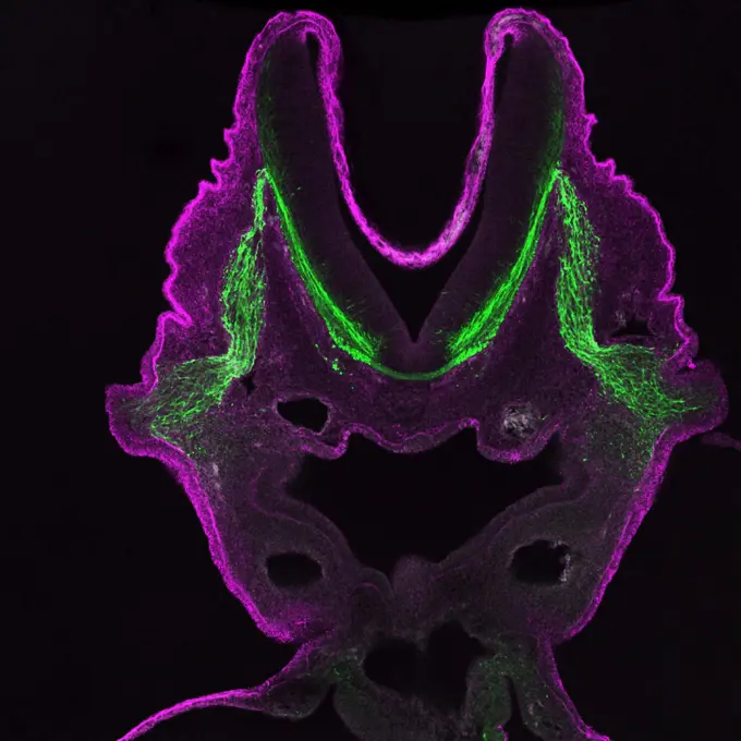
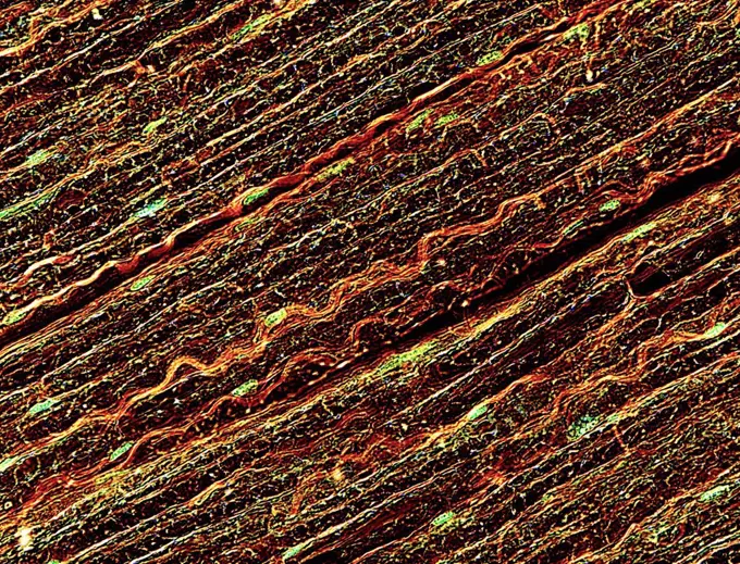
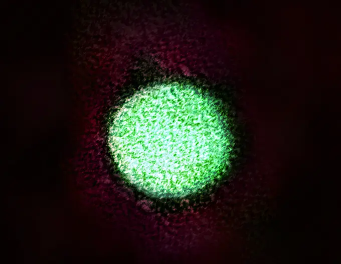
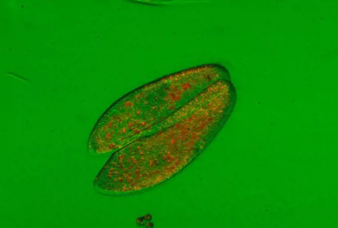
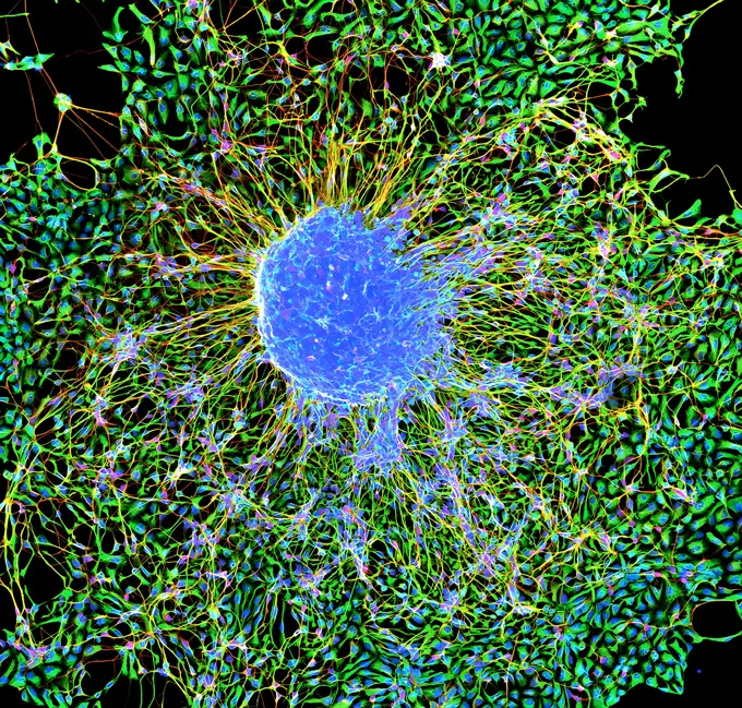
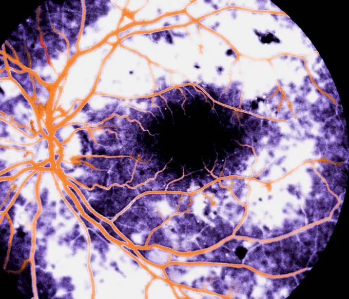
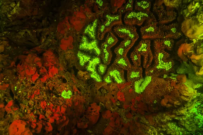
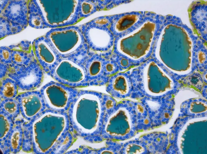
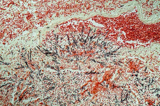
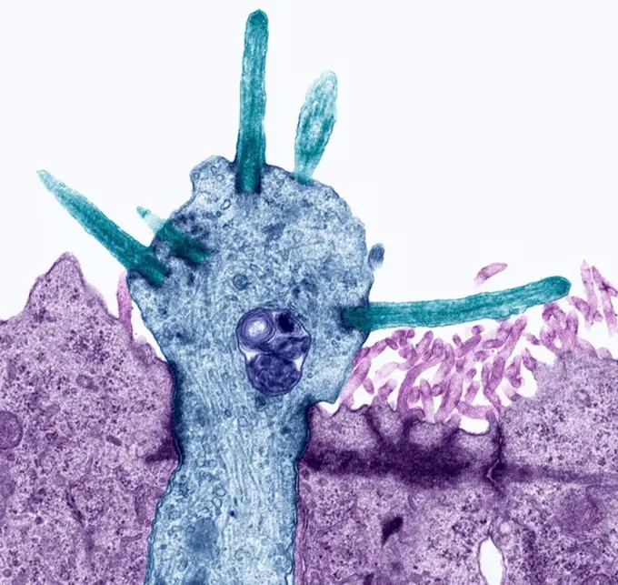
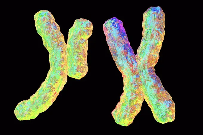
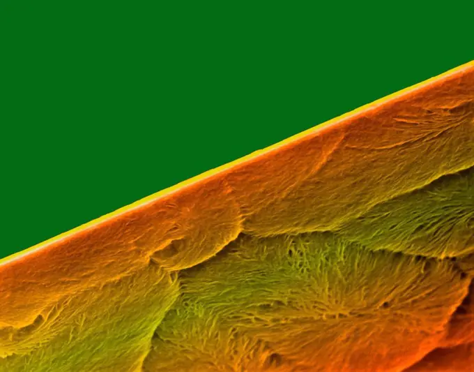
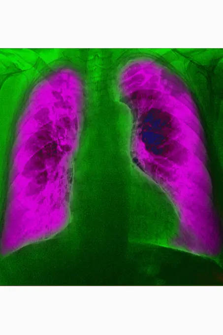
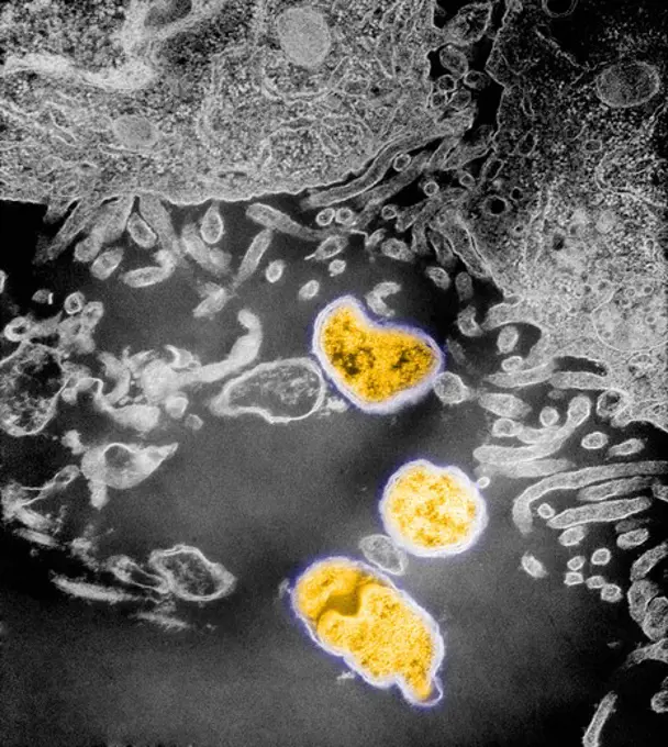
Fine ArtVintageFootageSubscriptionBoardsFree ResearchPricing
  • Fine Art
  • Vintage
  • Footage
  • Subscription
  • Boards
  • Free Research
  • Pricing

Microscopic Tissue Analysis

Close-up microscopic images of various tissues, including skin and cancerous cells, revealing intricate textures and colors.

Scroll Coral, Night Fluorescing. Bonaire, Caribbean
210 assets in this story
4269-24731
AIDS virus (VIH). Color enhanced transmission electron micrograph (TEM) of an HIV virus budding out of an infected T-lymphocyte cell.
4128R-7966
Tongue cancer. Light micrograph of a section through a squamous cell carcinoma of the tongue. Here large sheets of pleomorphic keratinocytes make up t...
1525-26942388
Marbleized effect. Ancient oriental drawing technique. Marble texture. Beautiful pattern. Marbling background. White, blue and ochre mixed oil paints.
6188-55644655
ZebraBärbling ZebraBärbling gills of the fish, 100x copyright: xzoonar.com/dr.xnorbertxlangex 13747287
4128-111493502
Illustration of RNA (ribonucleic acid) polymerase transcribing DNA (deoxyribonucleic acid) to messenger RNA (mRNA). The RNA polymerase enzyme (centre)...
824-63227058
Transmission electron micrograph of SARS-CoV-2 virus particles (UK variant B.1.1.7), isolated from a patient specimen and grown in cell culture. Image requested from the NIAID Integrated Research Facility (IRF) in Fort Detrick, Maryland. Credit: NIAID.
824-65830874
Transmission electron micrograph of HIV-1 virus particles (pink) replicating from the plasma membrane of an H9 T cell. 2024 : Vaginal Ring and Oral Pre-Exposure Prophylaxis Found Safe for HIV Prevention Throughout Pregnancy .
1899-65662469
Colorized transmission electron micrograph of mpox virus particles (yellow) found within an infected cell (red), cultured in the laboratory.
824-63225942
Small intestine at steady state.
824-63227390
Sensory nerve marker CGRP staining (green) of the inferior alveolar nerve in adult mouse incisors.
4298-1030
Marchantia liverwort archegonium, magnification 40x, image enhanced
824-737
Human Artery showing Atheroma
4384-109
This negatively-stained transmission electron micrograph (TEM) revealed the presence of numerous intracellular Group C RNA tumor virus particles, which were inside the central nervous system. The RNA tumor virus is a member of family Oncovirinae, and are linked to the initiation of tumors, otherwise known as oncogenesis.  Based upon virion morphology, and host range, Oncovirinae members are further classified into four groups: A, B, C, and D, whereupon, Groups A members are found located intra
824-63207412
Transluminal agioplasty of the right, subclavian artery, seen on a frontal chest x-ray.
1899-65662238
Streptococci - Bacteria with a rounded shape (gram-positive cocci). Bacteria involved in several pathologies: scarlet fever, angina, septicemia, meningitis, neonatal infections, endocarditis, pneumonia, erysipelas, erythema nodosum, rheumatoid purpura, urinary infection. Optical microscopy.
6188-55644945
Leaf cross -section leaf cross -section under the microscope in the dark field 100x copyright: xzoonar.com/DR.XNORBERBERTXLANGEX 13775985
4269-27163
Syndrome of the pyelo-ureteral junction. Color enhanced 3D computed tomography (CT) scan of the kidneys showing a syndrome of the pyelo-ureteral junction of the right kidney (at left).
1788-20761740
Tonsil with gram-positive bacteria, at x800 magnification.
824-63191264
The hypothalamus from a D2-eGFP (green) transgenic mouse stained for calretinin (red) and DAPI to show nuclei.
1439R-995037
Cavities in bone
7172-70194624
USA, Hawaii, The Big Island, Kilauea. Molten lava flowing at night.
4197-V71961792
4128R-11473210
Coloured scanning electron micrograph (SEM) of uric acid crystals. Magnified X 2000 at 10cm wide.
1848-50943776
Ebru marbling handmade art, traditional Ebru technique. abstract background of marble liquid ink art
824-63227350
This image shows pancreatic cancer cells (nuclei in blue) growing as a sphere enclosed in membranes (red). By growing cancer cells in the lab, researchers can study the factors that cause and prevent the formation of deadly tumors.
824-65830654
Transmission electron micrograph of West Nile virus particles (gold) replicating within the cytoplasm of an infected VERO E6 cell (pink/purple/blue).
1788-20761704
Thyroid follicles seen under a microscope.
4269-25160
AIDS virus. Color enhanced transmission electron micrograph (TEM) of an HIV virus budding out of an infected T-lymphocyte cell.
4128-19489287
Monkeypox virus, illustration
4128R-13622318
Coloured scanning electron micrograph (SEM) of Mould, hyphae, conidiophores (fruiting structures) and conidium (asexual spore) formation (Penicillium ...
4128R-1230
Animal cell, artwork.
6188-55643682
Gallenblasentumor Gallenblasentumor Gewebe unter dem Mikroskop 100x Copyright: xZoonar.com/Dr.xNorbertxLangex 13633615
4128-111580071
Measles virus particle, computer illustration. This virus, from the Morbillivirus group of viruses, consists of an RNA (ribonucleic acid) core surroun...
1848-20178398
Detail of the Rio Tinto (Red river) with its deep reddish hue due to oxidised iron minerals in its water, aerial view, drone shot, Huelva province, An...
1296R-218
Microscopic view of the cell Neospora
1848-58228098
Fluoresce hard coral, Coral fluorescenc
6123-V28555497
6188-55643702
chronische Myokarditis chronische Myokarditis Erkranktes Gewebe 100x Copyright: xZoonar.com/Dr.xNorbertxLangex 13644848
824-63223139
Colorized transmission electron micrograph of Ebola virus nucleocapsids (small orange circles) and virus particles (larger orange filamentous forms) within infected African green monkey kidney cells. Credit: NIAID.
4128-24795482
Healthy lungs, 3D CT scan
4128R-11474770
Rotavirus, computer artwork.
1848-49787197
Wild fruit found in the tree in nature
824-63227116
During normal bone development and fracture healing, cartilage turns into bone. Osteoclasts, a type of specialized cell, eat cartilage, provide passage for blood vessels, marrow and other bone cells. In this image of cartilage (purple and white) from a young mouse femur, osteoclasts (red) surround a blood vessel filled with red blood cells (yellow). Unlike normal osteoclasts, the cells present here have only one nucleus due to the absence of a gene involved in osteoclast development. This research program aims to understand how large bone-resorbing osteoclasts form and whether preventing them from fusing is a way to control bone loss in osteoporosis, arthritis or other conditions. This image won the 2015 Federation of American Societies for Experimental Biology (FASEB) BioArt competition.
824-63227315
Intense neural conversations thought to underpin learning and memory can be powered by an energy sensing flow loop. The scientist monitored energy levels in the form of ATP as the neurons talked to each other.
4128-18498425
Colon biopsy, light micrograph
4141-111428936
Light micrograph cross section abstract through the fruit of an Apple (Pyrus malus) CS, UK
824-63224706
MRI of pulmonary cystic fibrosis (viscous mucus disease, cystic fibrosis affecting glandular epithelia). Radial MRI of the lungs.
824-63224567
Fracture of the foot on the wedge bone. Foot scanner
824-63227100
An image of an adult zebrafish brain showing fluorescent granular perithelial cells (green) above blood vessels (purple).
6188-55645015
Wheat grain cross -section cross -section with starch grain 100x copyright: xzoonar.com/dr.xnorberberxlangeex 13782978
4128-V58561959
7203-70648783
Underwater Fluorescence, Blue Heron Bridge, Intracoastal Waterway, West Palm Beach, Florida, USA
824-65830967
Colorized transmission electron micrograph of mpox virus particles (orange) found within an infected cell (brown and yellow), cultured in the laboratory.
824-63225857
Transmission electron micrograph of SARS-CoV-2 virus particles (UK B.1.1.7 variant), isolated from a patient sample and cultivated in cell culture. Image captured at the NIAID Integrated Research Facility (IRF) in Fort Detrick, Maryland.
824-63226730
Cancer of the right kidney visualized by an abdominal scanner in radial section.
4292R-150178
Blue Ink in Water
4128-19489238
Monkeypox virus, illustration
4070-28891
Group of Jewel anemones (Corynactis viridis) photographed under blue light to show their fluorescence, Plymouth Sound, Devon, UK, July
824-63225811
Colorized electron micrograph showing malaria parasite (right, blue) attaching to a human red blood cell. The inset shows a detail of the attachment point at higher magnification.....
7203-70646313
Natural occurring fluorescence in underwater bubble coral (Euphyllia ancora), captured by using special UV blocking barrier filters. Night dive near Illi Api Island, Selat Boleng Strait, Indonesia
824-66066040
Colorized transmission electron micrograph of a West Nile virus particle (yellow/orange) found within the cytoplasm of a VERO E6 cell (green). Image captured at the Integrated Research Facility in Ft. Detrick, Maryland. Credit: NIAID.
1525-26931569
Small part of an infinite star field of space in the Universe. Elements of this image furnished by NASA".. Small part of an infinite star field.
4128R-13573026
Taste buds. Coloured light micrograph of a section through the tongue, showing taste buds (round, blue). The taste buds are within papillae (projectio...
4128R-8154
Cancer cell division, TEM
824-63188633
Vertebral edema seen on a saggital plane MRI scan of the lumbar vertebrae.
4269-24967
Stenosis. Stenosis of the left common iliac artery MRI (Magnetic Resonance Imaging) and angiography
6145-29256670
Laminin
4128R-21320
Nerve fibres, light micrograph
824-65830667
Colorized transmission electron micrograph of a hepatitis C virion.
4141-16147
protozoa pair conjugating paramaecium sp.
6123-V28554095
824-63227268
And quotWhy files' 2015 Cool Science Image Contest winning photo by Scott Vermilyea, Neuroscience Training Program, School of Medicine and Public Health and Scott Guthrie Undergraduate Neurobiology, with SCRMC members Ted Golos and Marina Emborg, professors at the School of Medicine and Public Health and Wisconsin National Primate Research Center. Golos is also a faculty member at the School of Veterinary Medicine.
4269-25473
Diabetic retinopathy. Diabetic Retinopathy Eye angiography Destruction of the retina (black mass in the center) Degeneration of the blood capillaries
7203-70647259
Natural occurring Red and Green Fluorescence in Stony Corals and Encrusting Corals, captured with special ultraviolet light-blocking filters, Palau, Micronesia, Rock Islands, World Heritage Site, Western Pacific
824-63211064
Fracture of a lumbar vertebra, with damage to the spinal cord with a bony fragment in the neuroforamen. Cross-section scanner.
4128R-13447206
Thyroid gland. Light micrograph (LM) of a thyroid gland showing the follicles. The follicles are lined by a single layer of cuboidal epithelial cells ...
824-63227284
New findings in a responsible mouse study that a loss of mossy cells may contribute to seizures and memory problems in a form of epilepsy.
824-63227378
Sagittal section of Nr4a1-eGFP transgenic mouse stained for CB1 cannabinoid receptor and DAPI to show nuclei. The image was awarded with a Zeiss Lumar stereomicroscope.
824-63227148
This image is the result of a research project aimed at visualizing which regions of the adult Drosophila brain derive from each neural stem cell.
6188-55644600
Lungs of an aid patient lung of an aid patient lung tissue 200x copyright: xzoonar.com/dr.xnorberbertxlangex 13735585
4413-81353
Mercury cristals by polarized light
4128R-8002
Smell receptors, TEM
6145-44710027
Microscopic Materials on a Magnet
4128R-13376645
Human chromosomes, illustration
1746-111944321
This photograph, made with the Wide Field Camera on NASA/ESA Hubble Space Telescope, reveals the detailed structure of a newly discovered jet of material streaming away from a young star in the Orion Nebula. While similar jets have been seen from the ground, the new observations reveal the structure of this jet with unprecedented clarity. Of particular significance is the fact that the jet is bright along its edges in the light of gaseous sulfur atoms (shown here in red). Experts believe this st
4128-18929755
Sacral vertebra and spinal cord, LM
4128R-34268
Rust fungus spore. Light micrograph of a section through an aeciospore from a Puccinia sp. rust fungus. Spores are the reproductive structures of a fu...
6188-55643136
Granite metamophes deep rock Audz, field spar and mica 100x granite metamophes deep rock aud quartz, feldspar and glow 100x copyright: xzoonar.com/dr.xlangeex 13451051
4128R-13619983
Human tooth fractured enamel, coloured scanning electron micrograph (SEM). Fracture in a tooth revealing the enamel rod morphology. Tooth enamel is th...
4128R-13620289
Coloured scanning electron micrograph (SEM) of Edge of a new razor blade (seamless double edge). A razor blade is a thin, sharp-edged piece of steel t...
824-63225962
Iliac artery stenosis before and after percutaneous angioplasty.
824-63206982
Pneumonia caused by a hospital acquired bacterian infection, seen on a frontal chest x-ray.
1788-20761601
Epithelial cell of the human tongue. Micrograph.
4269-25251
Prostate cancer. Ultrasound scan of a cancer of the prostate, seen in green.
4297-1448
Transmission electron micrograph of numbers of intraperitoneal Gram-negative Orientia tsutsugamushi rickettsial microorganisms which were inside the peritoneal cavity of an infected mouse
6145-45005740
NASA's NuSTAR is complementing previous observations of the Cassiopeia A supernova remnant (red and green) by providing the first maps of radioactive material forged in the fiery explosion (blue).
6145-44791641
The High Resolution Imaging Science Experiment camera on NASA's Mars Reconnaissance Orbiter captured winter images of NASA's Phoenix Mars Lander surrounded by dry-ice frost on Mars.
1746-21105979
Factor 8: Immunoperoxidase staining of formalin-fixed, paraffin-embedded mouse lung showing cytoplasm staining of the endothelial cells lining the arteries and veins
4141-32775
desmid alga dividing micrasterias apiculata magn x100 late binary fission dark background illumination
824-63227124
Additionally, a starved fly's brain turns sleepy orange due to Bruchpilot, a communication protein between brain cells. These bright orange areas of the brain are associated with learning. At the bottom, a well-rested fly shows lower levels of Bruchpilot, which could make the fly ready to learn after a good night's rest. These images illustrate the results of an April 2009 study showing that sleep reduces protein levels, suggesting that such downscaling and quot resets the brain to normal levels of synaptic activity and makes it ready to learn after a restful night.
PREVIOUS
of 3
NEXT
2401 S. Ervay, Suite 206
Dallas, Texas 75215
United States
Browse
CategoriesGalleriesFree Research
For Creators
License Your ContentSuperStock For StudentsFrame of Mind BlogContributor PortalNew Image SpotlightLicense AI Imagery
Resources
About UsLicensingTerms & ConditionsAPI AccessPrivacyPricing
Contact
+1 866 236 0087help@superstock.com Contact form
©2026 SuperStock. All Rights Reserved. -A
This site is protected by reCAPTCHA and Google Privacy Policy and Terms of Service apply.