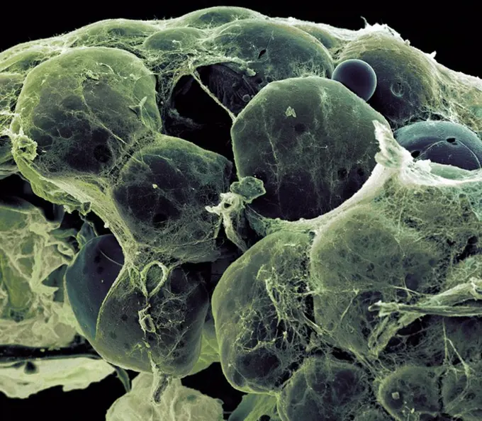
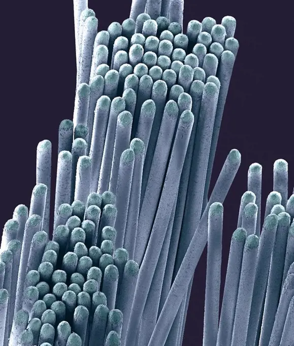
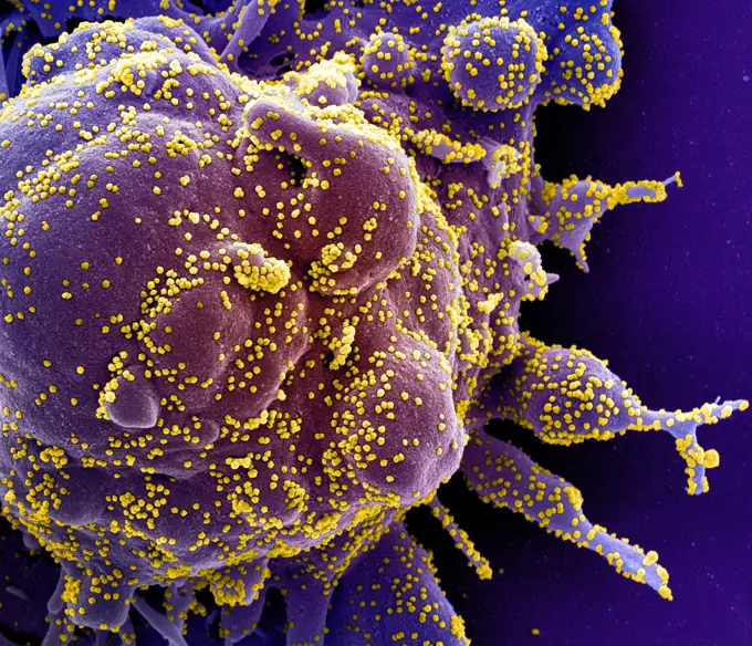
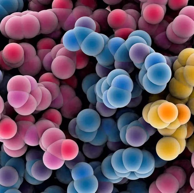
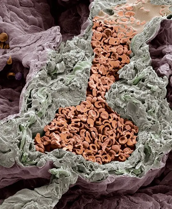
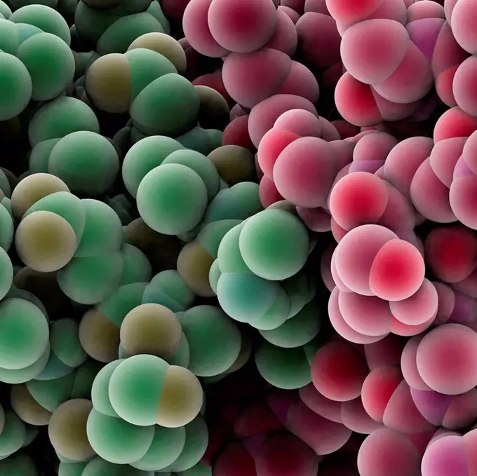
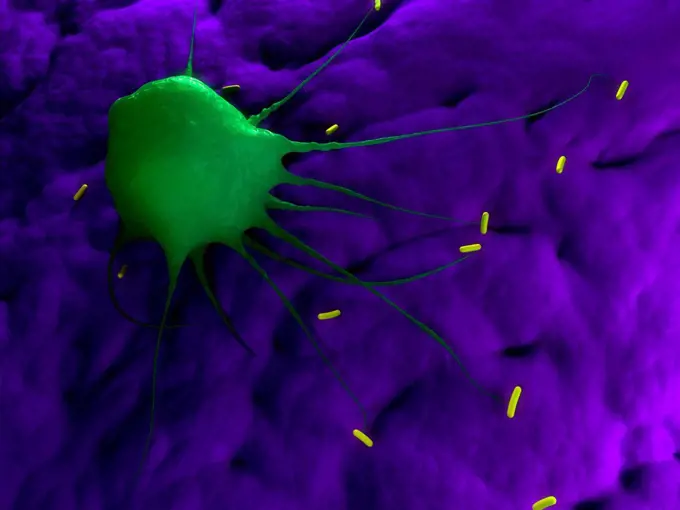
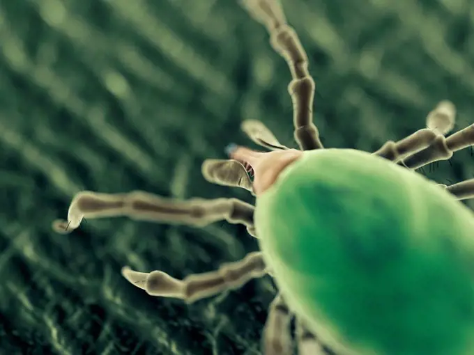
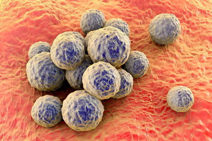
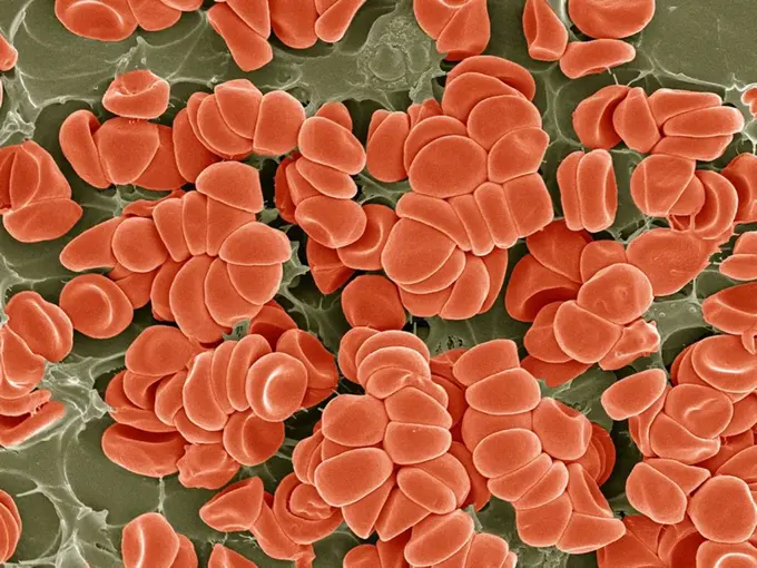
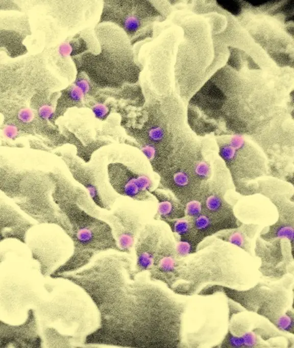
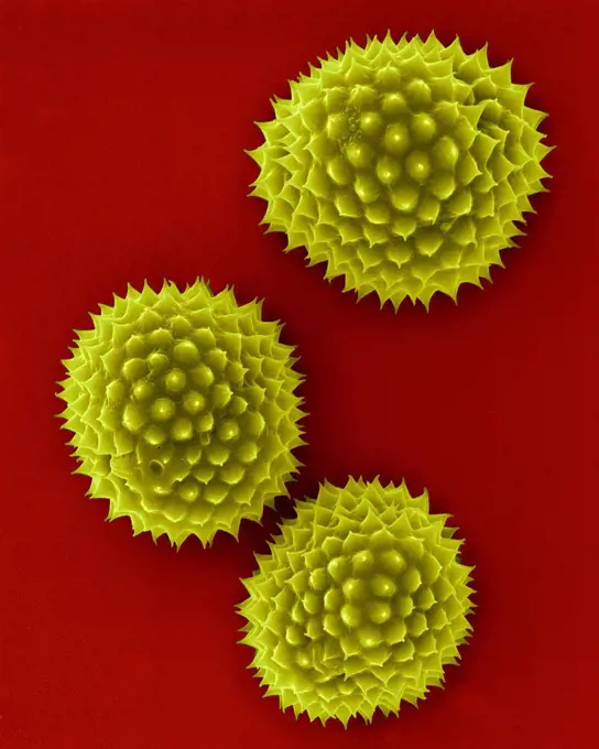
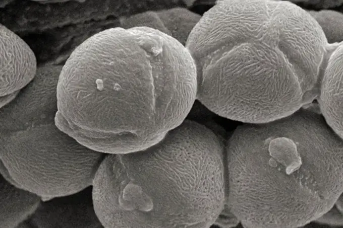
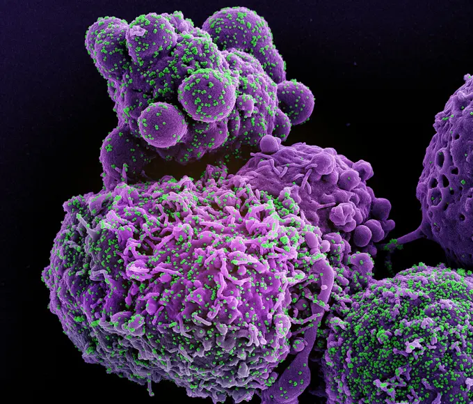
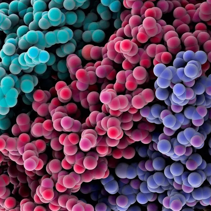
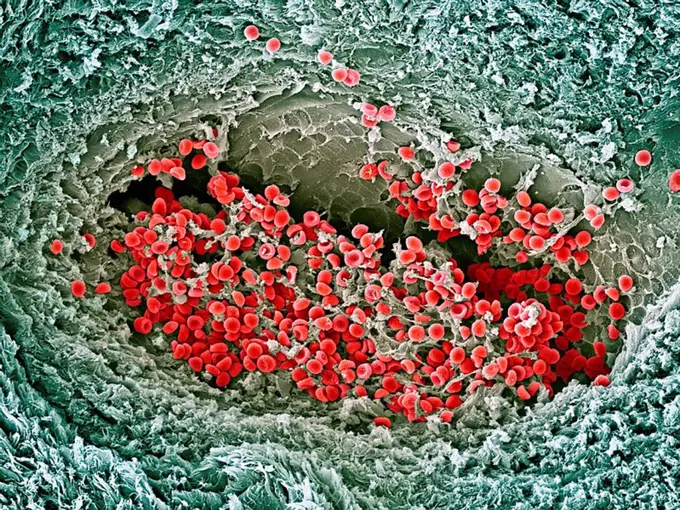
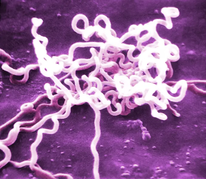
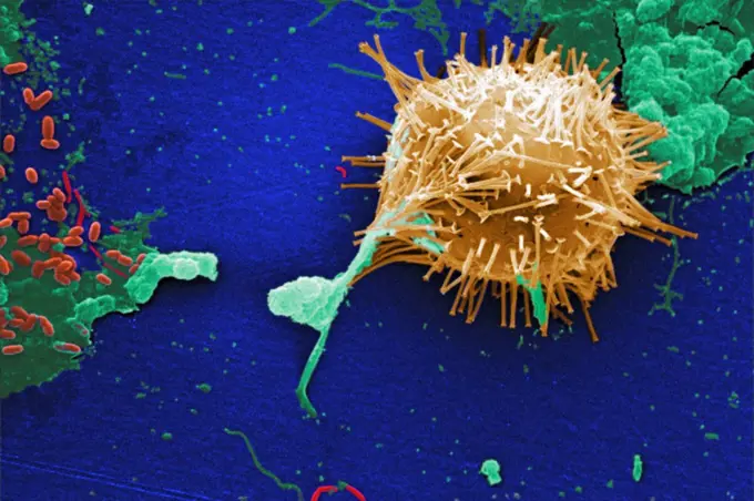
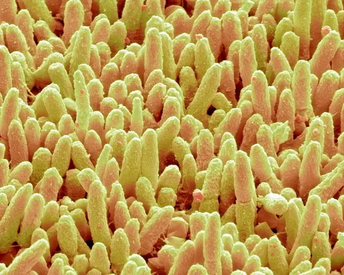
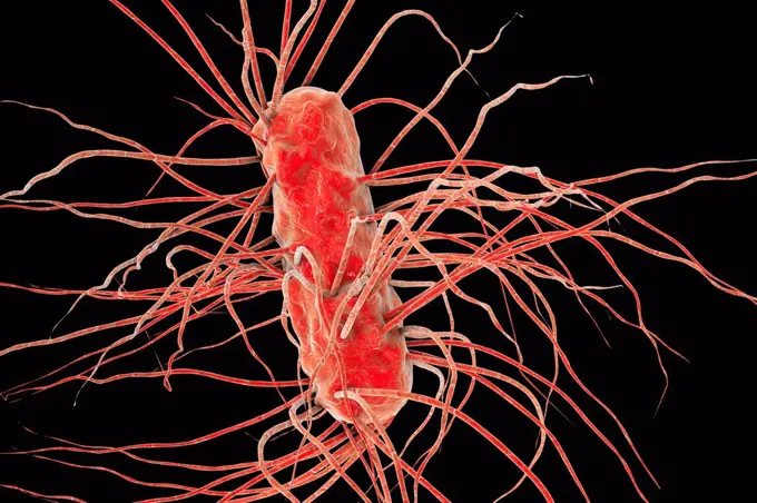
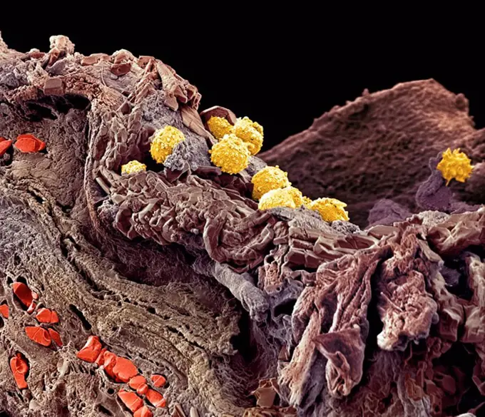
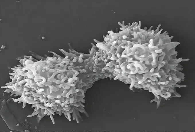
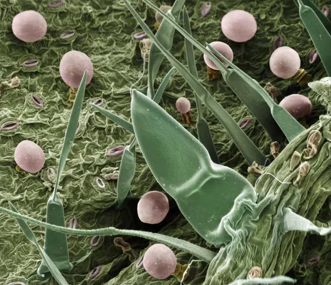
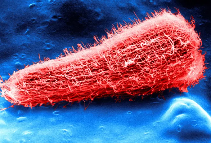
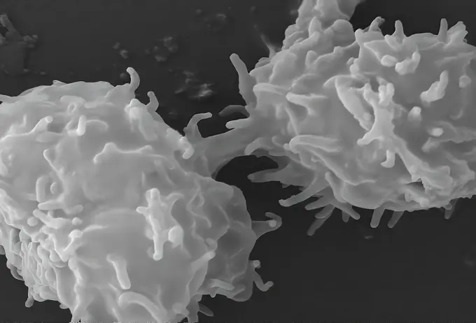
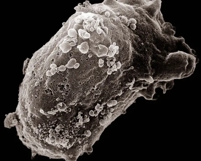
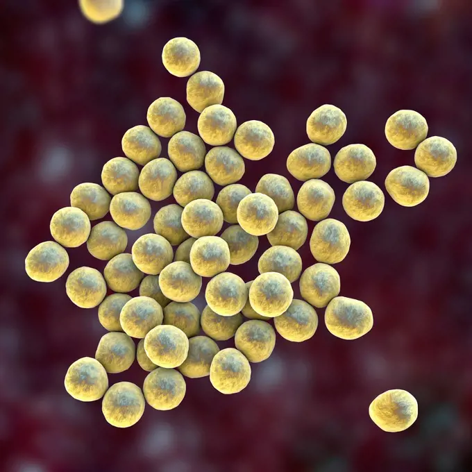
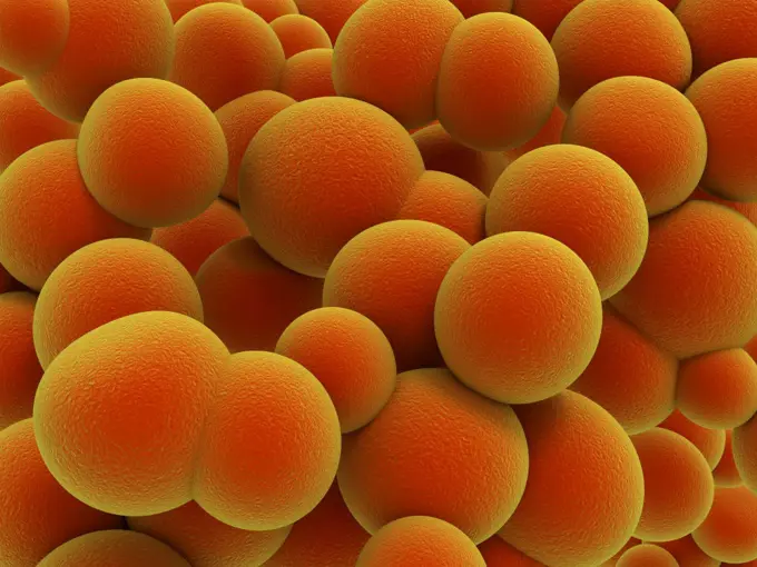
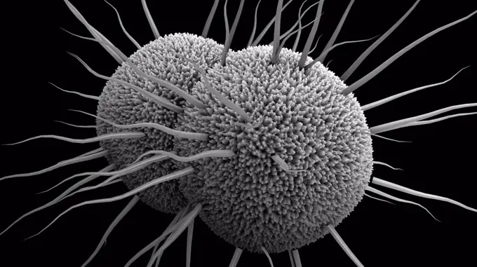
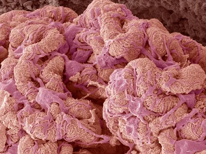
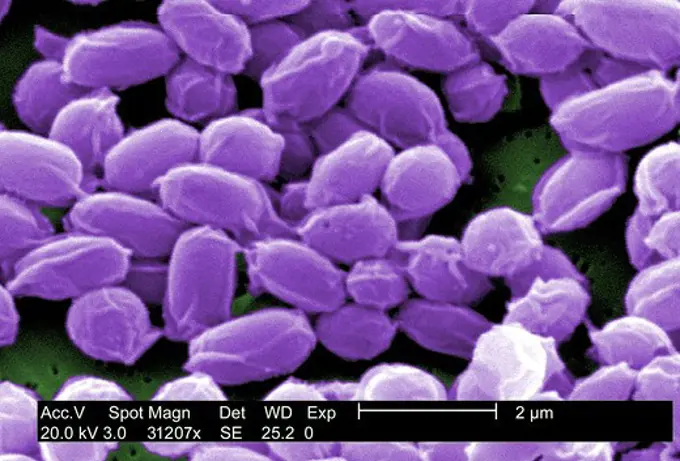
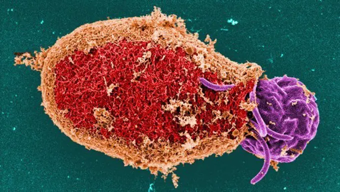
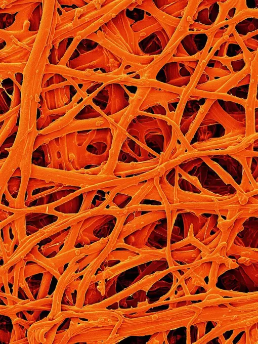
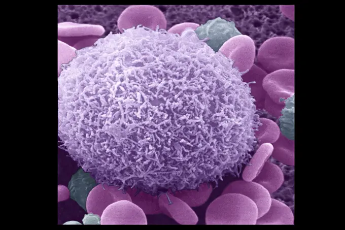
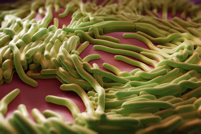
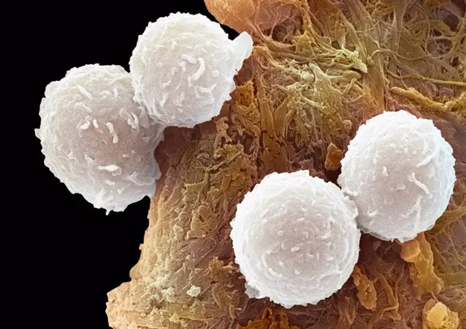
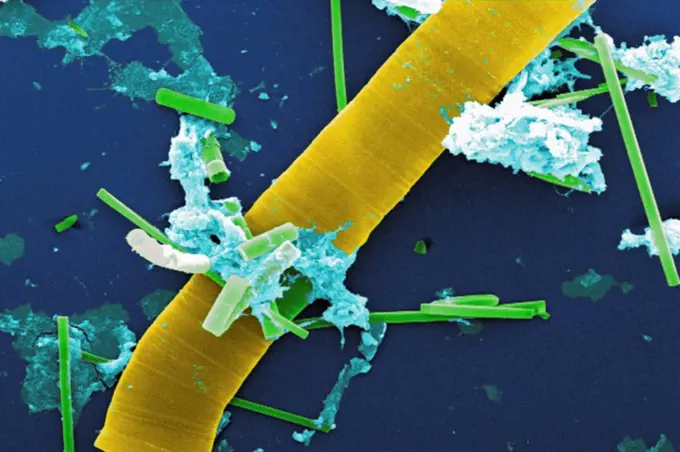
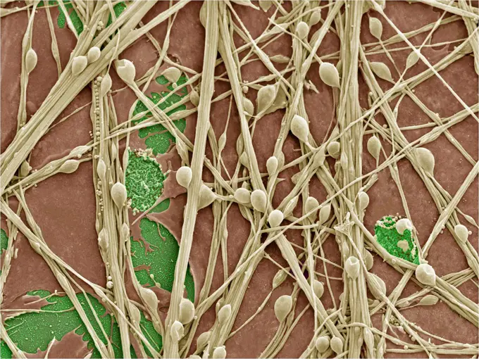
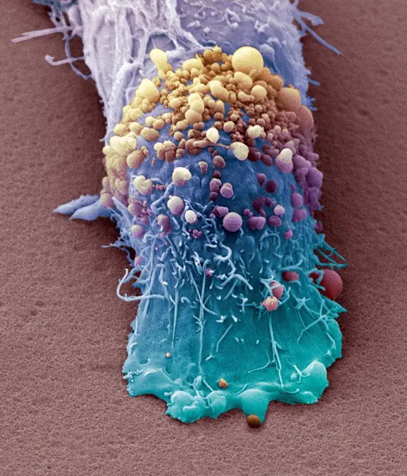
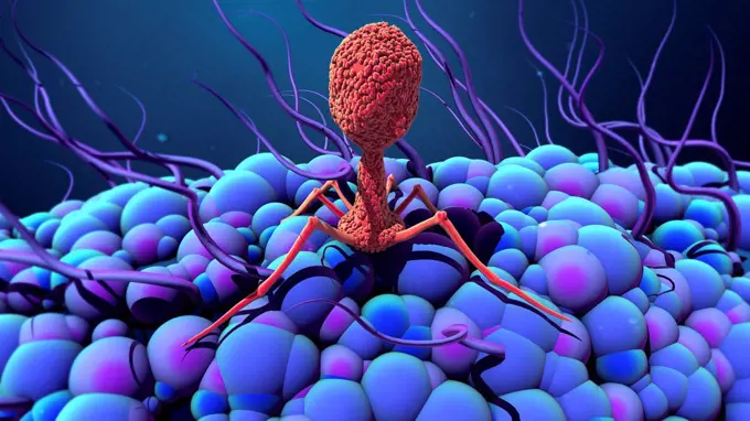
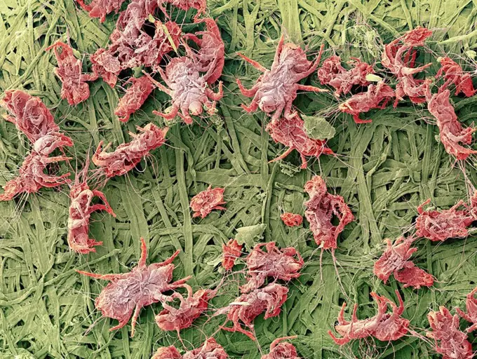
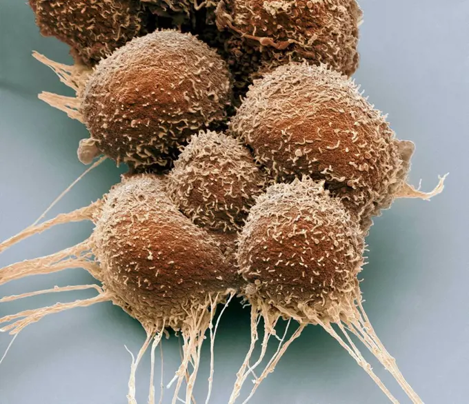
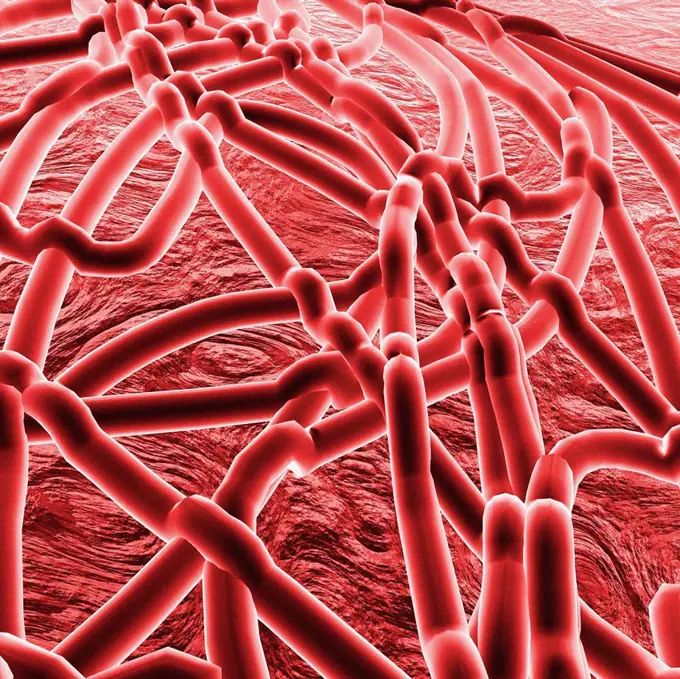
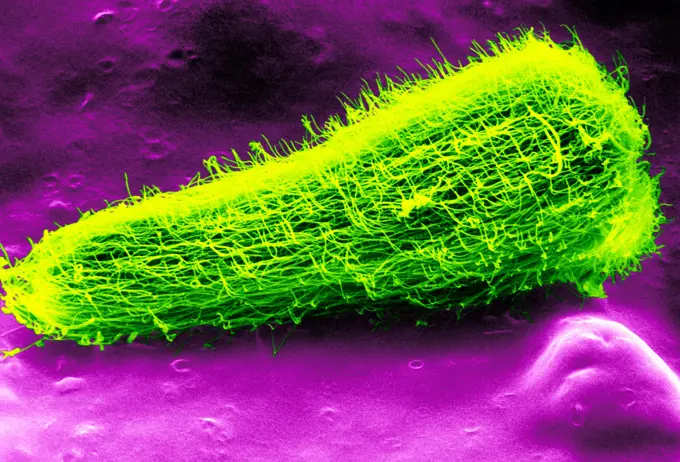
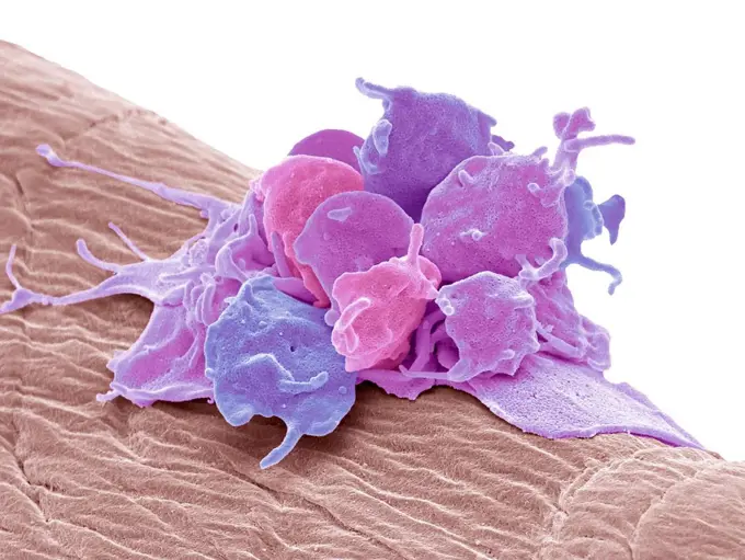
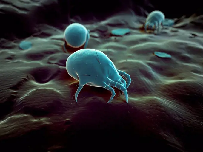
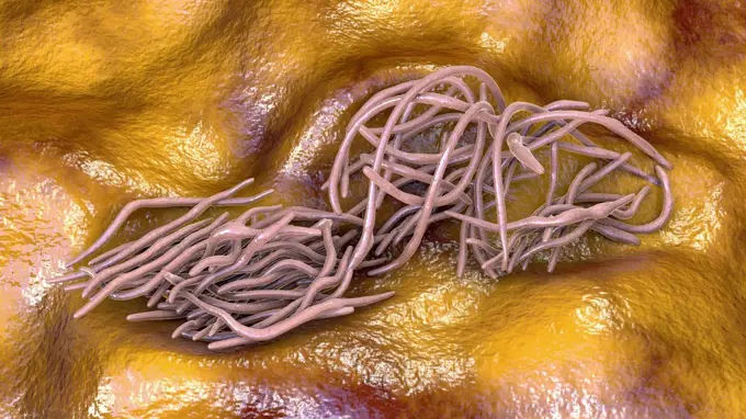
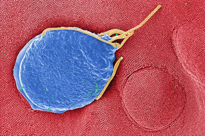
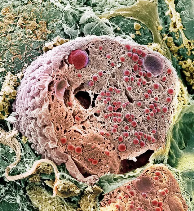
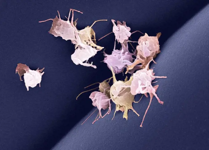
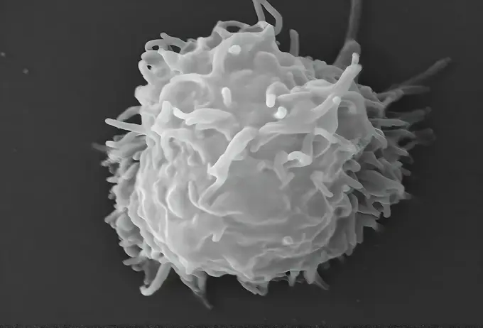
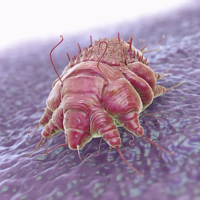
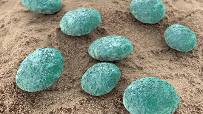
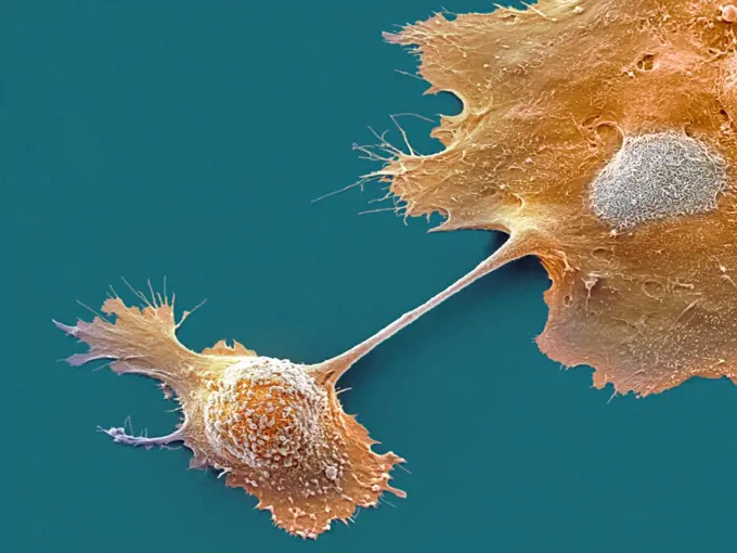
Fine ArtVintageFootageSubscriptionBoardsFree ResearchPricing
  • Fine Art
  • Vintage
  • Footage
  • Subscription
  • Boards
  • Free Research
  • Pricing

Scanning Electron Microscopy

Colorful close-up images captured by scanning electron microscopy showcasing bacteria and tissue at a microscopic level, revealing intricate structures and details.

Fat tissue, SEM
Fat tissue, SEM
201 assets in this story
4128R-4052
Toothbrush bristles, SEM
824-63223230
Colorized scanning electron micrograph of an apoptotic cell (purple) heavily infected with SARS-COV-2 virus particles (yellow), isolated from a patient sample. Image captured at the NIAID Integrated Research Facility (IRF) in Fort Detrick, Maryland. Credit: NIAID.
4128R-2331
Marjoram leaf surface, SEM
4128R-11476161
Protein molecules, computer artwork.
4128R-15465089
Tongue bacteria
4128R-5689
Blood vessel in the lung, SEM
4128R-14057539
Gonorrhoeae bacteria, illustration
6188-64776587
Ripe nutmegs levitate on a white background Ripe nutmegs levitate on a white background. Copyright: xZoonar.com/AlekseyxButenkovx 17585417
824-57657079
A scanning electron microscope picture of a nerve ending. It has been broken open to reveal vesicles (orange and blue) containing chemicals used to pass messages in the nervous system.
4128R-11476153
Protein molecules, computer artwork.
4128R-14541
Macrophage attacking bacteria, computer artwork.
4128R-15047186
Cancer cells, illustration
4128-15980098
Tick, computer illustration.
4128-18183812
Nasturtium leaf, SEM
4128-V58561798
4128R-13818102
MRSA bacteria. Computer artwork of methicillin-resistant Staphylococcus aureus (MRSA) bacteria.
4201-66341
Daisy (Chrysanthemum sp) SEM close-up view of pollen at 560x magnification
4128R-2569
Blood clot, SEM
4128-V58557896
4128-111494388
Starch grains. Coloured scanning electron micrograph (SEM) of starch grains (green) in the seed of a legume (bean). Starch is synthesised from sucrose...
4297-1222
Scanning electron micrograph of human immunodeficiency virus (HIV), grown in cultured lymphocytes. Virions are seen as small spheres on the surface of the cells
4297-1795
SEM image of Gram-positive Streptococcus pneumoniae bacteria
4128R-11476173
Protein molecules, computer artwork.
4128R-13622333
Ragweed pollen (Ambrosia psilostachya), coloured scanning electron micrograph (SEM). This pollen is an allergen. Ragweed is a main cause of weed aller...
4384-398
Fireworks Flower With Pollen
824-65831023
Colorized scanning electron micrograph of a cell (purple) infected with the Omicron strain of SARS-CoV-2 virus particles (green), isolated from a patient sample.
4128R-11476195
Protein molecules, computer artwork.
4378-5351
Legionella Pneumophila Bacteria
4128R-2141
Blood clot, SEM
4269-27379
Treponema pallidum. Scanning Electron Micrograph (SEM) of Treponema pallidum. This spirochaete bacterium is the agent of syphilis.
4384-149
This digitally-colorized scanning electron micrograph (SEM) of an untreated water specimen extracted from a wild stream mainly used to control flooding during inclement weather, revealed the presence of unidentified organisms, which included bacteria, protozoa, and algae. In this particular view, a microorganism is featured, the exterior of which is covered by numerous projections imparting an appearance of a sea urchin. This microscopic pin cushion was teathered to its surroundings by a biofi
4384-420
Tiny Mite on Lizard's Skin
4201-66354
Golden Calla (Zantedeschia elliottiana) SEM close-up view of pollen at 700x magnification
6188-58064850
Lillium anther Lillium anther under Optical Microscope Copyright: xZoonar.com/AlxCarrerax 21350948
4378-20013724
Legionella Pneumophila Bacteria
4128R-23745
Dental plaque. Coloured scanning electron micrograph SEM of dental plaque. Plaque consists of a film of bacteria embedded in a glycoprotein matrix. Th...
824-63227179
Ebola virus particles found both as extracellular particles and budding particles from chronically-infected African green monkey kidney cells .
4128R-13575534
Escherichia coli bacteria, computer illustration. E. coli is a gram-negative motile bacterium with peritrichous flagella that lives in the intestines ...
1899-53513331
Colorized scanning electron micrograph of Ebola virus particles (green) both budding and attached to the surface of infected VERO E6 cells (orange). Image captured and color-enhanced at the NIAID Integrated Research Facility in Fort Detrick, Maryland.
4384-137
This scanning electron micrograph revealed the presence of the human immunodeficiency virus (HIV-1), (spherical in appearance), which had been co-cultivated with human lymphocytes.
4128R-5643
Pyoderma skin disease, coloured scanning electron micrograph SEM. Lymphocytes yellow are present on the surface of the fractured skin and red blood ce...
824-63191250
Colorized image of HIV-infected H9 T-cell.
4269-6815
Scanning electron micrograph (SEM) of agglutinated red and white blood cells in a blood vessel.
4201-21258675
Upright Hedge-parsley (Torilis japonica) spines on seed enable animal dispersal, magnification 800x
824-57659309
This scanning electron microscope (SEM) image revealed a protozoan Acanthamoeba polyphaga, as it was about to complete the process of cell division, known as mitosis, becoming two separate organisms. Note the numerous pseudopodia, projecting from the surfaces of these organisms. These pseudopodia allow amoebas to move and grasp objects in their environment. CDC/Catherine Armbruster Margaret Williams 2009
4128-111581134
High cholesterol levels. Computer illustration of balls of cholesterol in a blood vessel. Cholesterol is a lipid, a fat-like substance. High blood lev...
4269-24575
Mycoplasma pneumoniae. Mycoplasma pneumoniae on cultured cells. Causes respiratory infections. SEM (Scanning Electron Microscope)
4201-21246325
Geranium (Pelargonium crispum) leaf, magnified 700 times, showing glands, stomata, and trichomes, Barcelona, Spain
824-63227072
This scanning electron microscope image shows SARS-CoV-2 (round orange particles) emerging from the surface of a cell cultured in the laboratory. SARS-CoV-2, also known as 2019-nCoV, is the virus that causes COVID-19. Image captured and colorized at Rocky Mountain Laboratories in Hamilton, Montana. Credit: NIAID
4269-7089
Scanning Electron Micrograph (SEM) of Streptococcus pneumoniae bacteria. This Gram-negative bacterium is a leading cause of pneumonia. It also causes many other pathologies like middle ear infections (otitis media), bacteremia and meningitis.
824-63128389
Miracidium larva of the liver fluke (fasciola hepatica) viewed in SEM.
4269-27731
Hartmanella vermiformis. Color enhanced scanning electron micrograph of cysts of the protozoan Hartmanella vermiformis on a laboratory-grown potable water biofilm. Aquatic bacteria were grown as biofilm on steel for one week. Hartmanella vermiformis protozoans were then added, and phagocytized the bacteria that multiplied within vesicles that became cysts in which the bacteria will live until they rupture.
4128R-11286531
Flu virus particles, computer artwork. In the virus' envelope are two types of protein spike, haemagglutinin (H, light green) and neuraminidase (N, da...
824-57659312
This scanning electron microscope (SEM) image revealed two Acanthamoeba polyphaga protozoa, as they interacted through their numerous pseudopodia, projecting from the surface of these organisms. These pseudopodia allow amoebas to move and grasp objects in their environment. In this particular case, it was not known whether these two amoebas were communicating, or coming to the conclusion of mitotic cell division, becoming separate organisms. CDC/Catherine Armbruster Margaret Williams 2009
4391-301
A scanning electron microscope image of a lymphocyte (white blood cell) with an HIV cluster.
4128-V58562025
4128-28575385
MRSA bacteria, illustration
1525-57024747
occi bacterium. Group of bacteria, closeup. This image may be used to describe are either spherical cocci bacterium, for example, streptococci, staphylococci, pneumococci causing pneumonia, meningitis, sinusitis, pharyngitis, dental caries, impetigo, scarlet fever.
4128-15745498
Gonorrhoea bacteria, illustration
4128R-1536
Kidney glomerulus, SEM
4384-103
This 2005 colorized scanning electron micrograph (SEM) depicted numerous clumps of methicillin-resistant Staphylococcus aureus  bacteria, commonly referred to by the acronym, MRSA; Magnified 4780. Recently recognized outbreaks, or clusters of MRSA in community settings have been associated with strains that have some unique microbiologic and genetic properties, compared with the traditional hospital-based MRSA strains, which suggests some biologic properties, e.g., virulence factors like toxins,
4297-1731
SEM of Anthrax bacteria spores (Bacillus anthracis)
4384-243
Scanning Electron Micrograph of Giardia Cyst
4128R-13620188
Coloured scanning electron micrograph (SEM) Chicken eggshell inner membrane (Gallus gallus domesticus). Shown here is the eggshell inner membrane that...
824-63123149
Scan electron micrography. 2 500x.
4378-4624
Legionella Pneumophila
4128R-6951
Leukaemia blood cells, SEM
4384-168
Under a relatively low magnification of 200X, this digitally-colorized scanning electron micrograph (SEM) of an untreated water specimen extracted from a wild stream mainly used to control flooding during inclement weather, revealed the presence of unidentified organisms, which included bacteria, protozoa, and algae. In this particular view, number of what appeared to be rod-shaped sections of various sizes were scattered throughout the field of view, which though unconfirmed, may have been vege
824-63191277
Cultured hippocampal neurons grown on a substrate of glial cells (astrocytes). The glial cells form the pink/brown underlayment in this image. The tan threads are the neurons. The round tan balls are synapses, the points where neurons meet and communicate with each other. The cover slip underlying the cells is green. Neurons in culture can be used to study synaptic plasticity, activity-dependent protein turnover, and other topics in neuroscience.
4128R-11476145
Protein molecules, computer artwork.
4128R-13047374
Skin cancer cell, coloured scanning electron micrograph (SEM). Cultured melanoma cell showing the numerous blebs and microvilli characteristic of canc...
4128-111494695
Bacteriophage infecting bacterium, illustration.
4220-21334681
Scanning Electron micrograph (SEM): Cactus Pollen grains.     Date:
4128R-2645
Dust mites, SEM
4128R-15466817
Prostate cancer cells, SEM
4128R-11476171
Protein molecules, computer artwork.
4269-27446
Escherichia coli. This scanning electron micrograph (SEM) of a Escherichia coli bacterium ; Magnification 3607x. E. coli are rod-shaped Gram-negative bacteria that normally colonize the digestive tract in humans and other animals. However, some strains can produce a toxin that leads to severe illness or even death.
4128-V58569879
4128R-13620535
Coloured scanning electron micrograph (SEM) of Komagataella phaffii - methylotrophic yeast. Komagataella phaffii is a newly identified yeast that has ...
4239-18641582
Microscopic visualization of grass pollen grains.
4128R-6769
Deformed sperm cell, SEM
4128R-11474540
Ebola virus, computer artwork.
824-63128387
Miracidium larva of the liver fluke (fasciola hepatica) viewed in SEM.
4128R-14409
Dust mites. Computer artwork of house dust mites Dermatophagoides pteronyssinus.
4128-15982701
Prostate cancer cell, Coloured scanning electron micrograph (SEM). of the surface of prostate cancer cells. The cells shows numerous processes and mic...
4128R-15465090
Activated platelets, SEM
4128R-12573088
Dust mite, computer illustration.
4384-240
Scanning Electron Micrograph of Termite
4128-17857612
Round worms, computer illustration. Roundworms, or nematodes, include numerous free-living and pathogenic species. Nematodes that commonly parasitise ...
4384-144
This digitally-colorized scanning electron micrograph (SEM) depicted a Giardia muris protozoan adhering itself to the microvillous border of an intestinal epithelial cell.  Each small circular profile under the protozoan represents the rounded tip of a single microvillous, and it is estimated that 2000 to 3000 microvilli cover the surface of a single intestinal epithelial cell.  The two circular lesions on the right side of the photograph are impressions made by the ventral adhesive disk of othe
4128R-7365
Pancreas cell, SEM
4128R-3250
Activated platelets, SEM
4128-V58562067
824-63191231
Scanning electron micrograph of an apoptotic HeLa cell. Zeiss Merlin HR-SEM.
1899-61460852
This scanning electron microscope (SEM) image revealed some of the ultrastructural features observed on the surface of a protozoan Acanthamoeba polyphaga. Note the numerous pseudopodia, protruding from the surface of the organisms. These allow the amoeba to move and grasp objects in its environment. CDC/Catherine Armbruster Margaret Williams 2009
4128-V58574927
4378-3141
A single Sarcoptes scabiei mite which is the cause of the contagious skin infection Scabies. The mite burrows under the host's skin, causing intense allergic itching.
4128-24795776
Nematode worm eggs, illustration
4128-111453072
Ovarian cancer cells, sem
4128R-8917
Pancreatic cancer cells, SEM
PREVIOUS
of 3
NEXT
2401 S. Ervay, Suite 206
Dallas, Texas 75215
United States
Browse
CategoriesGalleriesFree Research
For Creators
License Your ContentSuperStock For StudentsFrame of Mind BlogContributor PortalNew Image SpotlightLicense AI Imagery
Resources
About UsLicensingTerms & ConditionsAPI AccessPrivacyPricing
Contact
+1 866 236 0087help@superstock.com Contact form
©2026 SuperStock. All Rights Reserved. -B
This site is protected by reCAPTCHA and Google Privacy Policy and Terms of Service apply.