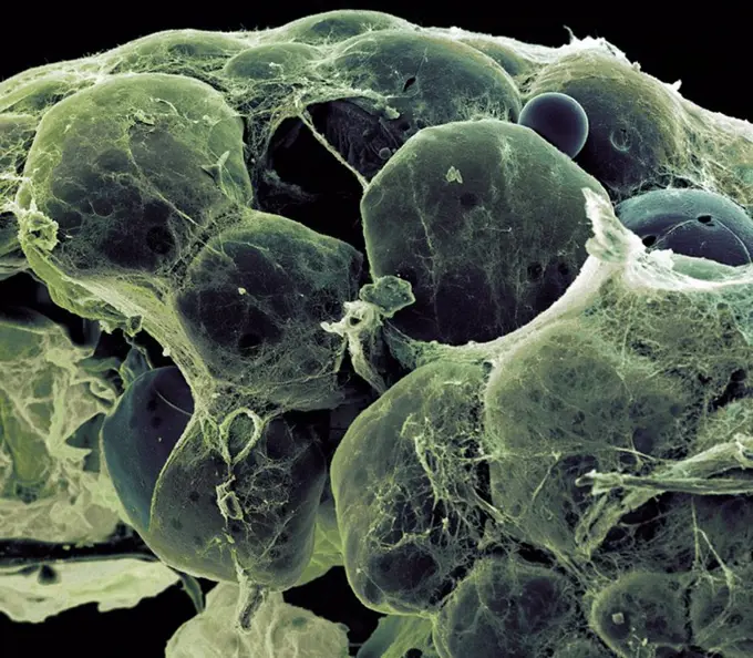
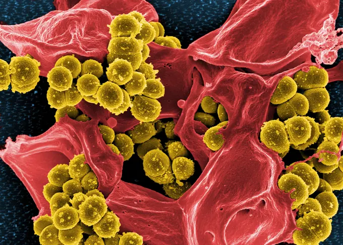
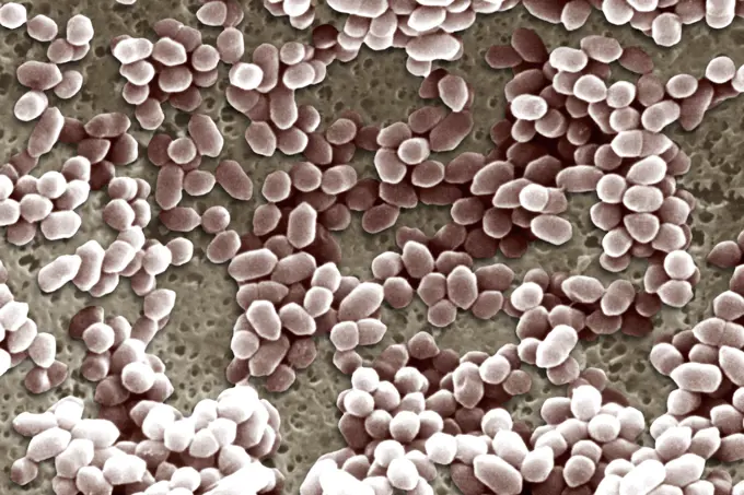
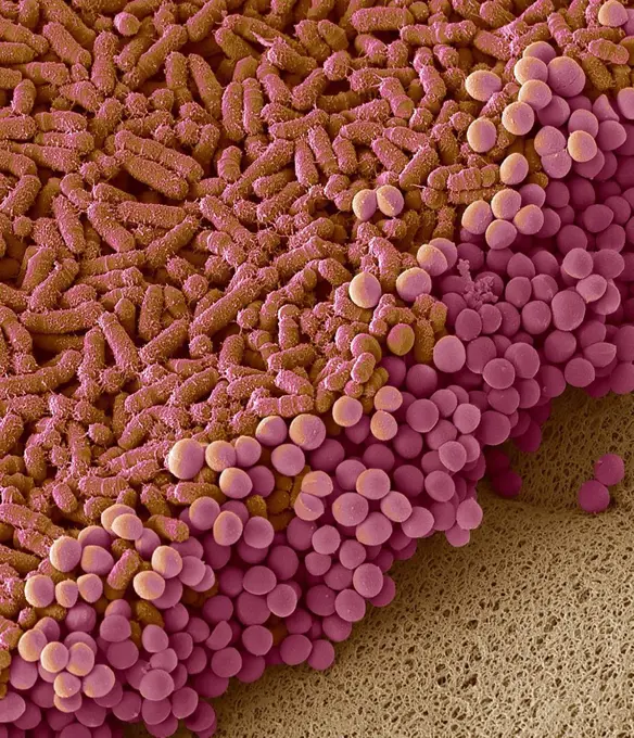
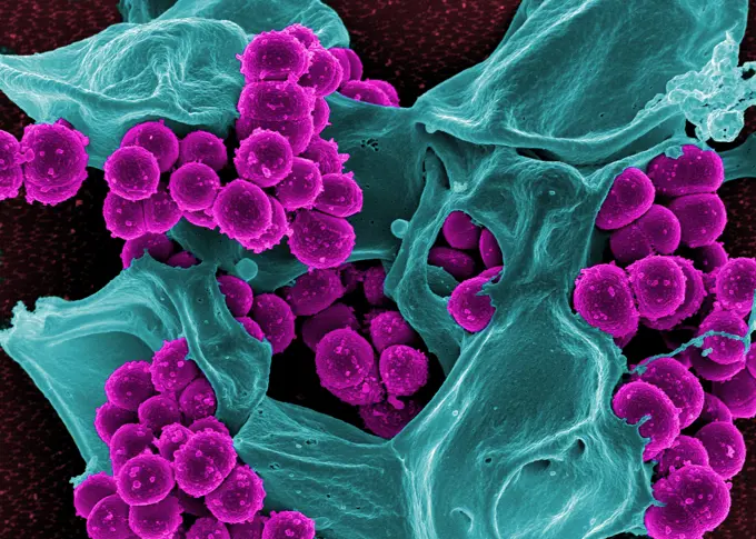
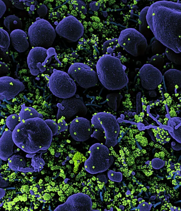
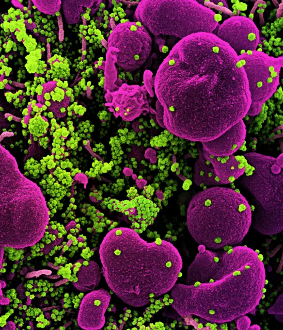
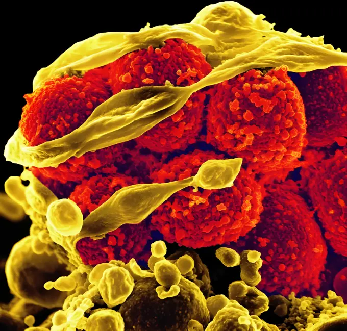
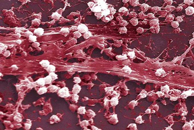
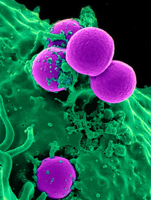
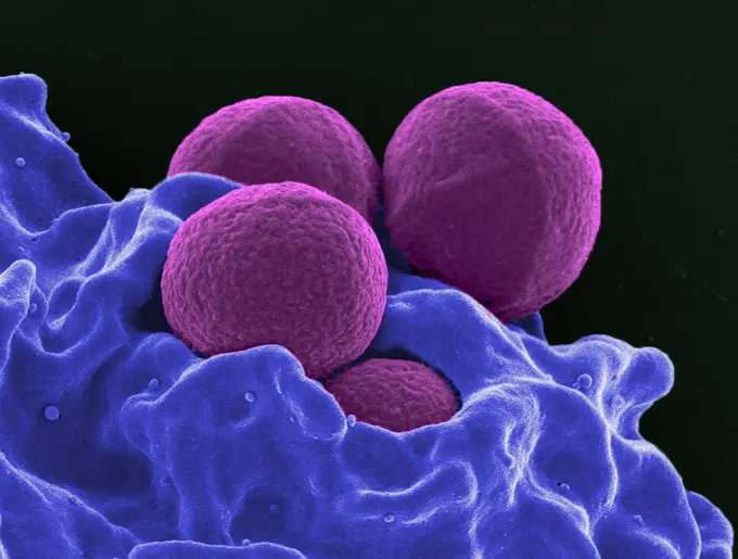
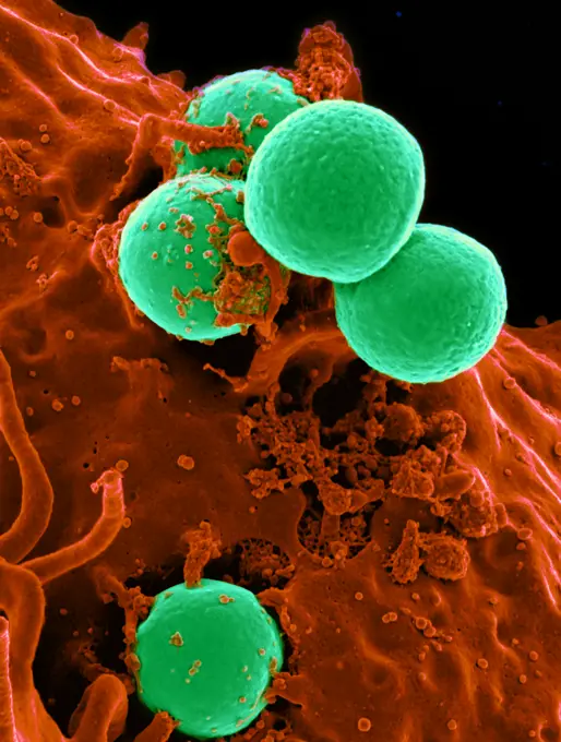
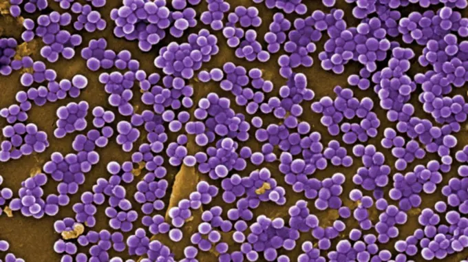
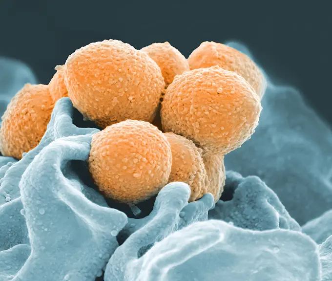
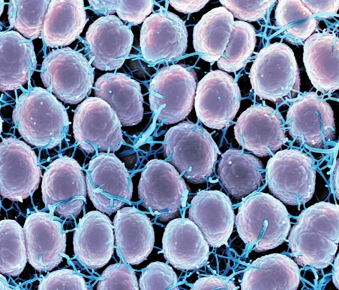
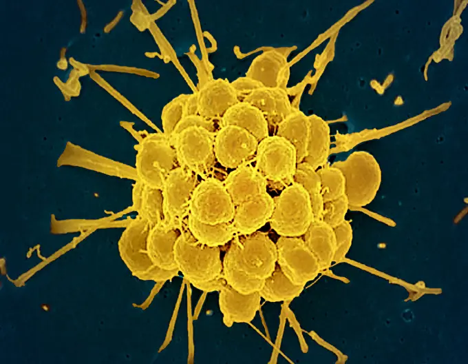
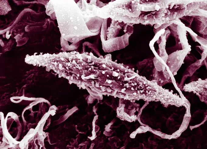
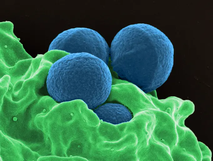
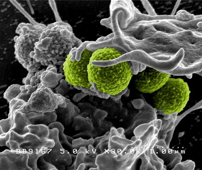
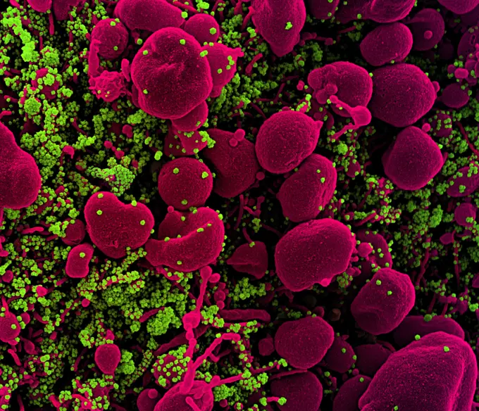
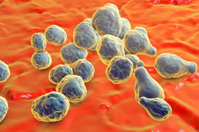
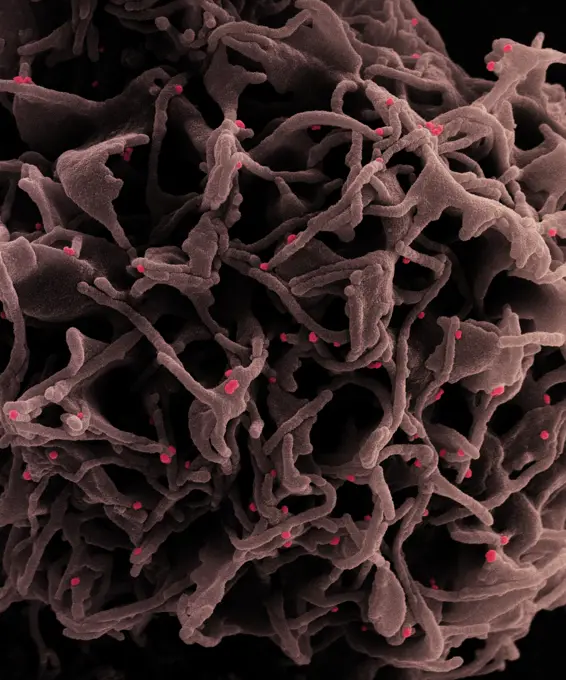
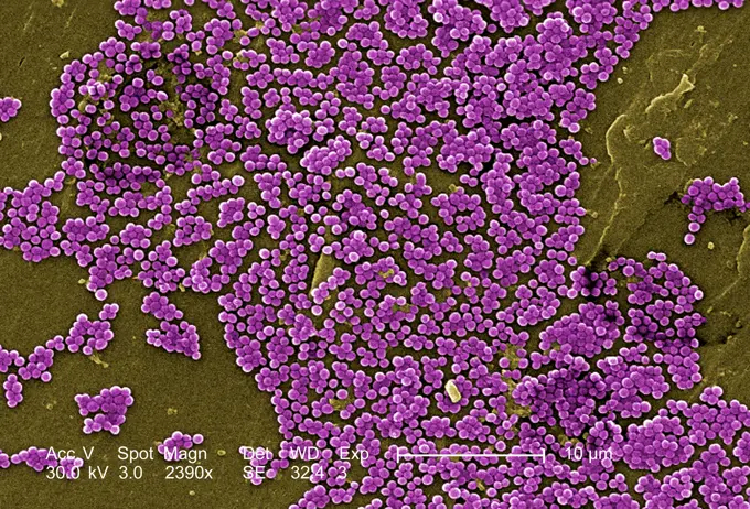
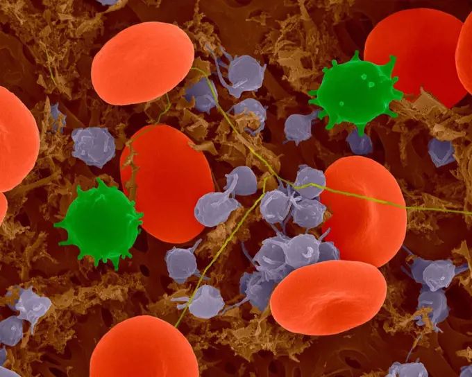
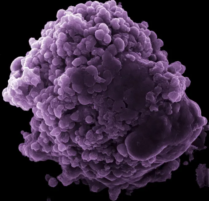
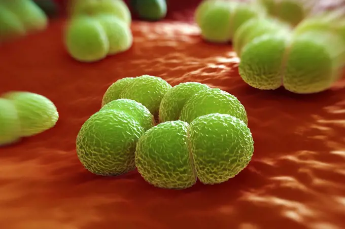
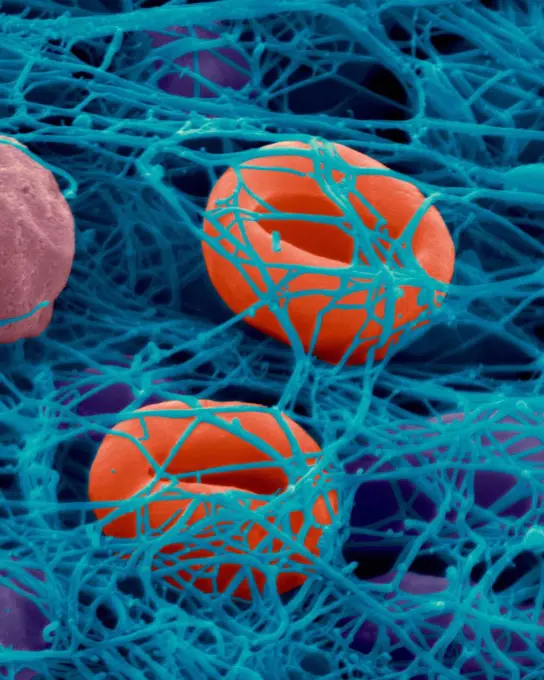
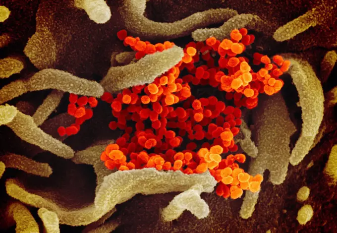
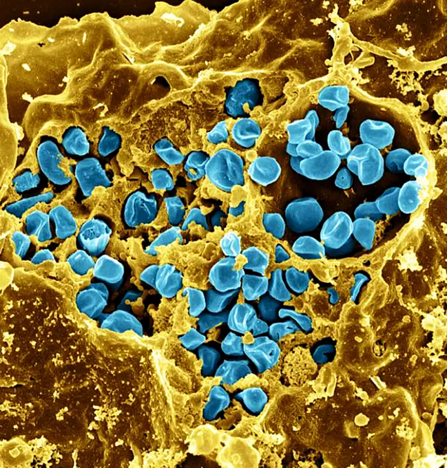
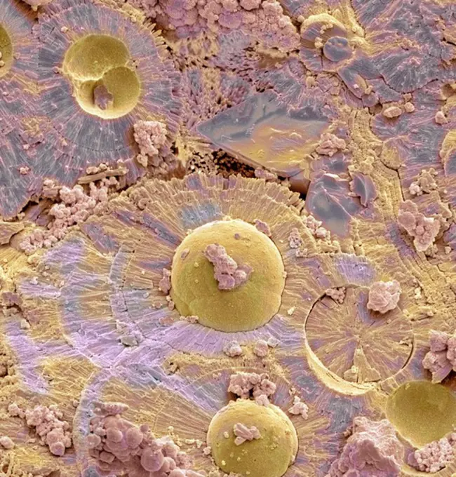
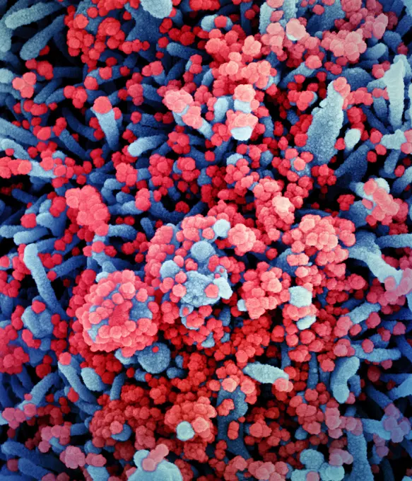
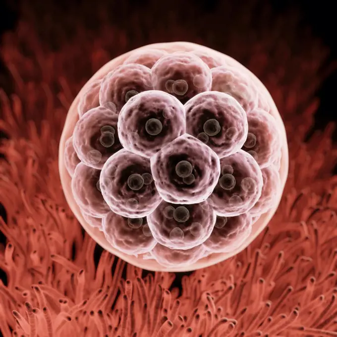
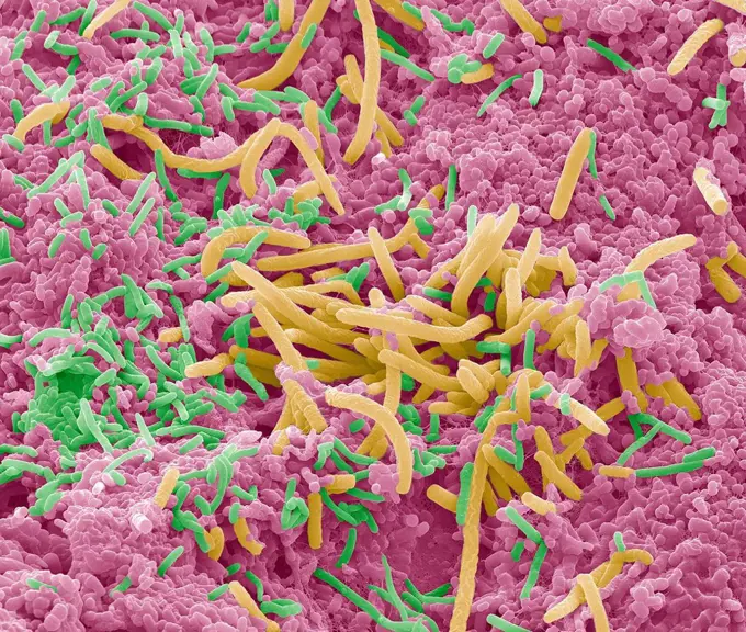
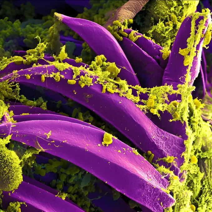
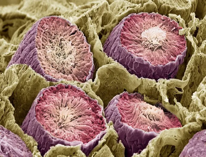
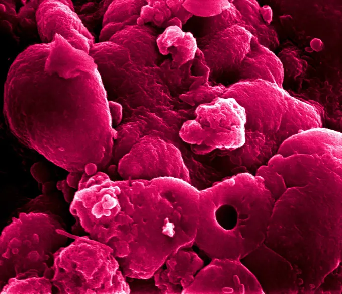
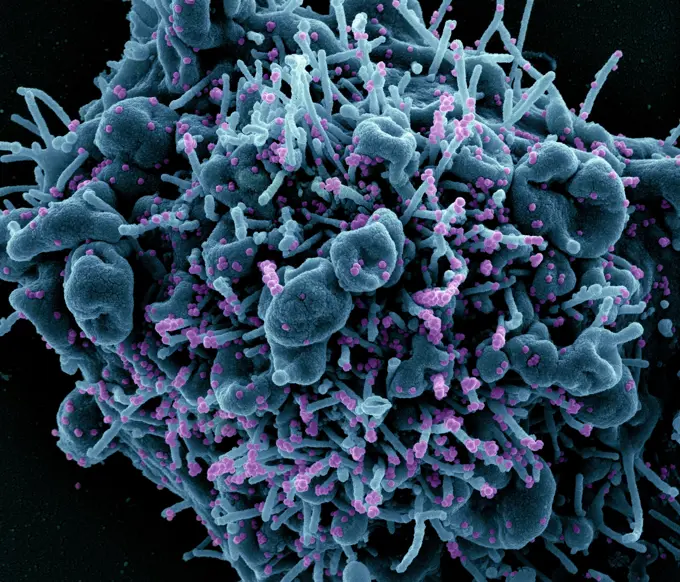
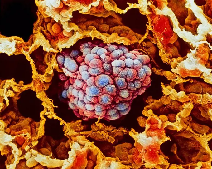
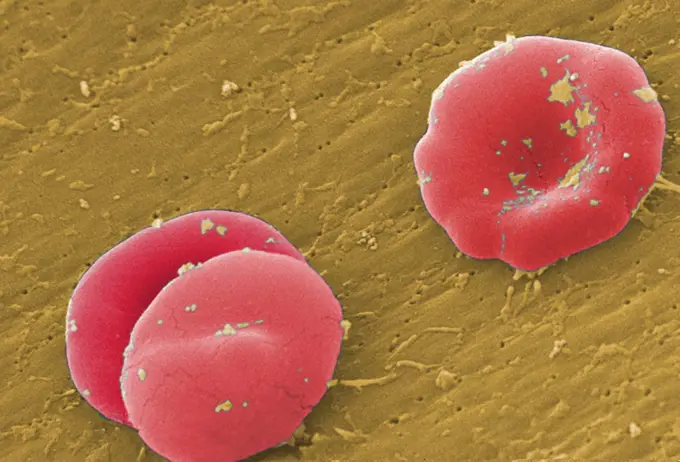
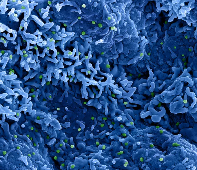
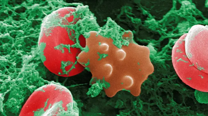
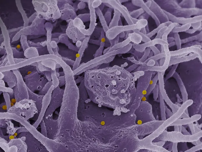
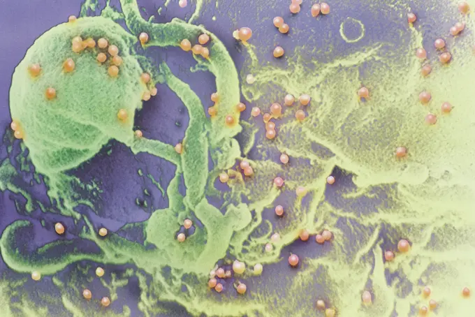
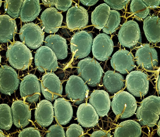
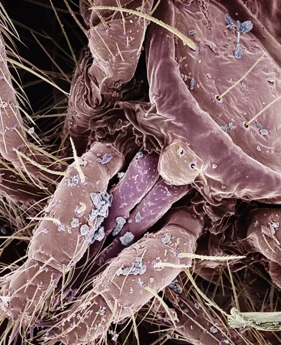
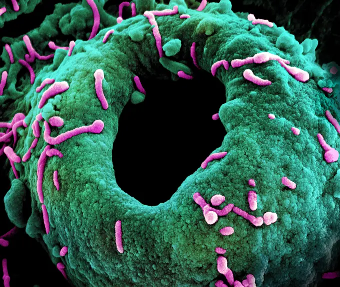
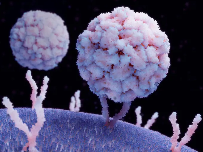
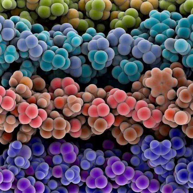
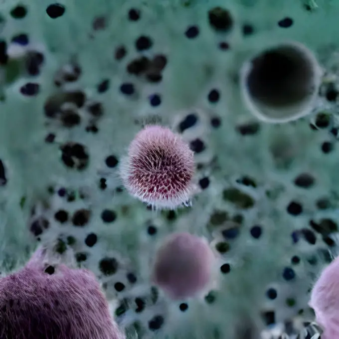
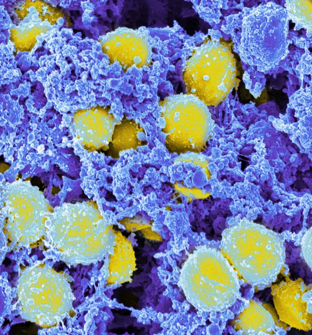
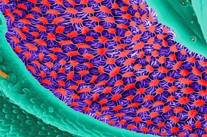
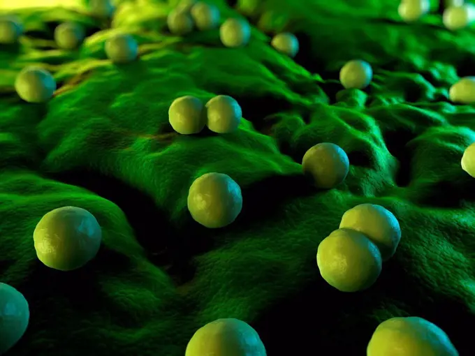
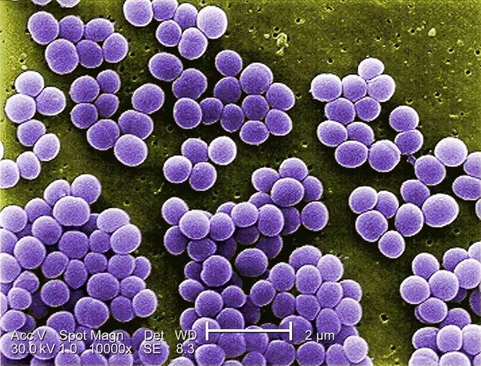
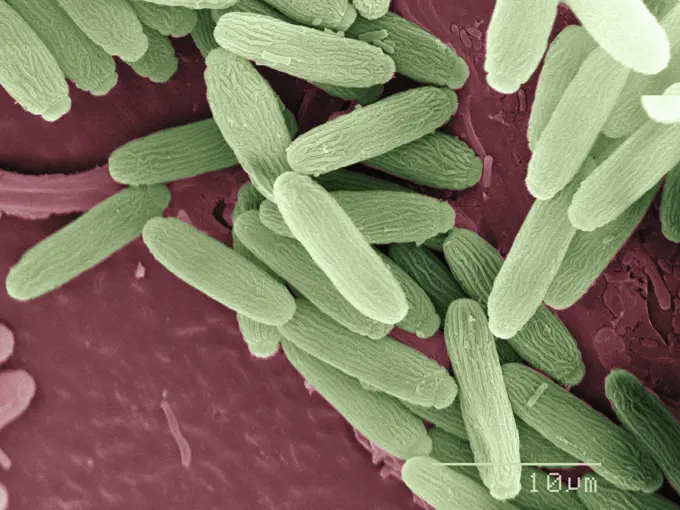
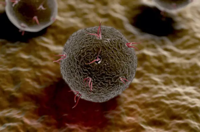
Fine ArtVintageFootageSubscriptionBoardsFree ResearchPricing
  • Fine Art
  • Vintage
  • Footage
  • Subscription
  • Boards
  • Free Research
  • Pricing

Scanning Electron Microscopy

Colorful close-up images captured by scanning electron microscopy showcasing bacteria and tissue at a microscopic level, revealing intricate structures and details.

Fat tissue, SEM
Fat tissue, SEM
201 assets in this story
4128R-396
Fat tissue, SEM
824-63191254
Scanning electron micrograph of methicillin-resistant Staphylococcus aureus and a dead human neutrophil.
824-63123132
Listeria ivanovii. A scanning electron microscope view.
4128R-15465082
Fecal bacteria, SEM
824-63191255
Scanning electron micrograph of methicillin-resistant Staphylococcus aureus and a dead human neutrophil.
824-63225881
Colorized scanning electron micrograph of an apoptotic cell (purple) heavily infected with SARS-CoV-2 virus particles (green), isolated from a patient sample. Image captured at the NIAID Integrated Research Facility (IRF) in Fort Detrick, Maryland.
824-63225878
Colorized scanning electron micrograph of an apoptotic cell (pink) heavily infected with SARS-CoV-2 virus particles (green), isolated from a patient sample. Image captured at the NIAID Integrated Research Facility (IRF) in Fort Detrick, Maryland.
824-63225815
Scanning electron micrograph of methicillin-resistant Staphylococcus aureus bacteria (red, round items) killing and escaping from a human white cell.
4269-5504
Electron micrograph (SEM) of methicillin-resistant Staphylococcus aureus bacteria (MRSA); Magnification 2363x. This strain of Gram-positif bacterium has developed wide-ranging antibiotic resistance such as methicillin, and are hospital pathogens.
824-63191211
Scanning electron micrograph of a human neutrophil ingesting MRSA.
824-63191225
Scanning electron micrograph of a human neutrophil ingesting MRSA.
4128-V58562065
824-63191213
Scanning electron micrograph of a human neutrophil ingesting MRSA.
4384-101
This 2005 colorized scanning electron micrograph (SEM) depicted numerous clumps of methicillin-resistant Staphylococcus aureus  bacteria, commonly referred to by the acronym, MRSA; Magnified 4780. Recently recognized outbreaks, or clusters of MRSA in community settings have been associated with strains that have some unique microbiologic and genetic properties, compared with the traditional hospital-based MRSA strains, which suggests some biologic properties, e.g., virulence factors like toxins,
4128R-15545695
Sputum culture, SEM
824-65831060
A colorized scanning electron microscope image of Group A Streptococcus (orange) during phagocytic interaction with a human neutrophil (teal).
824-65830999
Scanning electron micrograph of Neisseria gonorrhoeae bacteria, which causes gonorrhea.
824-65830722
Scanning electron micrograph of Neisseria gonorrhoeae bacteria, which causes gonorrhea. Exploring a Meningitis Vaccine for Gonorrhea Prevention
824-65831000
Scanning electron micrograph of Neisseria gonorrhoeae bacteria, which causes gonorrhea. 2024 Exploring a Meningitis Vaccine for Gonorrhea Prevention.
824-66065993
Group A streptococcal bacterial infections can range from a mild skin infection or a sore throat to severe, life-threatening conditions. Most people are familiar with strep throat, which along with minor skin infections, is the most common form of the disease. Health experts estimate that more than 10 million mild infections (throat and skin) occur every year. NIHs National Institute of Allergy and Infectious Diseases supports research to develop a group A streptococcus vaccine, and several candidate vaccines are in various phases of development. Image: A colorized scanning electron microscope image of Group A Streptococcus (orange) during phagocytic interaction with a human neutrophil (teal). Credit: NIAID/NIH.
824-63223235
Colorized scanning electron micrograph of an apoptotic cell (gray) heavily infected with SARS-COV-2 virus particles (yellow), isolated from a patient sample. Image captured at the NIAID Integrated Research Facility (IRF) in Fort Detrick, Maryland. Credit: NIAID.
1899-65662311
Colorized scanning electron micrograph of Marburg virus particles (purple) both budding and attached to the surface of an infected VERO E6 cell (green).
4128R-11476235
Protein molecules, computer artwork.
1899-65662359
Scanning electron micrograph of Neisseria gonorrhoeae bacteria, which causes gonorrhea.
4128-20041780
Faecal bacteria, sem
824-63225893
Colorized scanning electron micrograph of a cell (blue) infected with SARS-CoV-2 virus particles (yellow), isolated from a patient sample. Image captured at the NIAID Integrated Research Facility (IRF) in Fort Detrick, Maryland.
824-63191245
MRSA being ingested by neutrophil.
4128R-5160
Passion flower pollen, SEM
4269-20408839
Coloured scanning electron micrograph (SEM) of methicillin-resistant Staphylococcus aureus (MRSA) bacteria.
824-63123134
Listeria ivanovii. A scanning electron microscope view.
4128-V58562066
824-63128570
See image n0571405 on the site.
824-63191224
Scanning electron micrograph of a human neutrophil ingesting MRSA.
4128-V58562074
824-63191258
Interaction of MRSA (green bacteria) with a human white cell. The bacteria shown is strain MRSA252, a leading cause of hospital-associated infections in the United States and United Kingdom.
4384-210
Scanning Electron Micrograph of MRSA
4128R-11476193
Protein molecules, computer artwork.
824-63223242
Colorized scanning electron micrograph of an apoptotic cell (pink) heavily infected with SARS-CoV-2 virus particles (green), isolated from a patient sample. Image captured at the NIAID Integrated Research Facility (IRF) in Fort Detrick, Maryland. Credit: NIAID.
4128R-14060504
Cryptococcus neoformans fungus, illustration
1899-53513317
Scanning electron micrograph of Crimean-Congo hemorrhagic fever (CCHF) viral particles (yellow) budding from the surface of cultured epithelial cells from a patient.
824-63225898
Colorized scanning electron micrograph of a cell infected with SARS-CoV-2 virus particles (red), isolated from a patient sample. Image captured at the NIAID Integrated Research Facility (IRF) in Fort Detrick, Maryland.
824-63128425
Human gastric mucosa seen with SEM. See image n0572605 on the site.
824-63194409
This colorized scanning electron micrograph (SEM) depicted numerous clumps of methicillin-resistant Staphylococcus aureus bacteria, commonly referred to by the acronym, MRSA, Magnified 2390x. Recently recognized outbreaks, or clusters of MRSA in community settings have been associated with strains that have some unique microbiologic and genetic properties, compared with the traditional hospital-based MRSA strains, which suggests some biologic properties, eg virulence factors like toxins, may allow the community strains to spread more easily, or cause more skin disease.
4128R-13620340
Human red blood cells, monocytes (white blood cells), activated platelets and fibrin thread, composite coloured scanning electron micrograph (SEM). Re...
4378-20013923
Legionella Pneumophila Bacteria
4269-27597
Islet of Langerhans. SEM (Scanning Electron Microscope) of a islet of Langerhan, the endocrine component of the pancreas.
4128R-11315838
Virus particles, computer artwork.
824-63225884
Colorized scanning electron micrograph of an apoptotic cell (blue) infected with SARS-COV-2 virus particles (orange), isolated from a patient sample. Image captured at the NIAID Integrated Research Facility (IRF) in Fort Detrick, Maryland.
4378-4474
Neisseria meningitidis
4128R-13620360
Human red blood cells trapped in a fibrin blood clot, coloured scanning electron micrograph (SEM). Human red blood cells trapped in a fibrin blood clo...
4048-16067040
Novel Coronavirus SARS-CoV-2, causes 'coronavirus disease-2019', abbreviated as COVID-19. Scanning electron microscope image shows SARS-CoV-2 (orange) emerging from surface of cells (green) isolated from a patient in the US. Colors are artificially assigned to a monotone image for clarity. Image by National Institute of Allergy and Infectious Disease (NIAID)  (BSLOC_2020_2_245)
824-63194435
Scanning electron micrograph of a murine macrophage infected with Francisella tularensis strain LVS. Macrophages were dry-fractured by touching the cell surface with cellophane tape after critical point drying to reveal intracellular bacteria. Bacteria (colorized in blue) are located either in the cytosol or within a membrane-bound vacuole.
4128-V58562043
4128R-4345
Bladder stone, SEM
824-63223253
Colorized scanning electron micrograph of a cell (blue) heavily infected with SARS-CoV-2 virus particles (red), isolated from a patient sample. Image captured at the NIAID Integrated Research Facility (IRF) in Fort Detrick, Maryland. Credit: NIAID.
4378-1801
Morula development after 4-6 days.
4128-V58557895
4128R-13047353
Tongue bacteria. Coloured scanning electron micrograph (SEM) of bacteria on the surface of a human tongue. Large numbers of bacteria can form a visibl...
824-63225945
Scanning electron micrograph of Yersinia pestis, which causes bubonic plague, on proventricular spines of a Xenopsylla cheopis flea.
4128R-1089
Sperm production, SEM
824-63223134
Ebola virus isolated in November 2014 from patient blood samples obtained in Mali. The virus was isolated on Vero cells. Credit: NIAID.
4269-27596
Islet of Langerhans. SEM (Scanning Electron Microscope) of a islet of Langerhan, the endocrine component of the pancreas.
824-63225844
Colorized scanning electron micrograph of an apoptotic cell (teal) infected with SARS-COV-2 virus particles (purple), isolated from a patient sample. Image captured at the NIAID Integrated Research Facility (IRF) in Fort Detrick, Maryland.
4128R-3190
Coloured SEM of a cancerous tumour in the lung
1899-85550
Sickle Cell Anemia SEM
824-63219649
This scanning electron microscope image shows SARS-Cov-2 also known as 2019-nCoV, the virus that causes COVID-19 isolated from a patient in the United States, emerging from the surface of cells cultured in laboratory.
1899-61460448
Colorized scanning electron micrograph of monkeypox virus (green) on the surface of infected VERO E6 cells (blue)
4269-24586
Dust mite. SEM (Scanning Electron Microscope)
1899-53513332
Colorized transmission electron micrograph of Ebola virus particles (purple) attached to and budding from an infected VERO E6 cell (green). Image captured and color-enhanced at the NIAID Integrated Research Facility in Fort Detrick, Maryland.
824-63225880
Colorized scanning electron micrograph of an apoptotic cell (gray) heavily infected with SARS-CoV-2 virus particles (yellow), isolated from a patient sample. Image captured at the NIAID Integrated Research Facility (IRF) in Fort Detrick, Maryland.
824-63225902
Colorized scanning electron micrograph of a cell (teal) infected with a variant strain of SARS-CoV-2 virus particles (UK B.1.1.7 purple), isolated from a patient sample. Image captured at the NIAID Integrated Research Facility (IRF) in Fort Detrick, Maryland.
4384-126
This scanning electron micrograph (SEM) depicted a number of red blood cells found enmeshed in a fibrinous matrix on the luminal surface of an indwelling vascular catheter; Magnified 7766x. In this instance, the indwelling catheter was a tube that was left in place creating a patent portal directly into a blood vessel. The erythrocyte in the center had undergone the process of  crenation, whereupon, it developed a number of cell wall projections, thereby, transforming it into what is termed an
824-63227133
Scanning electron micrograph of Crimean-Congo hemorrhagic fever (CCHF) virus particles (yellow) budding from the surface of cultured epithelial cells from a patient.
4128-V58569647
4269-7100
Colored scanning electron micrograph of HIV viruses (red dots) budding from a T-lymphocyte white blood cell (yellow & green).
824-63194919
This digitally-colorized scanning electron micrograph (SEM) revealed some of the ultrastructural morphology displayed on the head region of a bedbug, Cimex lectularius. Of interest is one of the insect's compound eyes. The compound eye is given this name due to the fact that the single large eye is really made up of many repeating units known as ommatidia. Each ommatidium is composed of separate units made up of a photoreceptor cell, support cell, and pigment cells. Though each of these visual mechanisms functions as a separate organ, together they provide the organism with a compound picture of its environment. Due to what is referred to as the flicker effect, the compound eye is made very sensitive to movement, with each ommatidium turning on and off, as objects pass across its field of view. The bilateral anatomical placement of the insect's eyes provides the organism with a very wide range of visual sensitivity.
824-65830997
Scanning electron micrograph of Neisseria gonorrhoeae bacteria, which causes gonorrhea.
4201-21259090
Moss Mite (Oribatidae) gnathosoma magnified 240x
1899-61460447
Colorized scanning electron micrograph of monkeypox virus (blue) on the surface of infected VERO E6 cells (pink)
824-65830620
Colorized scanning electron micrograph of Marburg virus particles (pink) both budding and attached to the surface of an infected VERO E6 cell (green).
4128-19489269
Rhinovirus bound to human cell, illustration
4128-V58562046
824-57659299
This scanning electron microscopic (SEM) image of an untreated water specimen extracted from a wild stream mainly used to control flooding during inclement weather, revealed the presence of unidentified organisms, which included bacteria, protozoa, and algae. In this particular view, a mass of gelatinous biofilm had enmeshed numbers of microorganisms, including amoebae and bacteria. For a colorized version of this image, see PHIL 11707. CDC/ Janice Haney Carr 2009
4128R-11476163
Protein molecules, computer artwork.
4128-V58569646
4269-27740
Staphylococcus aureus. Color ehanced scanning electron micrograph (SEM) of Staphylococcus aureus bacteria. These Gram-positive bacteria are commonly carried on the skin or in the nose of healthy people. They are one of the most common causes of skin infections.
824-63225909
Colorized scanning electron micrograph of a cell infected with a variant strain of SARS-CoV-2 virus particles (yellow), isolated from a patient sample. Image captured at the NIAID Integrated Research Facility (IRF) in Fort Detrick, Maryland.
4269-27598
Islet of Langerhans. SEM (Scanning Electron Microscope) of a islet of Langerhan, the endocrine component of the pancreas.
1815R-13377840
Group of fat cells, 3D Rendering
4128R-13620213
Coloured scanning electron micrograph (SEM) Pet dander. dog hair, cat hair and dead skin (cat hair. blue, dog hair. dark yellow, dander. purple). Dog ...
6019-19983687
Microscopic view of floating influenza virus cells. Dangerous illness
4128R-20389
Nanoparticles, artwork
824-63194628
Scanning electromicrograph of Staphylococcus aureus bacteria.
4384-163
A digitally-colorized close-up scanning electron micrograph (SEM) of a beautiful diaphanous structure in the mesothoracic region of a bedbug (Cimex lectularius).  It is speculated that this wondrous ultrastructural organ is most probably a scent gland, or related to the dissemination of scent, which may be pheromonal in nature. Photo by Janice Carr.
4128R-15357
Bacteria, conceptual computer artwork.
824-63194418
Under a high magnification of 10,000x, this scanning electron micrograph (SEM) shows a strain of Staphylococcus aureus bacteria taken from a vancomycin intermediate resistant culture (VISA). Under SEM, one can not tell the difference between bacteria that are susceptible, or multidrug resistant, but with transmission electron microscopy (TEM), at least with VISA isolates one can see a thickening in the cell wall that may attribute to their reduced susceptibility to vancomycin. VISA and VRSA are specific types of antimicrobial-resistant staph bacteria. While most staph bacteria are susceptible to the antimicrobial agent vancomycin some have developed resistance. VISA and VRSA cannot be successfully treated with vancomycin because these organisms are no longer susceptibile to vancomycin. However, to date, all VISA and VRSA isolates have been susceptible to other Food and Drug Administration (FDA) approved drugs.
1439-57940061
Magnified bacteria under the microscope in hues of green and red
4378-1016
Stylized close up of generic virus particles.
4128-V58557943
824-63225833
A single elongated CCL-81 cell heavily infected with SARS-CoV-2 virus particles. The small spherical structures in the image are SARS-CoV-2 virus particles. The string-like protrusions from the cells are cell projections or pseudopodium. Image captured at the NIAID Integrated Research Facility (IRF) in Fort Detrick, Maryland.
PREVIOUS
of 3
NEXT
2401 S. Ervay, Suite 206
Dallas, Texas 75215
United States
Browse
CategoriesGalleriesFree Research
For Creators
License Your ContentSuperStock For StudentsFrame of Mind BlogContributor PortalNew Image SpotlightLicense AI Imagery
Resources
About UsLicensingTerms & ConditionsAPI AccessPrivacyPricing
Contact
+1 866 236 0087help@superstock.com Contact form
©2026 SuperStock. All Rights Reserved. -A
This site is protected by reCAPTCHA and Google Privacy Policy and Terms of Service apply.