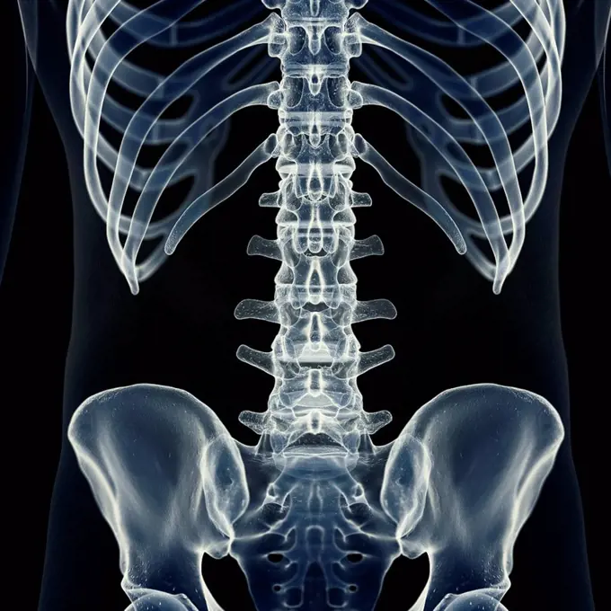
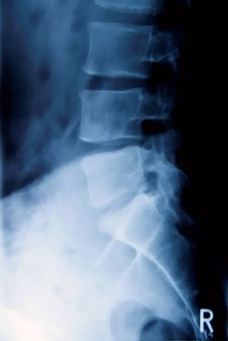
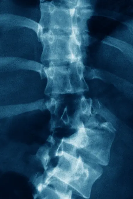
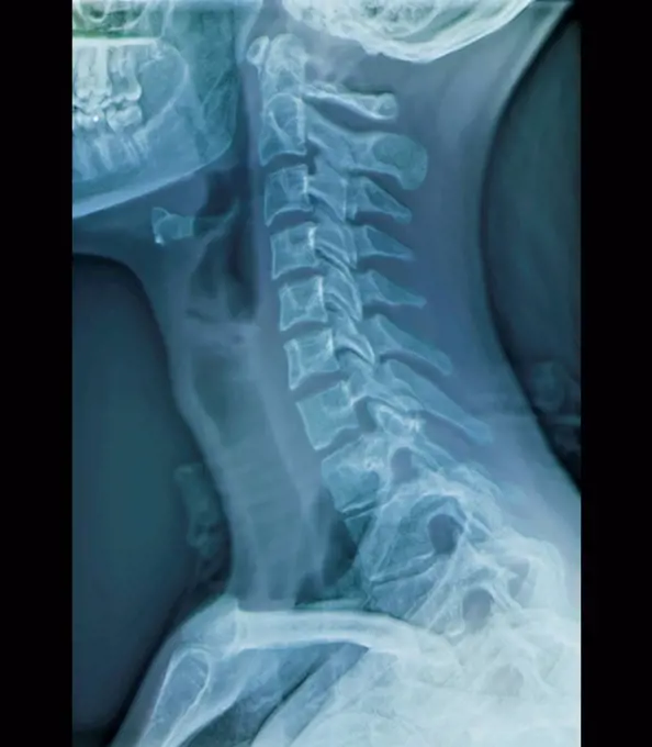
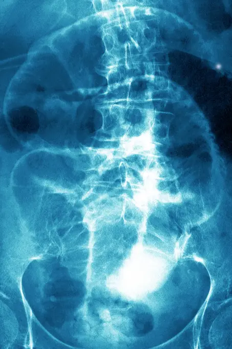
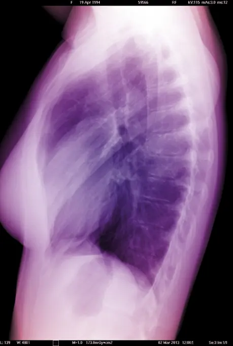
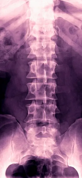
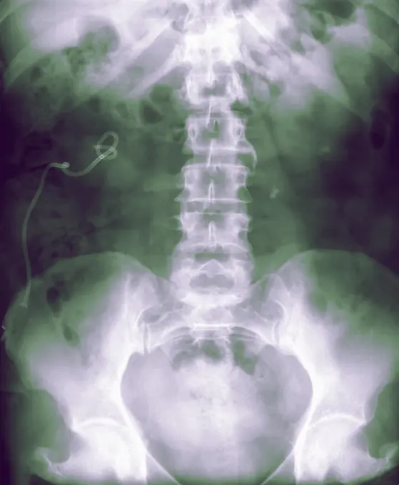
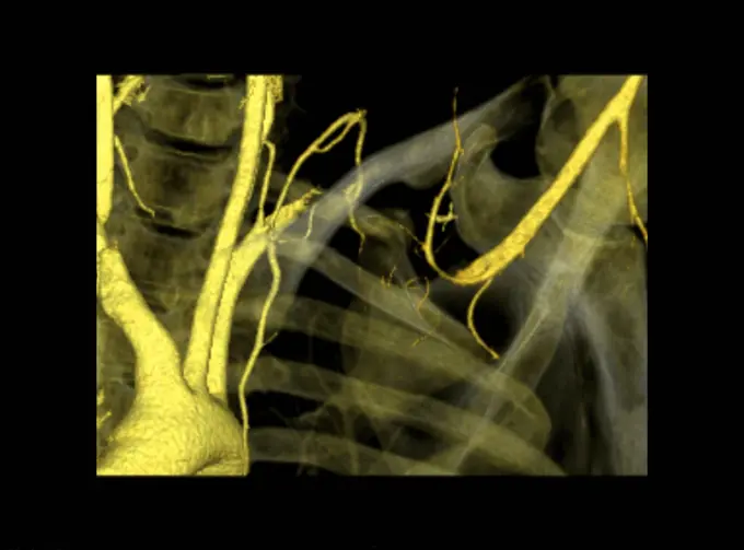
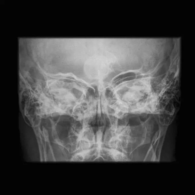
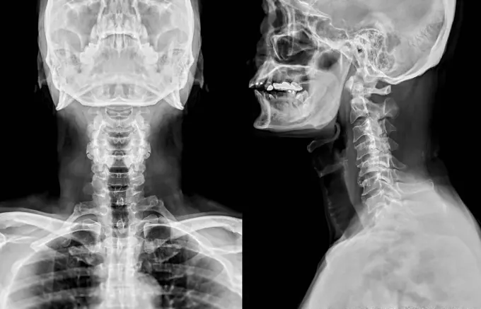
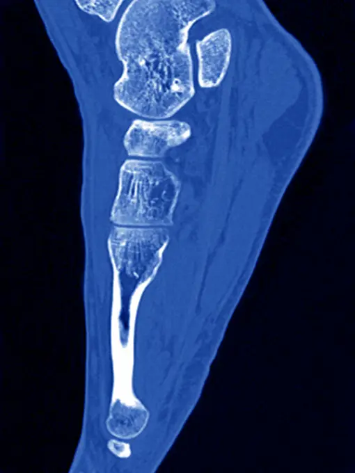
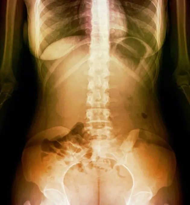
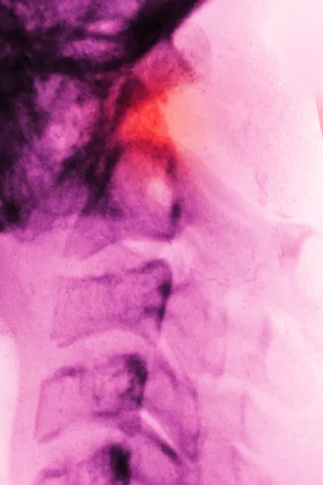
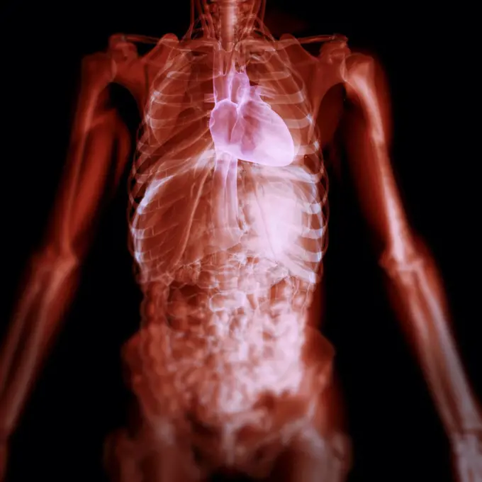
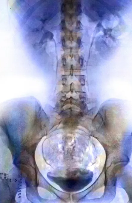
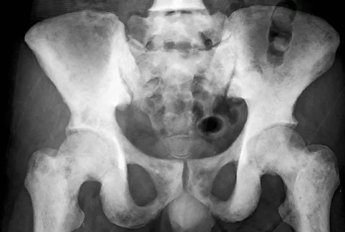
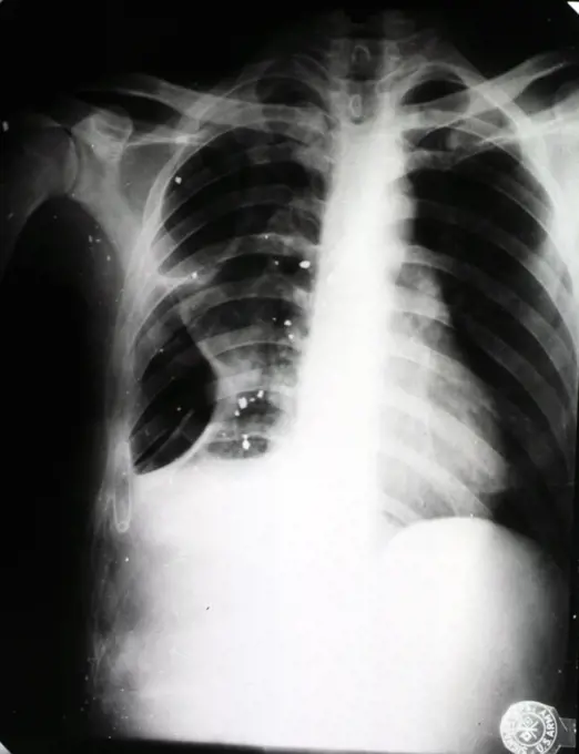
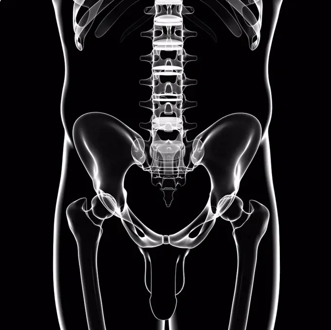
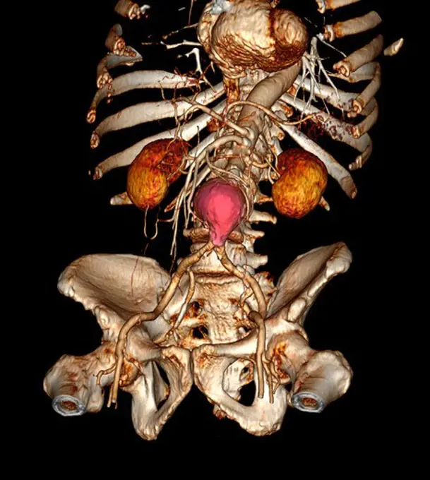
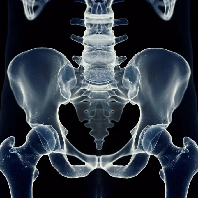
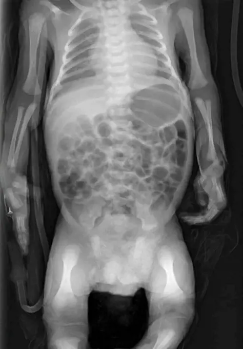
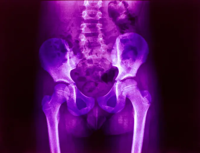
Fine ArtVintageFootageSubscriptionBoardsFree ResearchPricing
  • Fine Art
  • Vintage
  • Footage
  • Subscription
  • Boards
  • Free Research
  • Pricing

Spinal and Pelvic X-rays

X-ray images focusing on the human spine and pelvis, illustrating various medical conditions and anatomy in a clinical context.

Illustration of the lumbar spine
136 assets in this story
4128R-15536931
Illustration of the lumbar spine
5507-40438480
X-ray of the pelvis and spinal column
1815R-156186
Radiograph of lumbar spine
1899-54027389
X-ray of urinary schistosomiasis. Frontal pelvic x-ray. (Photo by: BSIP/UIG via Getty Images)
824-63226732
Rotational dislocation at the junction of the thoracic and lumbar vertebrae, visualized by an AP chest radiograph.
824-63128418
See image n0756005 on the site.
4128R-12660
Normal neck. Coloured X_ray of the cervical spine of a 27 year old.
1899-53511987
Chronic colitis (inflammatory bowel disease), revealed by a frontal abdominal x-ray.
824-63204759
Frontal x-ray of a 60-year old woman's trunk.
824-63193987
A healthy lung x-ray on an 18-year old patient.
824-63193985
A healthy lung x-ray on an 18-year old patient.
4128R-5152
Normal lumbar spine, X_ray
1439-57940808
Xray image revealing the human spine and pelvic bones.
4128-28970823
Invertogram X-ray
1439-57940360
Radiant xray image revealing the intricate bones of the human spine and pelvis.
4128R-15536886
Illustration of the shoulder bones
6188-55478595
film with X-ray image of human female pelvis film with X-ray image of human female pelvis isolated on white background Copyright: xZoonar.com/ValeryxVoennyyx 13808323
1899-85843
Thorax X Ray
4269-27164
Thoracic outlet syndrome. Color enhanced 3D computed tomography (CT) scan showing a thoracic outlet syndrome (TOS).
1439-57940805
Xray image revealing the intricate structures within a human head.
824-63175258
X-ray of the skull showing sinonasal polyposis.
4128-30420004
Head and neck bones, X-ray
4128R-36671
Male pelvic bones, computer artwork.
824-63184394
Sagittal plane scan of the right foot.
4128R-12678
Normal abdomen. Coloured X_ray of the abdomen of a 20 year old female.
1899-53511624
Dislocation between the first and second cervical vertebrae, visualized by a sagittal x-ray of the cervical spine.
1525-28007762
medical science image of human skeleton bones. Transparent Human Body with Visible Bones
1525-28468563
X ray film for human skull
4378-3631
Front view of the torso anatomy with heart highlighted.
824-63194008
A healthy lung x-ray on an 18-year old patient.
824-63178593
X-ray of the pelvis and lower back of a 25 years old woman. Light discopathy on L4-L5 and L5-S1
4128-30421464
Posterior skeletal anatomy of the hip, illustration
4128R-12771
Normal shoulder. Coloured X_ray of the shoulder of a 19 year old man.
1899-66335
X-ray image of female chest, sternam, rib cage and spine
4128R-30959
Scoliosis of the spine. Coloured X-ray of the chest of a 31 year old patient with scoliosis (sideways curvature) of the spine.
4128-19358123
X-ray showing a dislocated shoulder, where the head of the humerus is detached from the shoulder joint.
4128R-26338
Male skeleton, computer artwork.
1899-85878
Cervical Vertebra X Ray
4128R-12668
Normal neck. Coloured frontal X_ray of the joint between the skull and the neck.
1525-24031592
3d illustration - x-ray panels on black background
824-63224749
Hip asymmetry, visualized by frontal pelvic x-ray.
824-63198847
Degenerative disk disease. Vertebral osteoarthritis seen on an side x-ray of the thorax.
4128R-2925
Normal human torso, monochrome frontal magnetic resonance imaging MRI scan. The spinal bones vertebrae are seen running from the top of the chest to t...
4128-30415769
Atlas bone, illustration
1899-66326
X-ray image of female lumbar vertibrae
4128-30415797
Skeletal back, illustration
824-63193991
A healthy lung x-ray on an 18-year old patient.
4269-25313
Prosthetic hip. X-ray of prosthetic hip showing the shaft of steel inserted into the femur (pink), with the artificial ball articulating with the bone socket of the pelvis.
824-63204708
Frontal x-ray exmaining gastrointentinal transit.
824-63194002
A healthy lung x-ray on an 18-year old patient.
824-57655646
Profile x-ray of lungs.
4128-19357540
X-ray of the pelvis of a patient with prostate carcinoma. The X-ray shows an osteoblastic lesion on the pelvis bone and vertebra lumbalis indicating b...
1899-18977930
1944 - X-ray showing chest wound caused by a Japanese 50mm mortar shell.
4128R-34300
Male pelvic bones, computer artwork.
4297-1378
Lateral skull x-ray of a 18 year old man
1525-25424267
X-ray of a human male chest
824-63226737
Dislocation of the cervical vertebrae, with spinal trauma, between the sixth and seventh cervical vertebrae, visualized by sagittal section radiography.
1525-28468521
X ray film for human skull
4378-265
Close-up of stylized bones of the left hip within the skin.
824-63178554
X-ray of a child's sinuses. Normal
4269-25454
Hysterography. X-ray of female reproductive organs. A contrast medium was introduced into the uterus (center) and its Fallopian tubes (oviducts, thin, extending from uterus) through the uterine neck.
4269-27187
Human body. 3D Computed Tomography (CT) reconstruction scan of a human body revealing the skeleton, the heart, the kidneys (red), and the arterious system (yellow).
4128-19358120
X-ray of a 1 day old baby showing a normal chest, normal abdomen and bone structure.
4128-16375523
Oil drum skeleton, X-ray
4128-19358201
X-ray of a 1 day old baby showing a normal chest, normal abdomen and bone structure.
1899-53508427
Aortic coarctation of newborn, before surgery, aortic coarctation is a narrowing of the aorta, often considerable and sometimes complete, most commonly just past the point where the aorta and the subclavian artery meet. Aortic coarctation causes low blood flow, often leading to arterial hypertension. Surgery is often advised to establish normal anatomy of the aortic intersection and gain normal blood flow. 3D CT scan.
4128-24795785
Healthy stomach, barium X-ray
6188-55588298
Human teeth xray X ray of human mouth with teeth bones Copyright: xZoonar.com/ClaudioxDiviziax 8918967
1841R-111806
4128R-11323293
Human sacroiliac joint, computer artwork.
4128-19357514
X-ray of the lower (lumbarsacral) spine taken from a patient with ankylosis spondylitis and chronic lower back pain showing syndesmophytes.
6114-50976698
An x-ray of a fractured humerus.
1570R-132784
CAT Scan of a midsection of a person
4297-1141
CT scan images showing an abdominal aortic aneurysm which developed between the renal arteries and the common iliac arteries
1525-24739556
medical science image of human skeleton bones. Transparent Human Body with Visible Bones
4128R-15536938
Illustration of the sacrum
1525-26510343
Lungs ribs x-ray scan test result.
4128-30420595
Urinary system stent, X-ray
1899-53508430
Large intercostal arteries, visible in a CT scan, developed to compensate pulmonary blood flow due to a congenital heart defect characterized by atresia of pulmonary arteries (small or absent pulmonary arteries). 3D CT scan.
824-63165971
Large intercostal arteries, visible in a CT scan, developed to compensate pulmonary blood flow due to a congenital heart defect characterized by atresia of pulmonary arteries (small or absent pulmonary arteries). 3D CT scan.
4128-18680909
Intussusception of the intestines, X-ray
4128-19358031
X-ray of a 1 day old baby showing a normal chest, normal abdomen and bone structure.
4269-27534
Dislocation of the hip. 3D Computed Tomographic (CT) reconstruction scan of the pelvis showing a posterior hip luxation. The head of the femur (bone of the thigh) has totally slipped out on its insertion point  in the pelvis bone.
1815R-12499504
Pelvis x-ray
6188-55478592
film with X-ray image of human jaws isolated film with X-ray image of human jaws isolated on white background Copyright: xZoonar.com/ValeryxVoennyyx 13808297
1899-61460135
Spontaneous vertebral fracture due to oteoporosis (bone disease characterized by a decrease in bone mass, as well as bone deformation by compression and the risk of spontaneous fracture.). Lumbar vertebral x-ray
824-63225990
Lung cancer located in the upper right lobe. Frontal chest x-ray.
1899-66332
X-ray image of female wrist
4128-19489633
Colon diverticula, X-ray
4297-1342
normal shoulder x-ray of a 20 year old woman, showing the upper humerus and scapula
4128-19358080
X-ray showing a dislocated shoulder, where the head of the humerus is detached from the shoulder joint.
4128-30420028
Sinuses, X-ray
4269-5166
X-ray of a normal pelvis of a child.
824-33581
LUNG METASTASIS, X_RAY. Pulmonary metastases secondary cancer, carcinoma. Thoracic x_ray in front view.
4128R-12788
Arthritis of the hip. Coloured X_ray of an arthritic hip.
4356-319
An x-ray of a human shoulder from the front, showing the humerus (the upper arm bone), the shoulder blade or scapula, the collarbone or clavicle, and the rib cage.
824-63178682
X-ray of the stomach of a young child. Transit without particular anomaly
4128-16375226
Pig skull, X-ray
4128-19357553
X-ray of a pinned femur fracture with internal fixation plate and screw.
1899-66343
Dentistry X-ray image of female jaw and teeth
PREVIOUS
of 2
NEXT
2401 S. Ervay, Suite 206
Dallas, Texas 75215
United States
Browse
CategoriesGalleriesFree Research
For Creators
License Your ContentSuperStock For StudentsFrame of Mind BlogContributor PortalNew Image SpotlightLicense AI Imagery
Resources
About UsLicensingTerms & ConditionsAPI AccessPrivacyPricing
Contact
+1 866 236 0087help@superstock.com Contact form
©2026 SuperStock. All Rights Reserved. -A
This site is protected by reCAPTCHA and Google Privacy Policy and Terms of Service apply.