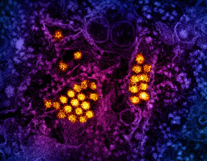
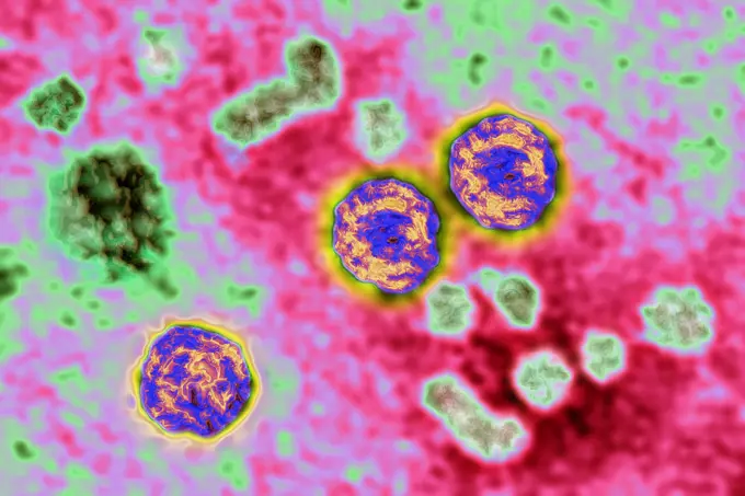
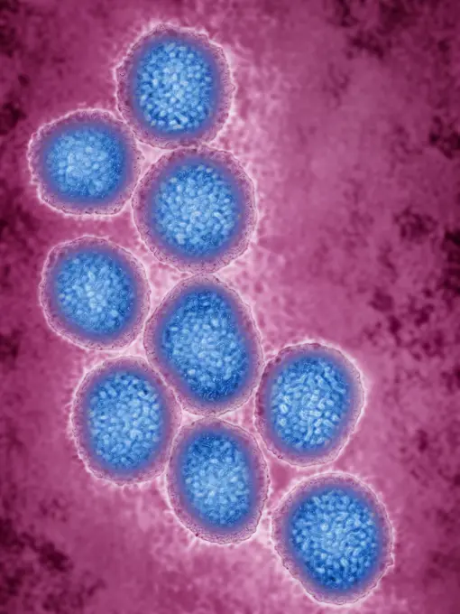
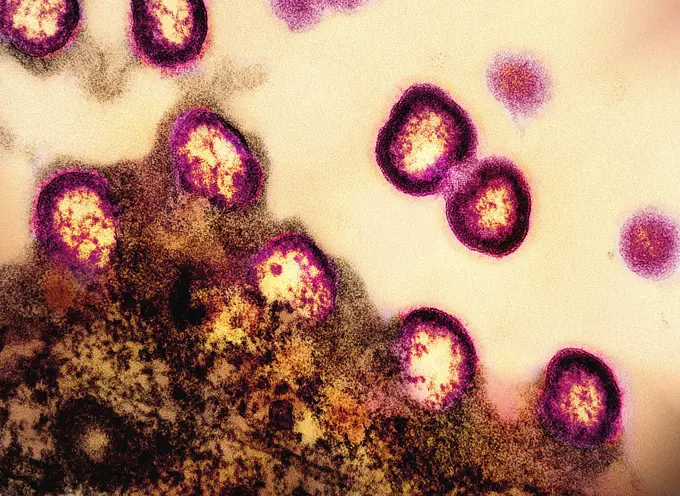
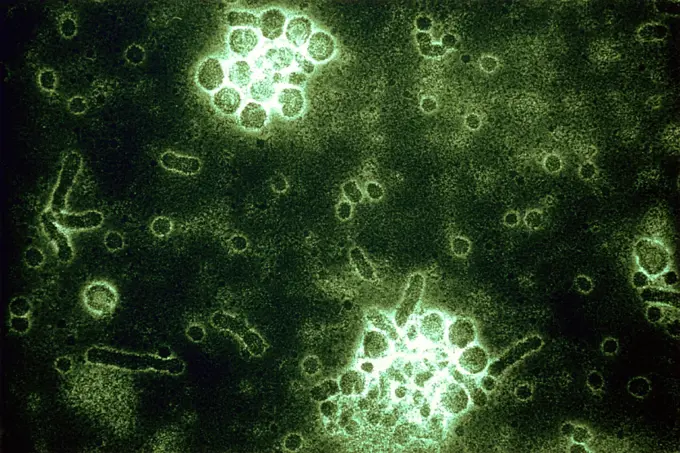
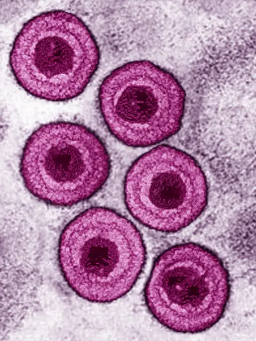
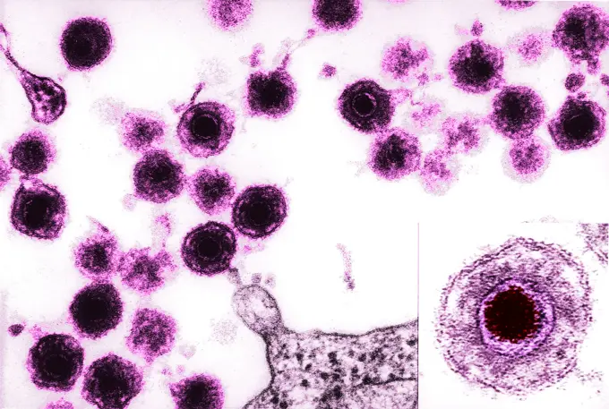
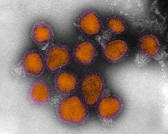
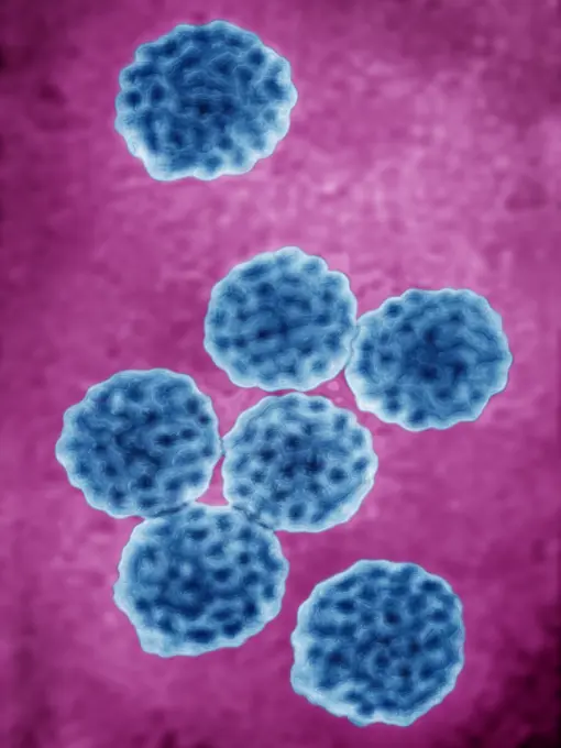
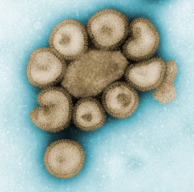
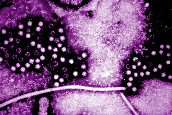
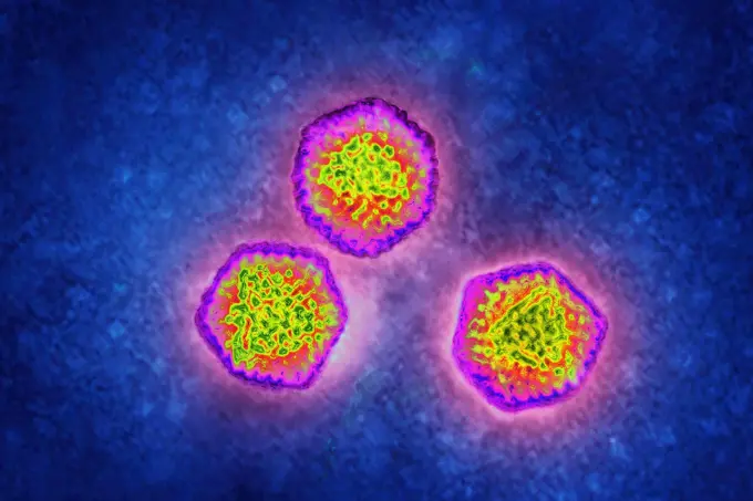
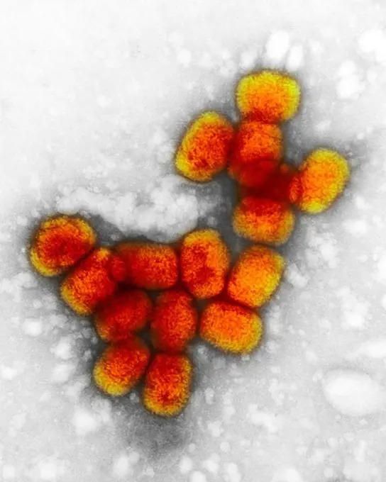
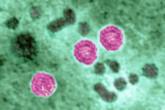
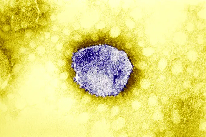
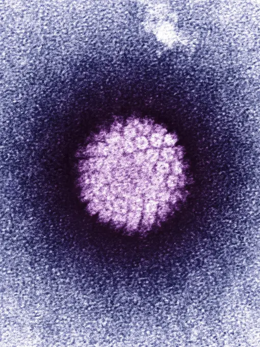
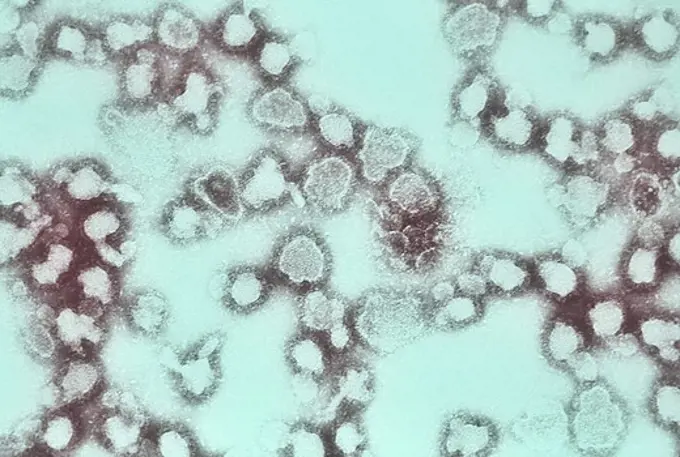
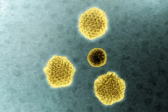
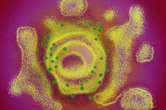
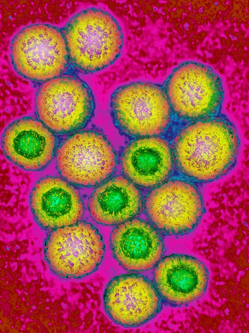
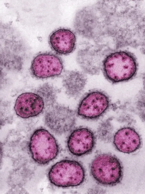
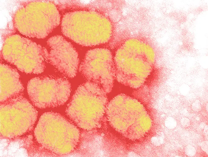
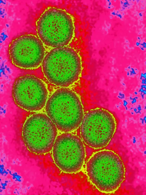
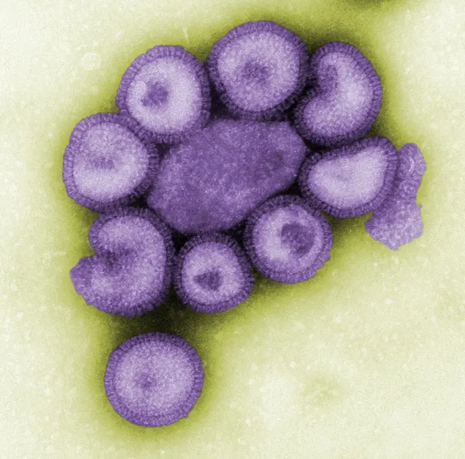
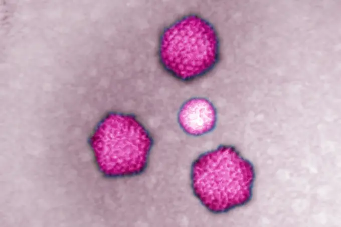
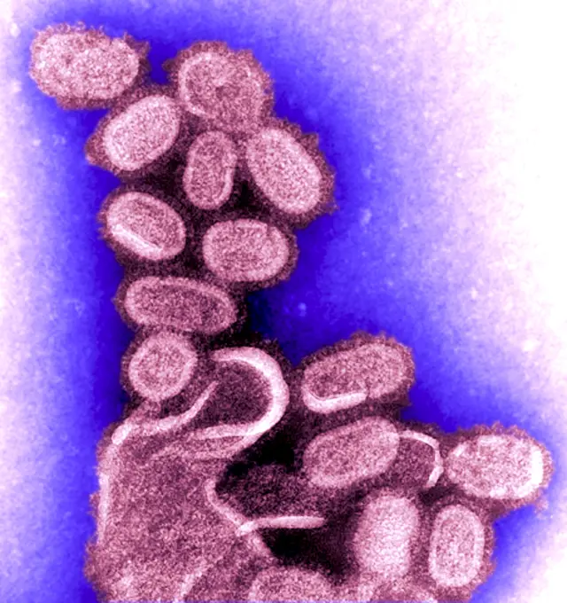
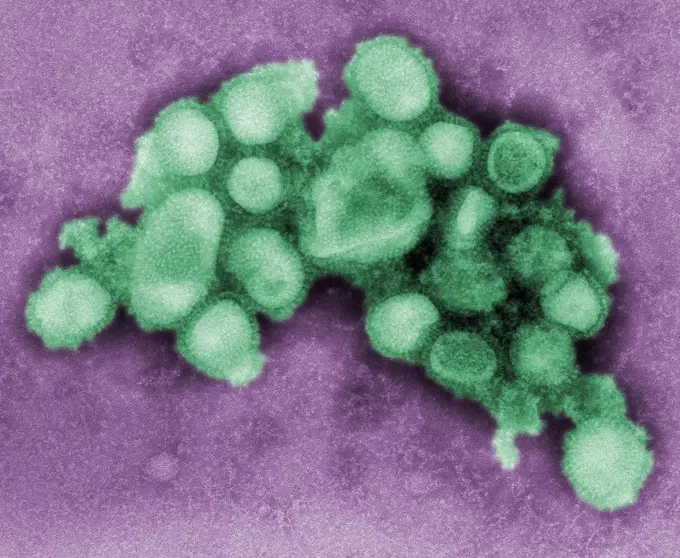
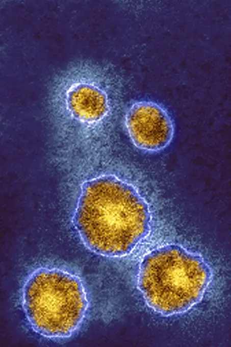
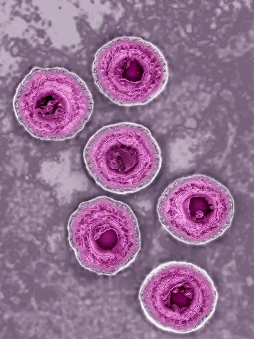
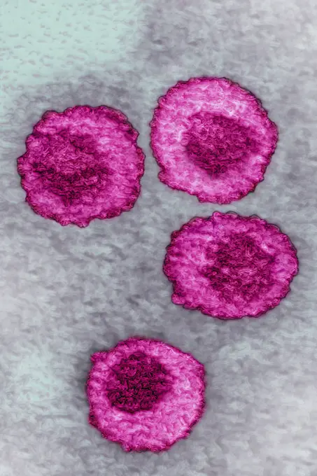
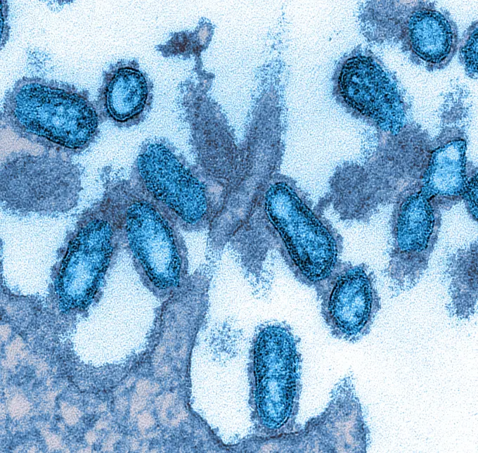
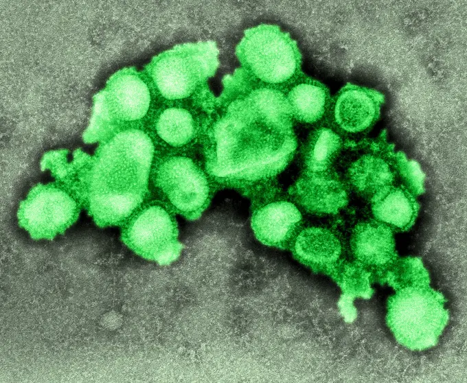
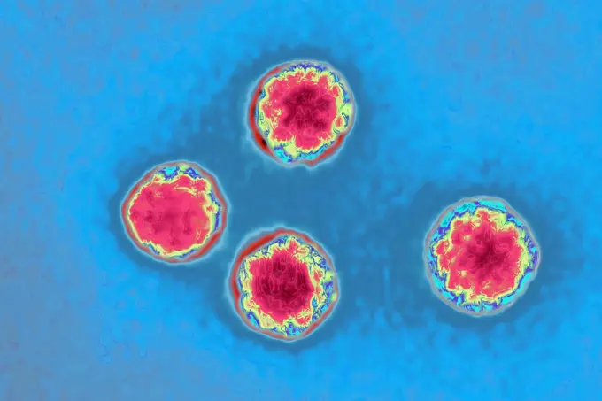
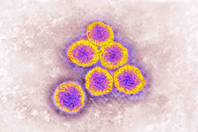
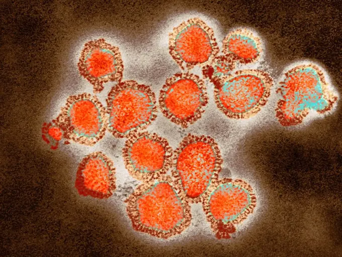
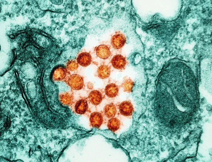
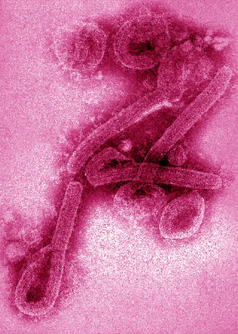
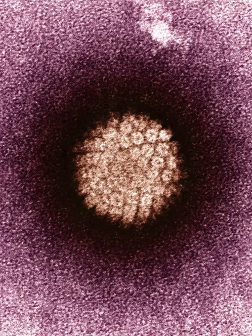
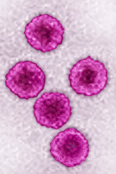
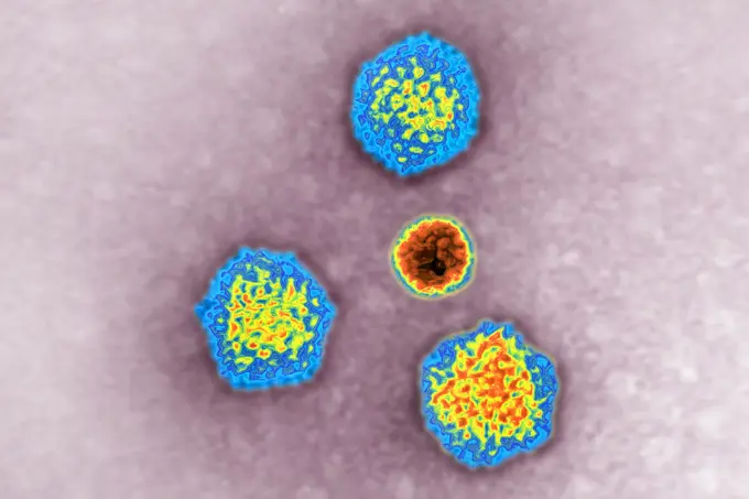
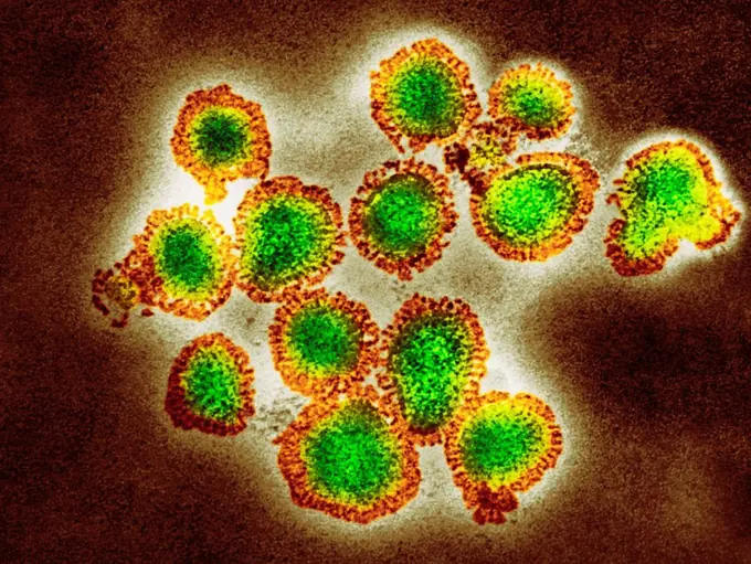
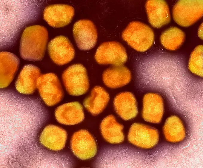
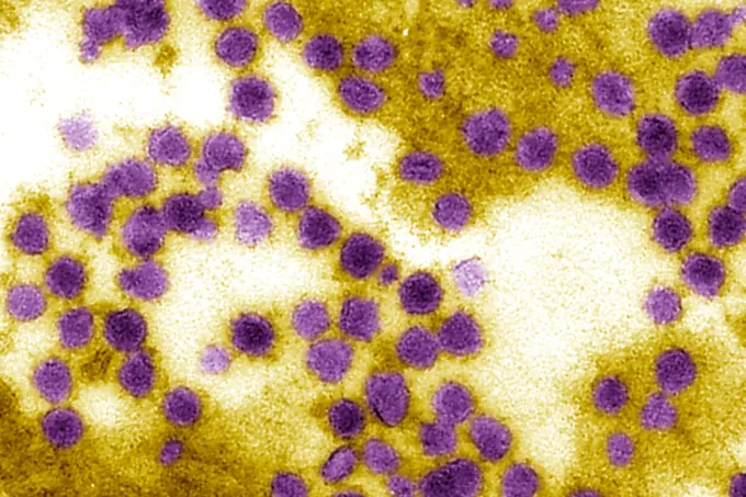
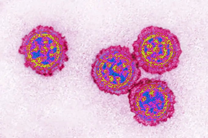
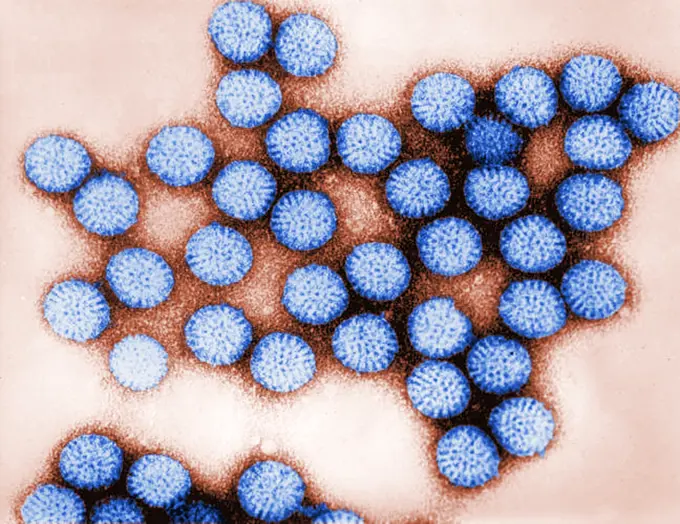
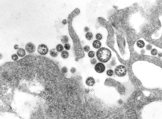
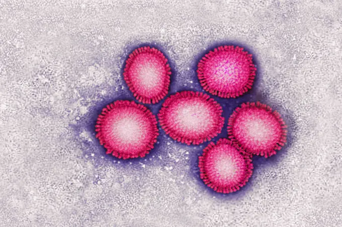
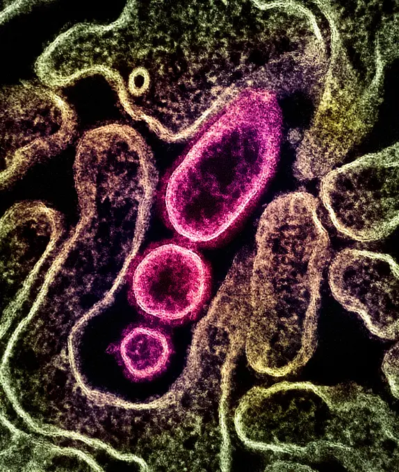
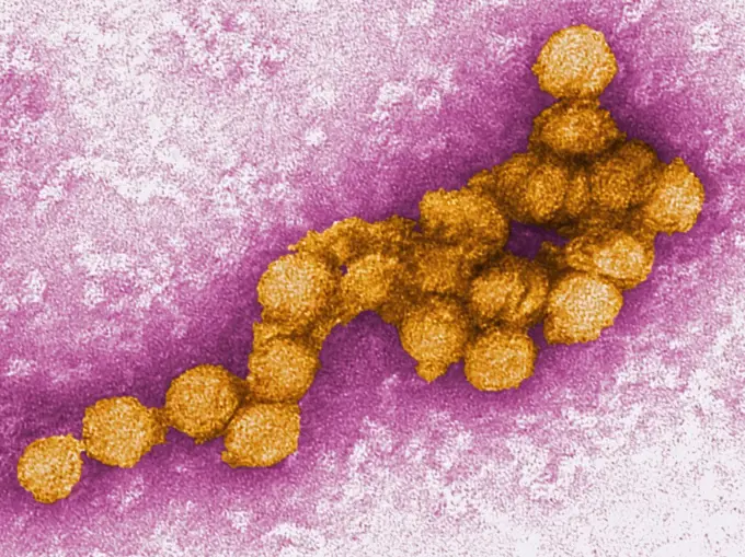
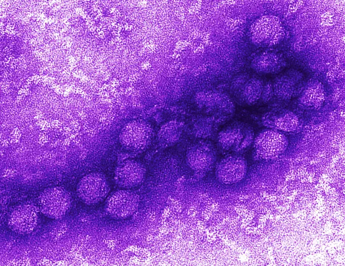
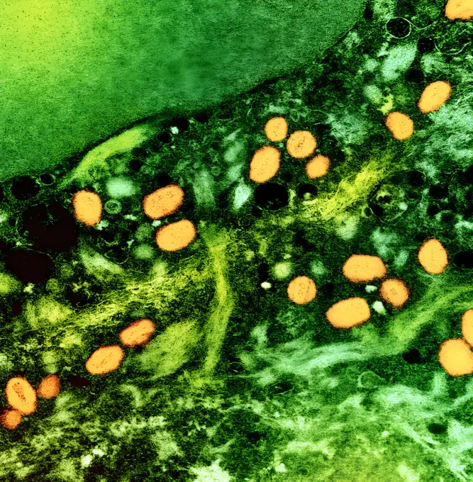
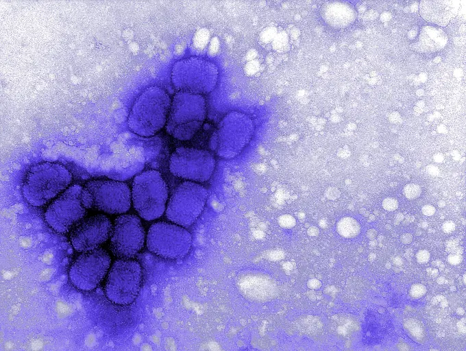
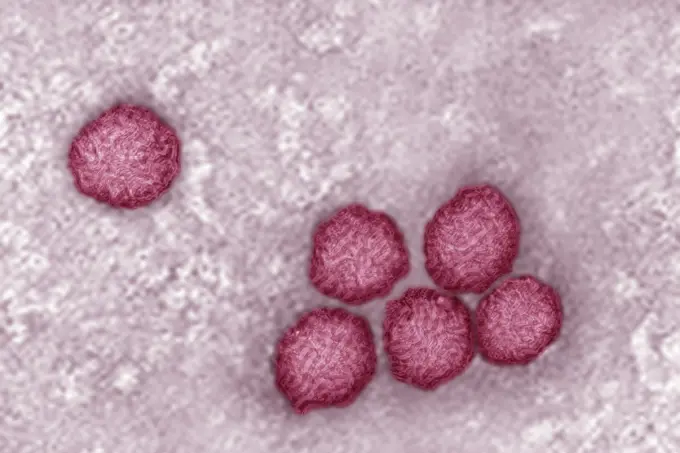
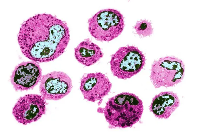
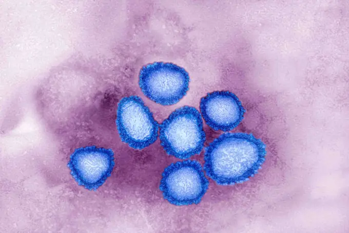
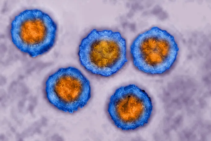
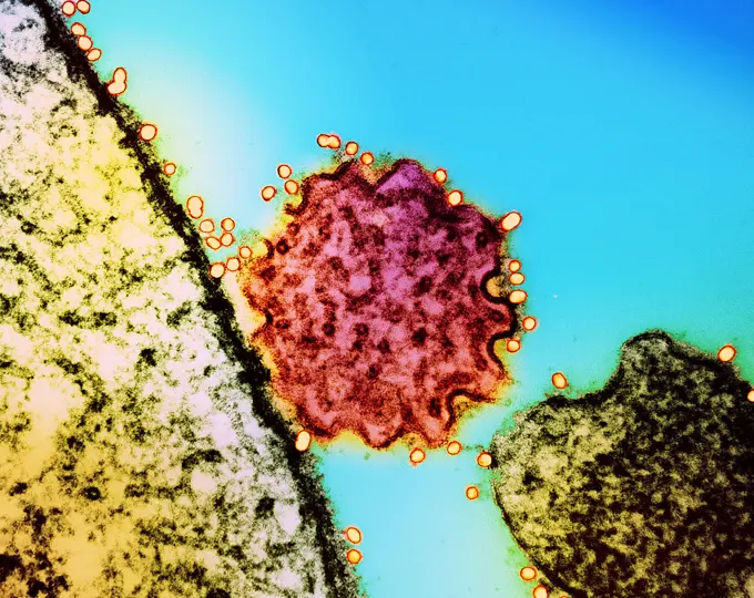
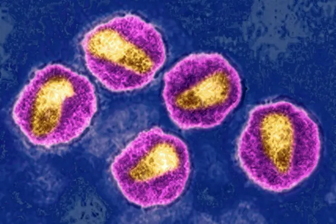
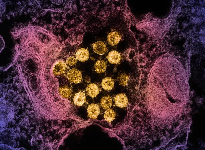
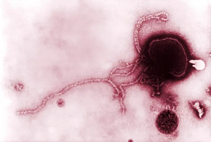
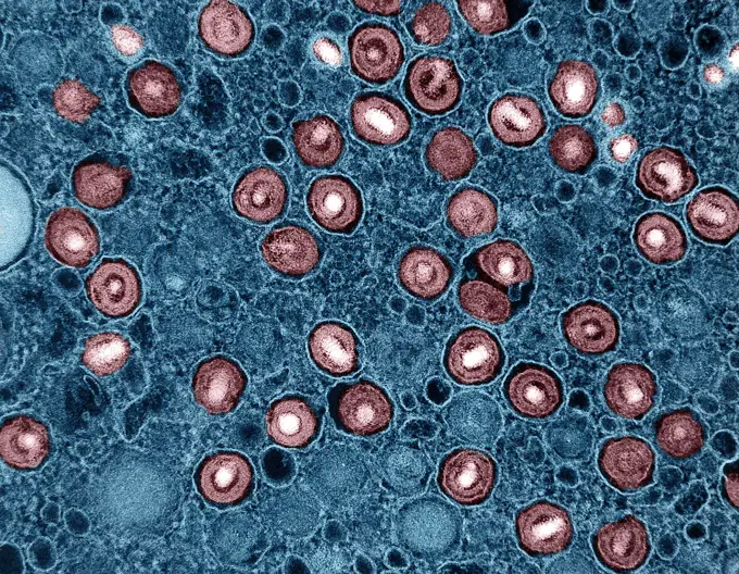
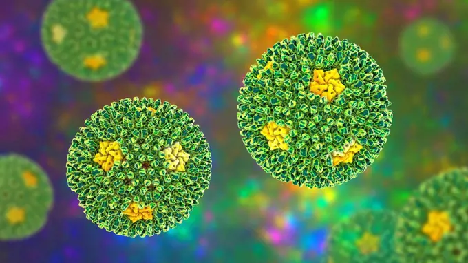
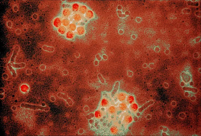
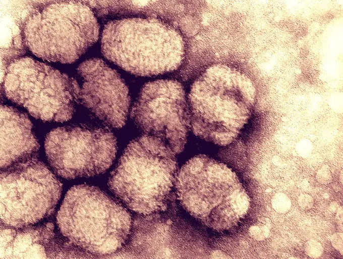
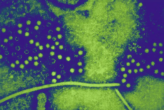
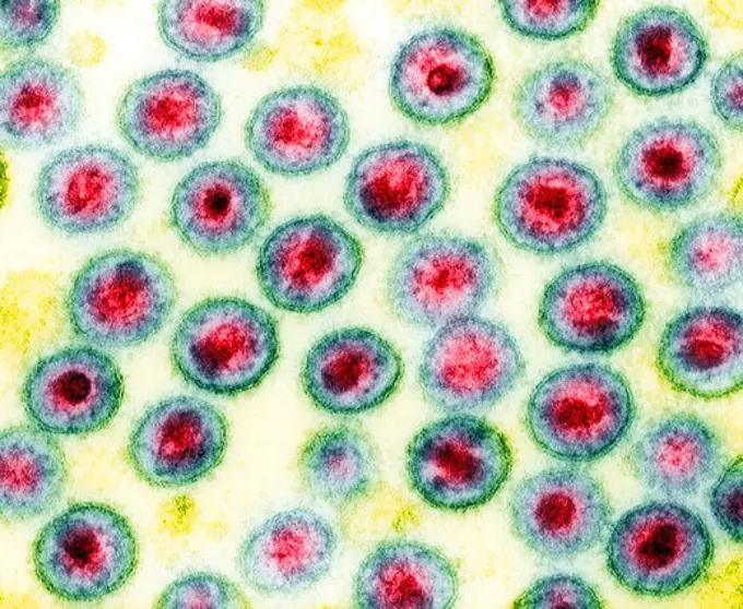
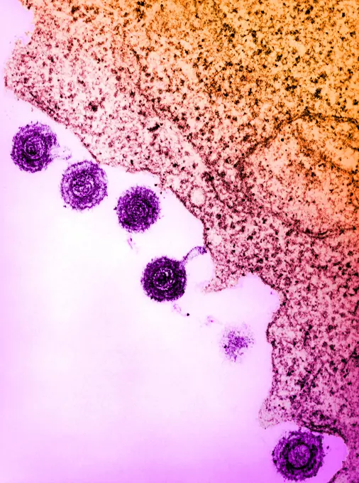
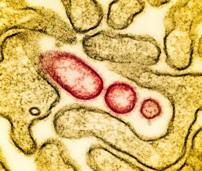
Fine ArtVintageFootageSubscriptionBoardsFree ResearchPricing
  • Fine Art
  • Vintage
  • Footage
  • Subscription
  • Boards
  • Free Research
  • Pricing

Viral Particles in Microscopy

Colorized transmission electron micrographs showcasing various viruses, including Hepatitis B, HIV-1, and SARS-CoV-2, against vibrant, abstract backgrounds.

Transmission electron micrograph of dengue virus particles (gold).
210 assets in this story
824-63218227
Hepatitis B virus (Hepadnavirid family). HBV causes sexually transmitted or blood-borne hepatitis. Cirrhosis occurs in 20% of cases 15 to 20 years after the onset of infection. Image made from transmission electron microscopy. Approximate viral diameter: 22 nm.
824-63183648
Togavirus. Image produced using high-dynamic-range imaging (HDRI) from an image taken with transmission electron microscopy. Viral diameter around 100 nm.
824-65830641
Transmission electron micrograph of HIV-1 virus particles (colorized pink/yellow) replicating from an HIV-infected H9 T-cell (brown).
824-63183627
Epstein-Barr virus (human herpesvirus 4, HHV-4). It causes infectious mononucleosis and Burkitt's lymphoma. View taken with HDRI image processing from a transmission electron microscopy image. Viral diameter about 200 nm.
824-63225918
Transmission electron micrograph of SARS-CoV-2 virus particles (green) within endosomes of a heavily infected nasal Olfactory Epithelial Cell. Image captured at the NIAID Integrated Research Facility (IRF) in Fort Detrick, Maryland.
4269-24754
Hepatitis B virus. Color enhanced Transmission Electron Micrograph (TEM) of the Hepatitis B virus (HBV, Hepadnaviridae).
824-65830781
Transmission electron micrograph of dengue virus particles (gold).
824-63195707
This transmission electron micrograph (TEM) depicted numbers of intact rotavirus double-shelled particles. Note the distinctive rim of radiating capsomeres.
824-63218236
Hepatitis C virus (close to the Flavivirid family). HCV causes blood-borne hepatitis, cirrhosis occurs in 25% of cases, 10 to 20 years after the onset of infection, with a risk of hepatic carcinoma. Image made from transmission electron microscopy. Approximate viral diameter: 50 to 60 nm.
824-63183623
Varicella zoster virus (human herpesvirus type 3, HHV-3). It causes chicken pox and shingles (herpes zoster). Image produced using high-dynamic-range imaging (HDRI) from an image taken with transmission electron microscopy. Viral diameter around 200 nm.
824-65830819
Transmission electron micrograph of HIV-1 virus particles (pink) replicating from the plasma membrane of an infected H9 T cell.
4269-6704
Transmission electron micrograph (TEM) of human herpes virus-6 (HHV-6) previously called HBLV (human b-lymphotropic virus). It infects lymphocyte T cells as well. It is the cause of the childhood rash roseola and some cases of mononucleosis.
824-63183595
Coronavirus. Image produced using high-dynamic-range imaging (HDRI) from an image taken with transmission electron microscopy. Viral diameter ranges from around 80 to 160 nm.
4297-1225
Negatively-stained transmission electron micrograph of Hong Kong flu virus virions
824-63183636
Human papilloma virus (HPV). Image produced using high-dynamic-range imaging (HDRI) from an image taken with transmission electron microscopy. Viral diameter around 55 nm.
824-63194474
This colorized negative-stained transmission electron micrograph (TEM) depicts the ultrastructural details of a number of influenza virus particles, or virions. A member of the taxonomic family Orthomyxoviridae, the influenza virus is a single-stranded RNA organism. The flu is a contagious respiratory illness caused by influenza viruses. It can cause mild to severe illness, and at times can lead to death. The best way to prevent this illness is by getting a flu vaccination each fall.
4269-27644
Heptatitis E virus. Electron micrograph of Hepatitis E viruses (HEV). Formerly a RNA virus member of the Caliciviridae, HEV are now classified as Hepeviridae.
1899-53511259
Adenovirus (highly contagious virus causing lymphadenopathy symptomatizing swelling of the glands in the neck, accompanied by a cold, or pharyngitis, or bronchitis, sometimes also conjunctivitis, keratitis, or a combination of the two (keratoconjunctivitis). View produced from a transmission electron microscopy view. Viral diameter approximately: 80 to 110 nm.
824-63224489
Hepatitis C virus (close to the Flavivirid family). HCV causes blood-borne hepatitis, cirrhosis occurs in 25% of cases, 10 to 20 years after the onset of infection, with a risk of hepatic carcinoma. View produced from a transmission electron microscopy image. Viral diameter approximately: 50 to 60 nm.
4297-1439
A transmission electron micrograph of smallpox viruses using a negative stain technique. Smallpox is a serious, highly contagious, and sometimes fatal infectious disease. There is no specific treatment for smallpox disease
824-63218226
Hepatitis B virus (Hepadnavirid family). HBV causes sexually transmitted or blood-borne hepatitis. Cirrhosis occurs in 20% of cases 15 to 20 years after the onset of infection. Image made from transmission electron microscopy. Approximate viral diameter: 22 nm.
824-63123707
Tacaribe virus (New World Arenavirus). Electron micrograph of Arenaviridae. Arenaviridae are RNA viruses whose particles are spherical and have an average diameter of 110-130 nanometers. Arenaviridae members are zoonotic, which means that in nature they are found in reservoir animal hosts.
4269-27755
Human papilloma virus. Colored transmission electron micrograph (TEM) of human papillomavirus (HPV). The HPV is involved in the development of cervical cancers.
4297-1818
Negatively-stained transmission electron micrograph, TEM, of La Crosse, LAC, encephalitis virus Ribonucleoprotein Particles
824-63217781
Adenovirus (highly contagious virus), responsible for: Colds, pharyngitis, bronchitis, pneumonia, gastroenteritis, conjunctivitis and keratitis. Image made from a transmission electron microscopy view.
824-743
Varicella Virus
824-63194905
This colorized transmission electron micrograph (TEM) revealed some of the ultrastructural morphology displayed by norovirus virions, or virus particles.
824-63183646
Rotavirus. Rotavirus is the primary cause of severe diarrhoea in young children worldwide. Image produced using high-dynamic-range imaging (HDRI) from an image taken with transmission electron microscopy. Viral diameter ranges from around 60 to 80 nm.
824-63183590
Arenavirus. Arenaviruses cause several types of viral hemorrhagic fever (New World fevers). The viruses are transmitted to humans by rodents and bats. Image produced using high-dynamic-range imaging (HDRI) from an image taken with transmission electron microscopy. Viral diameter ranges from around 110 to 130 nm.
1899-53511290
Coronavirus (CoV). viruses of the Coronaviridae family and the Orthocoronavirinae subfamily. It is a pathogen of respiratory syndromes. Viewed from a transmission electron microscopy (TEM) image. Viral diameter 80nm to 100nm.
824-57657668
Colorized transmission electron micrograph of monkeypox virus particles (gold) cultivated and purified from cell culture.
824-63128587
Virus responsible for smallpox. A transmission electron micrograph of smallpox viruses. Smallpox is a serious, highly contagious, and sometimes fatal infectious disease. There is no specific treatment for smallpox disease, and the only prevention is vaccination. See image n0641905 on the site.
824-63183649
Togavirus. Image produced using high-dynamic-range imaging (HDRI) from an image taken with transmission electron microscopy. Viral diameter around 100 nm.
824-63194472
This colorized negative-stained transmission electron micrograph (TEM) depicts the ultrastructural details of a number of influenza virus particles, or virions. A member of the taxonomic family Orthomyxoviridae, the influenza virus is a single-stranded RNA organism. The flu is a contagious respiratory illness caused by influenza viruses. It can cause mild to severe illness, and at times can lead to death. The best way to prevent this illness is by getting a flu vaccination each fall.
824-63217780
Adenovirus (highly contagious virus), responsible for: Colds, pharyngitis, bronchitis, pneumonia, gastroenteritis, conjunctivitis and keratitis. Image made from a transmission electron microscopy view.
4269-14238
Transmission Electron Micrograph (TEM) of Spanish flu virus. This pandemic virus infected a fifth of the world population and killed between 20 and 50 million. This Influenza virus type a, strain H1N1 is very close to the swine flu (strain H1N1) and the avian flu virus (strain H5N1).
824-63183637
Human papilloma virus (HPV). Image produced using high-dynamic-range imaging (HDRI) from an image taken with transmission electron microscopy. Viral diameter around 55 nm.
4048-6182
This colorized negative stained transmission electron micrograph of the A/CA/4/09 swine flu virus. 2009 photo by C. S. Goldsmith and A. Balish.
824-63217280
The mumps virus (paramyovirus). Image produced with transmission electron microscope (viral diameter around 200 nm).
1899-61460440
Colorized transmission electron micrograph of monkeypox virus particles (gold) cultivated and purified from cell culture
824-63183620
Herpes virus (HSV 1 and 2). View taken with HDRI image processing from a transmission electron microscopy image. Viral diameter about 200 nm.
824-63208136
Varicella zoster virus (VZV or HHV 3). Image taken with transmission electron microscopy.
824-63197015
Hepatitis A virus (HAV). Image produced using high-dynamic-range imaging (HDRI) from an image taken with transmission electron microscopy. Viral diameter ranges from around 20 to 30 nm.
824-63194513
This colorized transmission electron micrograph (TEM) of an ultra-thin specimen revealed some of the ultrastructural morphologic features seen in 1918 influenza virus virions. The prominent surface projections on the virions are composed of either the hemagglutinin, or neuraminidase type of glycoproteins. Composed of what looked like dots or tubules, was a dense envelope known as a capsid, which surrounded each virion's nucleic acid constituents.
4269-14234
2009 C. S. Goldsmith and A. Balish This preliminary negative stained transmission electron micrograph (TEM) depicted some of the ultrastructural morphology of the A/CA/4/09 swine flu virus. See PHIL 11213 for a black and white version of this image.  What is Swine Influenza   Swine Influenza (swine flu) is a respiratory disease of pigs caused by type A influenza virus that regularly causes outbreaks of influenza in pigs. Swine flu viruses cause high levels of illness and low death r
824-63194505
This colorized negative stained transmission electron micrograph (TEM) depicted some of the ultrastructural morphology of the A/CA/4/09 swine flu virus. Swine Influenza (swine flu) is a respiratory disease of pigs caused by type A influenza virus that regularly causes outbreaks of influenza in pigs. Swine flu viruses cause high levels of illness and low death rates in pigs. Swine influenza viruses may circulate among swine throughout the year, but most outbreaks occur during the late fall and winter months similar to outbreaks in humans. The classical swine flu virus (an influenza type A H1N1 virus) was first isolated from a pig in 1930.
824-63217837
Picornavirus (picornavirid), responsible for: Colds, gastroenteritis, poliomyelitis and meningitis. Image made from a transmission electron microscopy view.
1899-53511294
Flavivirus (family Flaviviridae), these viruses are responsible for yellow fever, dengue fever, Japanese encephalitis, Zika virus, West Nile encephalitis. They are transmitted by mosquitoes or ticks. image produced from transmission electron microscopy (viral diameter approximately 40 to 60 nm).
4128R-14768298
H3N2 influenza virus particles, TEM
1899-61460443
Colorized transmission electron micrograph of monkeypox virus particles (blue) cultivated and purified from cell culture
824-65830710
Transmission electron micrograph of SARS-CoV-2 virus particles (colorized orange), isolated from a patient sample.
1746-30019487
Transmission electron microscopic (TEM) image of Marburg virus virions.
824-63183612
Hepatitis D virus (HDV). Image produced using high-dynamic-range imaging (HDRI) from an image taken with transmission electron microscopy. Viral diameter around 35 nm.
824-63194576
This highly-magnified, digitally-colorized transmission electron micrograph (TEM) depicted numbers of virions from a Novel Flu H1N1 isolate. 2009 H1N1 (sometimes called swine flu) is a new influenza virus causing illness in people. This new virus was first detected in people in the United States in April 2009. This virus is spreading from person-to-person worldwide, probably in much the same way that regular seasonal influenza viruses spread. On June 11, 2009, the World Health Organization (WHO) reported that a pandemic of 2009 H1N1 flu was underway.
4269-3241
Transmission electron micrograph (TEM) of measles virus. This RNA virus is a paramyxovirus, of the genus Morbillivirus.
4269-27754
Human papilloma virus. Colored transmission electron micrograph (TEM) of human papillomavirus (HPV). The HPV is involved in the development of cervical cancers.
824-63208083
Cytomegalovirus (CMV), it causes a mononucleosis syndrome. Image taken from transmission electron microscopy (viral diameter of 200 nm).
824-63197030
Human papilloma virus (HPV). Image produced using high-dynamic-range imaging (HDRI) from an image taken with transmission electron microscopy. Viral diameter around 55 nm.
824-63217783
Adenovirus (highly contagious virus), responsible for: Colds, pharyngitis, bronchitis, pneumonia, gastroenteritis, conjunctivitis and keratitis. Image made from a transmission electron microscopy view.
4128R-14768296
H3N2 influenza virus particles, TEM
824-63224479
Fluvirus - Influenza virus or Influenza virus. The influenza virus causes the seasonal flu, with in humans, a set of symptoms associating: fever, headache, cough, pharyngitis, myalgia, asthenia and anorexia. View produced from a transmission electron microscopy view. Viral diameter approximately: 80 to 120 nm.
824-63194503
This colorized negative stained transmission electron micrograph (TEM) depicted some of the ultrastructural morphology of the A/CA/4/09 swine flu virus. Swine Influenza (swine flu) is a respiratory disease of pigs caused by type A influenza virus that regularly causes outbreaks of influenza in pigs. Swine flu viruses cause high levels of illness and low death rates in pigs. Swine influenza viruses may circulate among swine throughout the year, but most outbreaks occur during the late fall and winter months similar to outbreaks in humans. The classical swine flu virus (an influenza type A H1N1 virus) was first isolated from a pig in 1930.
824-65830918
Influenza B virus particles, colorized red, gold, and green, isolated from a patient sample and then propagated in cell culture. Both influenza A and B can cause seasonal flu however, unlike influenza A virus, which can also infect animals, influenza B only infects humans.
1899-61460357
Colorized transmission electron micrograph of monkeypox virus particles (gold) cultivated and purified from cell culture
824-63224456
Coronavirus (CoV). viruses of the Coronaviridae family and the Orthocoronavirinae subfamily. It is a pathogen of respiratory syndromes. Viewed from a transmission electron microscopy (TEM) image. Viral diameter 80nm to 100nm.
824-63123742
West Nile virus is transmitted to humans through the bite of an infected mosquito. An electron micrograph of the West Nile virus. West Nile virus is a flavivirus commonly found in Africa, West Asia, and the Middle East. It is closely related to St. Louis encephalitis virus found in the United States. The virus can infect humans, birds, mosquitoes, horses and some other mammals.
824-57657550
Colorized transmission electron micrograph of monkeypox virus particles (gold) cultivated and purified from cell culture.
824-63217807
Influenza virus (from the Orthomyxoviridae family and the Influenzavirus genus). Image made from a transmission electron microscopy view.
824-63195703
This transmission electron micrograph (TEM) depicted numbers of intact rotavirus double-shelled particles. Note the distinctive rim of radiating capsomeres.
824-63218220
Hepatitis A virus (picornavirus). HAV causes a hepatitis that is almost always benign. Image made from transmission electron microscopy. Approximate viral diameter: 30 nm.
4269-27422
Lassa virus. Transmission electron micrograph (TEM) of the Lassa virus. The single-stranded RNA virus (Arenaviridae) causes Lassa fever which occurs in West Africa.
1899-53511298
Fluvirus - Influenza virus or Influenza virus. The influenza virus causes the seasonal flu, with in humans, a set of symptoms associating: fever, headache, cough, pharyngitis, myalgia, asthenia and anorexia. View produced from a transmission electron microscopy view. Viral diameter approximately: 80 to 120 nm.
824-57656523
Transmission electron microscopic image of an isolate from the first U.S. case of COVID-19, formerly known as 2019-nCoV. The spherical extracellular viral particles contain cross-sections through the viral genome, seen as black dots.
824-65830688
Colorized transmission electron micrograph of mature extracellular Nipah virus particles (pink) near the periphery of an infected VERO cell (green).
4384-114
An electron micrograph of the West Nile virus. West Nile virus is a flavivirus commonly found in Africa, West Asia, and the Middle East. It is closely related to St. Louis encephalitis virus found in the United States. The virus can infect humans, birds, mosquitoes, horses and some other mammals. Photo by Cynthia Goldsmith.
4269-27616
West Nile virus. Electron micrograph of the West Nile virus. This RNA virus is a flavivirus (flaviviridae) responsible for the West Nile fever.
824-63123708
Marburg virus seen under a transmission electron microscope.
824-57657574
Colorized transmission electron micrograph of monkeypox particles (yellow) found within an infected cell (green), cultured in the laboratory.
4269-27628
Smallpox virus. Transmission Electron Micrograph (TEM) of smallpox viruses.
824-63218201
Flavivirus (family Flaviviridae), these viruses are responsible for yellow fever, dengue fever, Japanese encephalitis, Zika virus, West Nile encephalitis. They are transmitted by mosquitoes or ticks. image produced from transmission electron microscopy (viral diameter approximately 40 to 60 nm).
4297-1813
Colorized TEM image of human white blood cells
1899-53511277
Coronavirus (CoV). viruses of the Coronaviridae family and the Orthocoronavirinae subfamily. It is a pathogen of respiratory syndromes. Viewed from a transmission electron microscopy (TEM) image. Viral diameter 80nm to 100nm.
824-63194681
This digitally-colorized transmission electron micrograph (TEM) revealed the presence of a number of infectious bronchitis virus (IBV) virions, which are Coronaviridae family members, and members of the genus Coronavirus. IBV is a highly contagious pathogen, which infects poultry of all ages, affecting a number of organ systems including the respiratory and urogenital organs. IBV is categorized as a Group 3 coronavirus member, with a helical genome composed of positive-sense single-stranded RNA ((+) ssRNA). This is an enveloped virus, which means that its outermost covering is derived from the host cell membrane. The coronavirus derives its name from the fact that under electron microscopic examination, each virion is surrounded by a corona or halo. This is due to the presence of viral spike peplomers emanating from its proteinaceous capsid.
824-63208108
Human herpes virus (HHV), image made from transmission electron microscopy. VHH 1: causes cold sores. VHH 2: causes genital herpes. VHH 3: causes chickenpox and shingles. VHH 4 or EBV: causes infectious mononucleosis and Burkitt's lymphoma. VHH 5 or cytomegalovirus (CMV): causes mononucleosis syndrome. VHH 6: causes roseola. VHH 7: also causes roseola. VHH 8: causes Kaposi's sarcoma and lymph node cancer.
4297-1214
TEM image of coronavirus, the causative agent of SARS
824-65831010
Colorized transmission electron micrograph of a single Nipah virus particle (center, colorized red) that has budded from the surface of an infected cell (green). Nipah virus surface glycoproteins have been labeled using a technique called immunogold labeling in which gold particles (yellow) are bound to antibodies that recognize a specific viral antigen or epitope, which then bind to that individual virus species both within and on the surfaces of infected cells, allowing researchers to see the antibodies and confirm the virus type in the image.
824-63208102
HIV virus. Image produced from an image taken with transmission electron microscopy. Viral diameter around 110 to 125 nm.
824-65830713
Transmission electron micrograph of SARS-CoV-2 virus particles (colorized gold), isolated from a patient sample.
4269-5513
Transmission electron micrograph (TEM) of parainfluenza virus. This RNA virus of the paramyxovirus class causes minor respiratory infections.
1899-65662381
Transmission electron micrograph of cytomegalovirus (CMV) particles (red) found within intracellular vesicles of an infected macrophage (blue).
4128-20239432
Reovirus, illustration
1525-24460463
Transmission electron micrograph of soy bean root
4269-24753
Hepatitis B virus. Color enhanced Transmission Electron Micrograph (TEM) of the Hepatitis B virus (HBV, Hepadnaviridae).
1899-61460216
Transmission electron micrograph of SARS-CoV-2 virus particles, isolated from a patient. Image captured and color-enhanced at the NIAID Integrated Research Facility (IRF) in Fort Detrick, Maryland
824-63123788
Virus responsible for smallpox. A transmission electron micrograph of smallpox viruses. Smallpox is a serious, highly contagious, and sometimes fatal infectious disease. There is no specific treatment for smallpox disease, and the only prevention is vaccination.
824-63127916
This electron micrograph depicts hepatitis E viruses (HEV), provisionally classified as members of the Caliciviridae family. Hepatitis E virus HEV the major etiologic agent of enterically transmitted non A non B hepatitis worldwide, is a spherical, non enveloped, single stranded RNA virus that is approximately 32nm-34nm in diameter. See image n1301705 on the site.
1899-65662324
Colorized transmission electron micrograph of immature mpox virus particles (pink with orange viral envelope) found within an infected VERO E6 cell (green), cultured in the laboratory.
4297-1410
HIV-1. Transmission electron micrograph. Cone-shaped cores are sectioned in various orientations
4269-27683
Human herpes virus-6. Transmission electron micrograph (TEM) of human herpes virus-6 (HHV-6) previously called HBLV (human b-lymphotropic virus). It infects lymphocyte T cells as well. It is the cause of the childhood rash 'roseola' and some cases of mononucleosis.
1899-65662372
Colorized transmission electron micrograph of mature extracellular Nipah virus particles (red) near the periphery of an infected VERO cell (yellow).
PREVIOUS
of 3
NEXT
2401 S. Ervay, Suite 206
Dallas, Texas 75215
United States
Browse
CategoriesGalleriesFree Research
For Creators
License Your ContentSuperStock For StudentsFrame of Mind BlogContributor PortalNew Image SpotlightLicense AI Imagery
Resources
About UsLicensingTerms & ConditionsAPI AccessPrivacyPricing
Contact
+1 866 236 0087help@superstock.com Contact form
©2026 SuperStock. All Rights Reserved. -A
This site is protected by reCAPTCHA and Google Privacy Policy and Terms of Service apply.