Fine ArtVintageFootageSubscriptionBoardsFree ResearchPricing
  • Fine Art
  • Vintage
  • Footage
  • Subscription
  • Boards
  • Free Research
  • Pricing

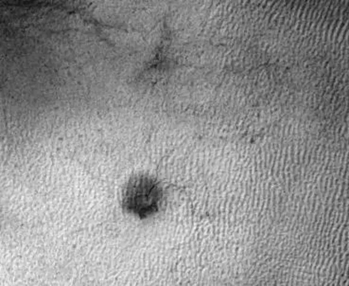
Viruses Under Electron Microscope

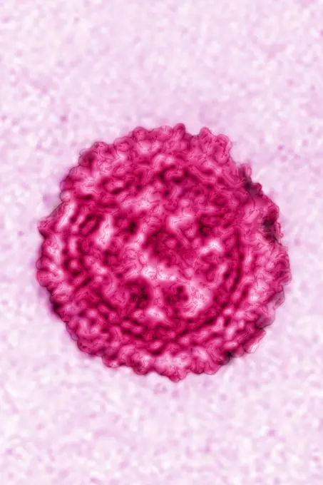
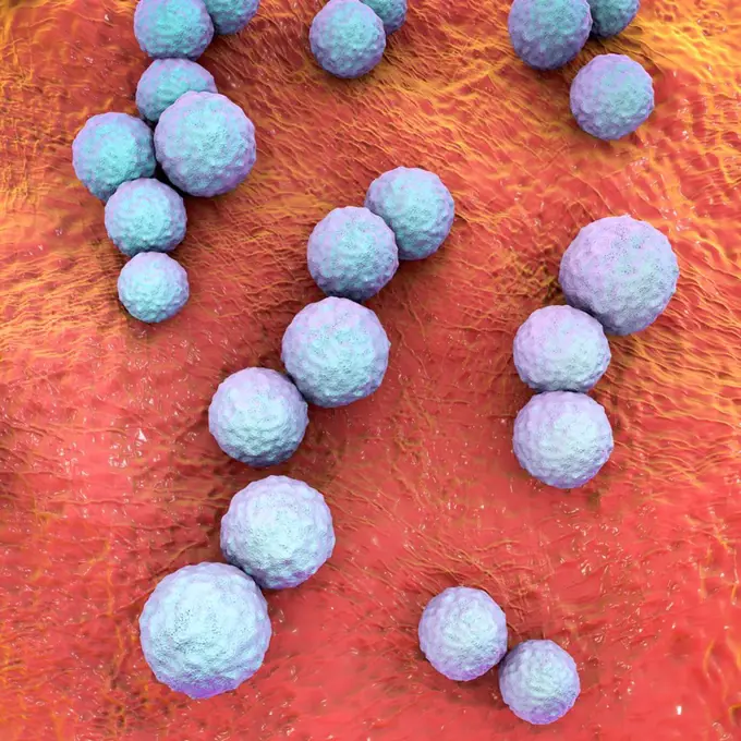
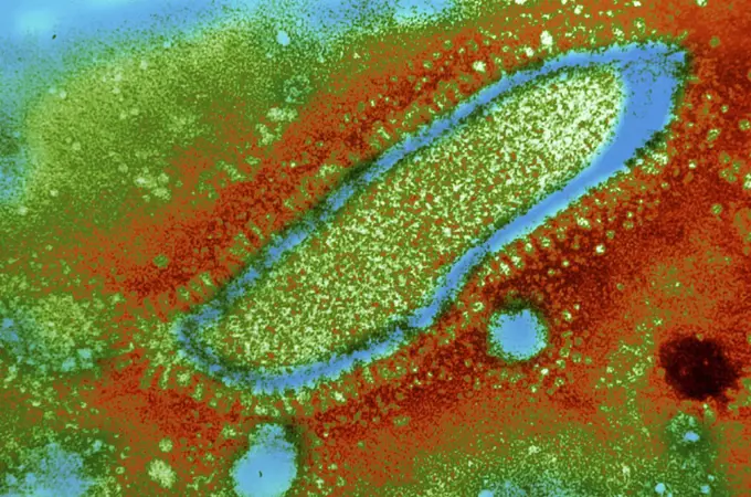
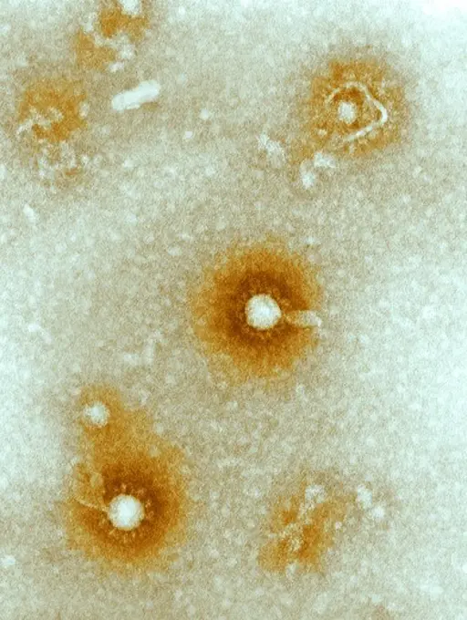
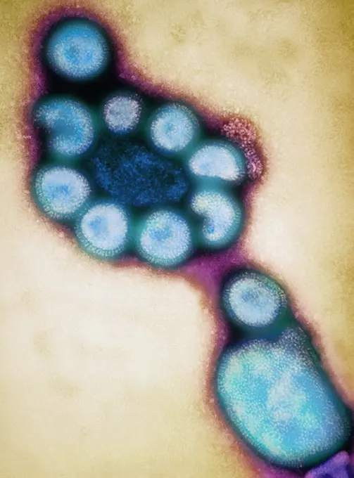
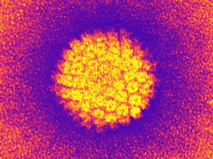
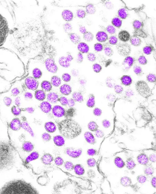
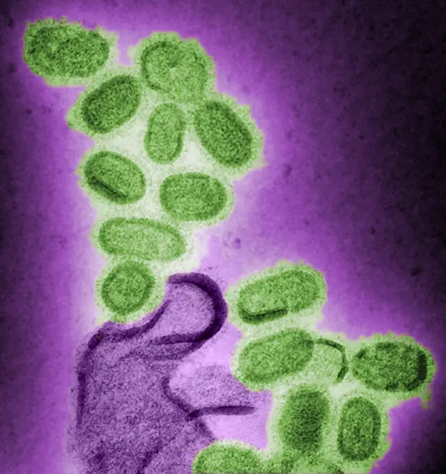
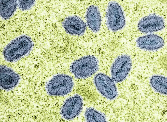
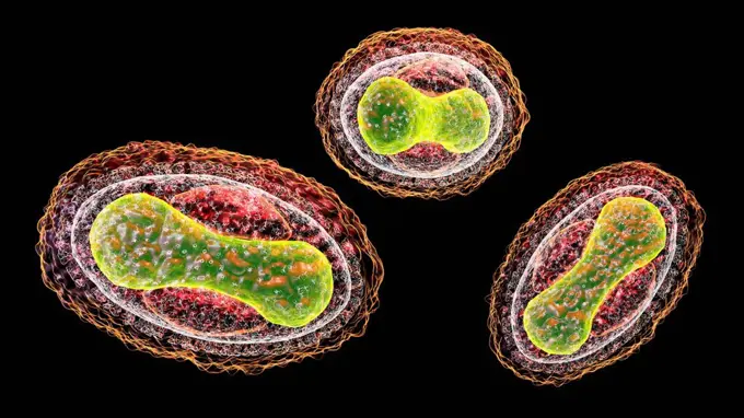
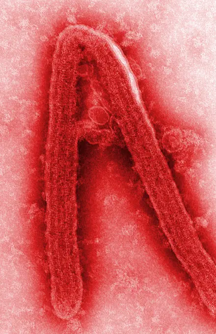
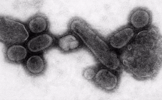
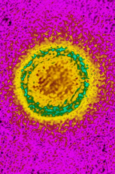
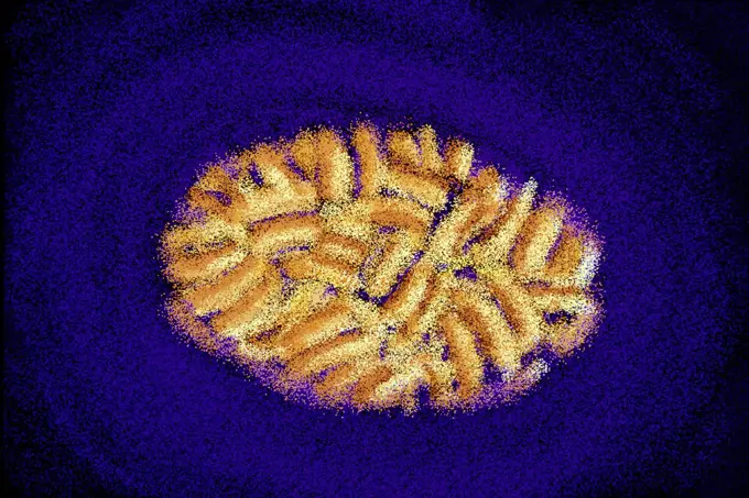
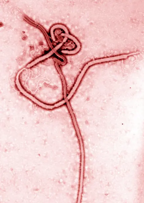
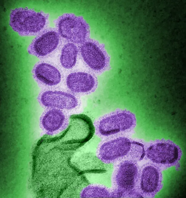
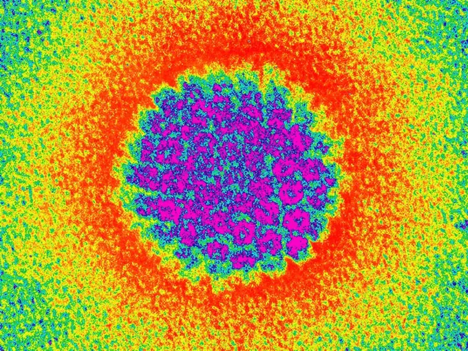
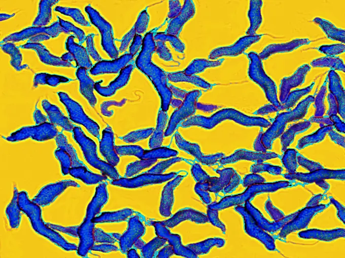
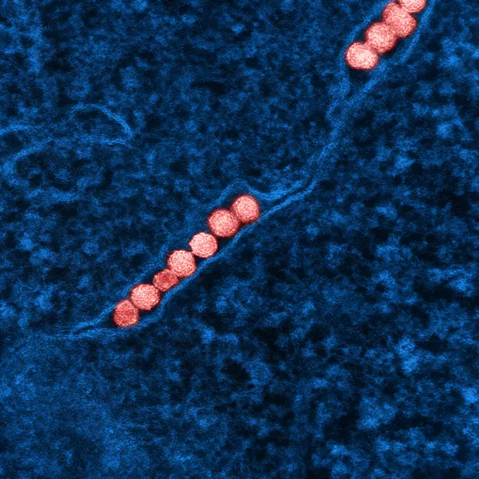
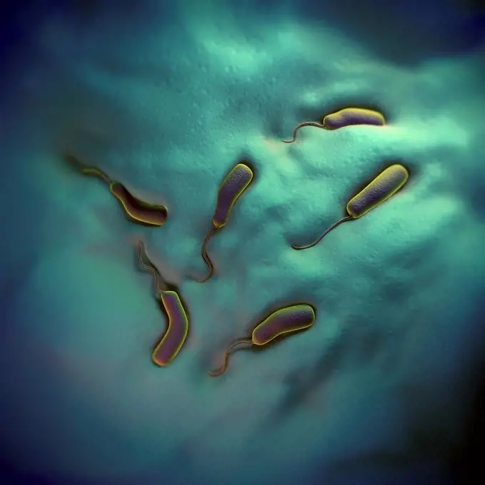
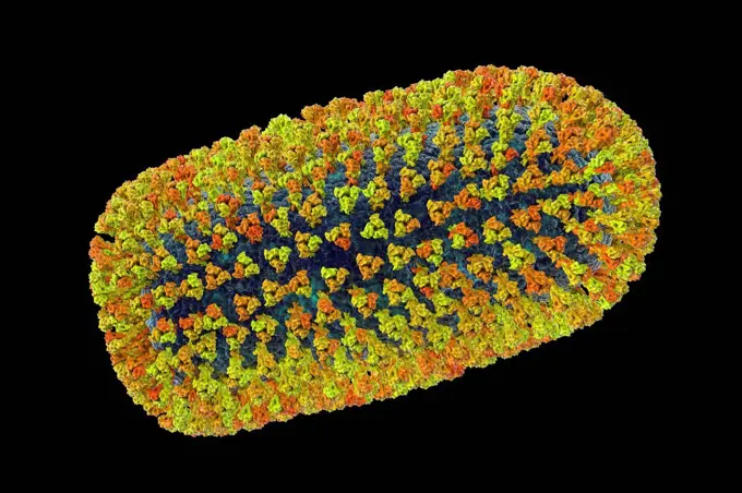
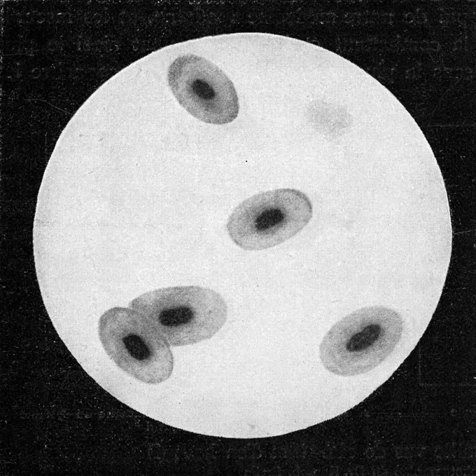
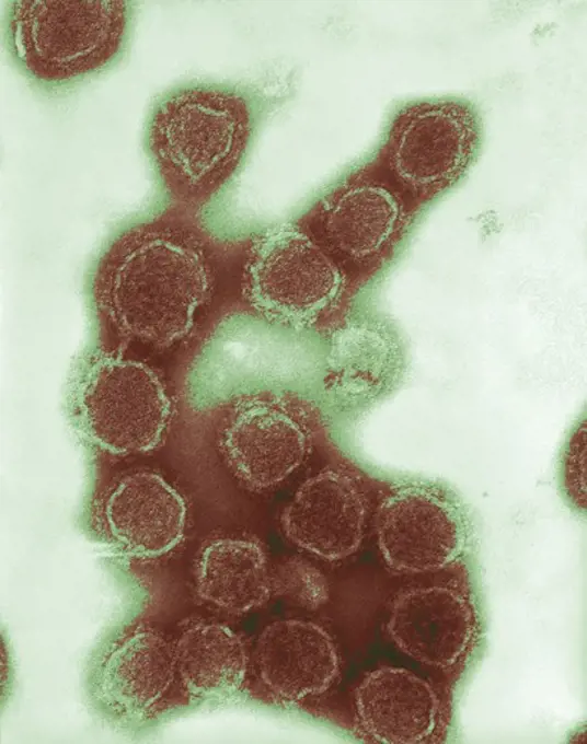
Variety of colorized transmission electron micrographs showcasing different viruses, including HIV, SARS-CoV-2, and influenza A, highlighting their structure and characteristics.

Negatively-stained transmission electron micrograph of California encephalitis virus virion
252 assets in this story
824-63197007
Coronavirus. Image produced using high-dynamic-range imaging (HDRI) from an image taken with transmission electron microscopy. Viral diameter ranges from around 80 to 160 nm.
4269-27636
Parvovirus. Electron micrograph of parvovirus H-1 virions of the Parvoviridae family of DNA viruses.
1899-61460457
Colorized transmission electron micrograph of monkeypox virus particles (red) found within an infected cell (blue), cultured in the laboratory
4297-1223
Transmission electron micrograph, human immunodeficiency virus (HIV), co-cultivated with human lymphocytes
1899-54028000
Influenza virus (from the Orthomyxoviridae family and the Influenzavirus genus). Image made from a transmission electron microscopy view.
4128R-13818136
Streptococcus mutans bacteria, computer illustration. These are Gram-positive, non-motile, spherical bacteria (cocci) which typically form chains of c...
824-63197020
Hepatitis C virus (HCV). Image produced using high-dynamic-range imaging (HDRI) from an image taken with transmission electron microscopy. Viral diameter around 22 nm.
4269-6789
Transmission electron micrograph (TEM) of a section of axone. It is protected by a layer of myelin around.
4128-V58570027
824-661B
Syncytium Virus  Family Paramyxovirus
4297-1765
transmission electron micrograph of Eastern Equine Encephalitis (EEE) virus virions that happened to be in a specimen of central nervous system tissue
4269-7099
Colored transmission electron micrograph (TEM) of Hong Kong influenza (flu) viruses. Between 1968 and 1970, this pandemic flu killed 800 000 persons. The Hong Kong influenza virus is an Influenza type A virus, strain H3N2. It is very close from the Spanish or swine flu (strain H1N1) or avian flu (strain H5N1). Magnification: x54,000 at 6x4,50cm size. x30,000 at 35mm.
4128-17857704
Helianthus stem. Light micrograph (LM) of the stem of a perennial sunflower (Helianthus sp.) in cross-section showing the vascular bundles (green, yel...
4128R-3682
Human papilloma virus, TEM
4239-69148151
Detail of the mantle of a giant clam.
4297-1213
TEM image of coronavirus, the causative agent of SARS
824-63194493
This colorized negative stained transmission electron micrograph (TEM) shows recreated 1918 influenza virions that were collected from supernatants of 1918-infected Madin-Darby Canine Kidney (MDCK) cells cultures 18 hours after infection. To separate these virions, the MDCK cells are spun down (centrifugation), and the 1918 virus in the fluid is immediately fixed for negative staining. The solid mass in lower center contains MDCK cell debris that did not spin down during the procedure.
824-65830650
Colorized transmission electron micrograph of mpox virus particles (blue) found within an infected cell (green), cultured in the laboratory.
4128-20044979
Smallpox virus, illustration
824-63222936
Coronavirus (CoV). viruses of the Coronaviridae family and the Orthocoronavirinae subfamily. It is a pathogen of respiratory syndromes. Viewed from a transmission electron microscopy (TEM) image. Viral diameter 80nm to 100nm.
4269-27409
Marburg virus. Transmission electron micrograph (TEM) of filamentous Marburg virus.This RNA virus of the Filovirus (like Ebola) is known to cause Marburg hemorrhagic fever, which can be fatal.
4269-12651
Onchocerca volvulus is a parasitic worm transmitted to human by a bite of the black fly (Simulium sp.) They cause onchocerciasis, a type of filariasis caracterized by skin or eyes lesion leading to blindness. Transmission Electronic Micrograph (TEM).
4048-6179
1918 influenza virions (virus particles).
824-63184615
This highly-magnified negatively-stained transmission electron micrograph (TEM) revealed ultrastructural morphology of the Middle East Respiratory Syndrome Coronavirus (MERS-CoV), which was formerly known as novel coronavirus. (MERS-CoV) was identified in 2012 as the cause of respiratory illness in people. Investigations are being done to figure out the source of MERS-CoV and how it spreads. So far, there are no reports of anyone in the US getting infected with MERS-CoV. Most people who got infected with MERS-CoV developed severe acute respiratory illness with symptoms of fever, cough, and shortness of breath. About half of them died. Some people were reported as having a mild respiratory illness. MERS-CoV has been shown to spread between people who are in close contact. Transmission from infected patients to healthcare personnel has also been observed. Clusters of cases in Saudi Arabia, Jordan, the UK, France, Tunisia, and Italy are being investigated.
824-63197018
Hepatitis B virus (HBV). Image produced using high-dynamic-range imaging (HDRI) from an image taken with transmission electron microscopy. Viral diameter ranges from around 50 to 60 nm.
824-63205995
Dengue virus (DENV) and Zika virus (ZIKV). It is an arbovirus member of the Flavivirus family. Visualization by transmission electron microscopy. Viral diameter: 75-100 nm.
1899-61461198
Bacillus Koch (Bk) or Mycobacterium tuberculosis, it is responsible for tuberculosis. Tuberculosis is making a comeback due to the appearance of multi-resistant bacterial strains. Pulmonary tuberculosis (phthisis) is the most widespread, but there are bone disorders (Pott's disease, white tumor of the knee), renal, intestinal, genital, meningeal and skin disorders (tuberculomas). Image taken from an X 1000 optical microscopy
824-656
Pox Virus
1746-111525041
Transmission electron Micrograph of the Ebola Virus Hemorrhagic Fever RNA Virus
824-63194496
This colorized negative stained transmission electron micrograph (TEM) shows recreated 1918 influenza virions that were collected from supernatants of 1918-infected Madin-Darby Canine Kidney (MDCK) cells cultures 18 hours after infection. To separate these virions, the MDCK cells are spun down (centrifugation), and the 1918 virus in the fluid is immediately fixed for negative staining. The solid mass in lower center contains MDCK cell debris that did not spin down during the procedure.
4128R-6358
Human papilloma virus, TEM
824-63178974
Campylobacter is a bacterium causing food poisoning which is the cause of: diarrhea, enteritis, fevers and abdominal pain. Optical microscopy
824-63227178
Colorized scanning electron micrograph of filamentous
4355-22243325
The White Pocket is an isolated,  patch of sandstone hidden within the desert expanse of Vermillion Cliffs National Monument near the Arizona/Utah border.
1899-61460430
Variola virus (poxviruses are large viruses of 300/200/100 nm. They are pathogenic for humans such as smallpox and vaccinia viruses as well as animal viruses that infect humans such as viruses monkeypox, cowpox, milker's nodule, pustular dermatitis in sheep Transmission electron microscopy
824-63194537
This colorized negatively-stained transmission electron micrograph (TEM) captured some of the ultrastructural details exhibited by the new influenza A (H7N9) virus. On April 1, 2013, the World Health Organization (WHO) first reported 3 human infections with a new influenza A (H7N9) virus in China. Since then, additional cases have been reported. Most reported cases have severe respiratory illness and, in some cases, have died. At this time, no cases of H7N9 outside of China have been reported. This new H7N9 virus is an avian (bird) influenza (flu) virus. Human infections with avian influenza (AI, or bird flu) are rare but have occurred in the past, most commonly after exposure to infected poultry. However, this is the first time that this bird flu subtype (H7N9) has been found in people. This virus is very different from other H7N9 viruses previously found in birds.
824-65830640
Scanning electron micrograph of a human H9 T cell (blue/green) infected with HIV virus particles (yellow).
1899-61460469
Transmission electron micrograph of West Nile virus particles (red) replicating within the cytoplasm of an infected VERO E6 cell (blue)
6145-44125415
Inflorescence of the plant Yellow Ageratum (Lonas Inodora)
1848-49569053
C.U. of Penicillin notatum Mold Growing on Agar
1788-111177012
Microphotograph of cultures of a fungus from the genus Penicillium, Trichocomaceae.
6145-29606219
Collection Dutch Heidematschappij. Cross-section female organ. undated
4378-2629
Group of vibrio cholerae bacteria which causes cholera.
4297-1441
Transmission electron micrograph showing eight virions of a newly-discovered virus which was determined to be a member of the genus Arenavirus
4297-1442
Transmission electron micrograph showing eight virions of a newly-discovered virus which was determined to be a member of the genus Arenavirus
4269-27434
Influenza virus. Transmission electron micrograph (TEM) of an influenza virus. Influenza virus are RNA virus (orthomyxoviridae).
824-63205994
Dengue virus (DENV) and Zika virus (ZIKV). It is an arbovirus member of the Flavivirus family. Visualization by transmission electron microscopy. Viral diameter: 75-100 nm.
4128-20239411
Mokola virus, illustration
1525-28067494
Blood cells of the frog, vintage engraved illustration. From the Universe and Humanity, 1910.
6145-46709771
Skiopticon image from the Department of Photography at the Royal Institute of Technology. Use by Professor Helmer Bäckström as lecture material. Bäckström was Sweden's first professor in photography at the Royal Institute of Technology in Stockholm 1948-1958.Mikro photograph of pollen mother cells. Possibly photographed in ultraviolet light. For more info see: Bäckström, Helmer. Photographic manual. Other revised edition. Nature and Culture. Stockholm. 1948. p. 1217.
6145-29606544
Collection Dutch Heidematschappij. Insects, damages, red forest mier. undated
6145-45124232
This image from NASA's Mars Reconnaissance Orbiter shows the growth of a branching network of troughs carved by thawing carbon dioxide over the span of three Martian years.
PREVIOUS
of 3
NEXT
2401 S. Ervay, Suite 206
Dallas, Texas 75215
United States
Browse
CategoriesGalleriesFree Research
For Creators
License Your ContentSuperStock For StudentsFrame of Mind BlogContributor PortalNew Image SpotlightLicense AI Imagery
Resources
About UsLicensingTerms & ConditionsAPI AccessPrivacyPricing
Contact
+1 866 236 0087help@superstock.com Contact form
©2026 SuperStock. All Rights Reserved. -A
This site is protected by reCAPTCHA and Google Privacy Policy and Terms of Service apply.