Fine ArtVintageFootageSubscriptionBoardsFree ResearchPricing
  • Fine Art
  • Vintage
  • Footage
  • Subscription
  • Boards
  • Free Research
  • Pricing

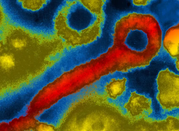
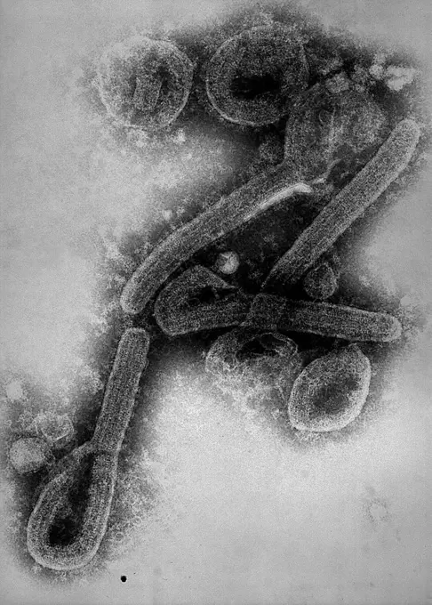
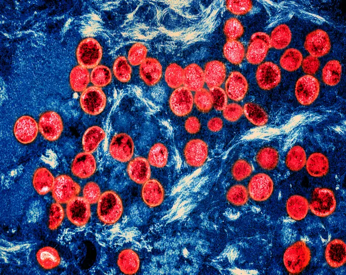
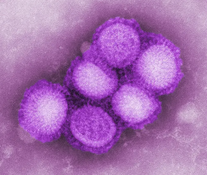
Viruses Under Microscopy

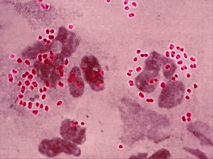
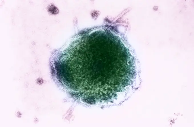
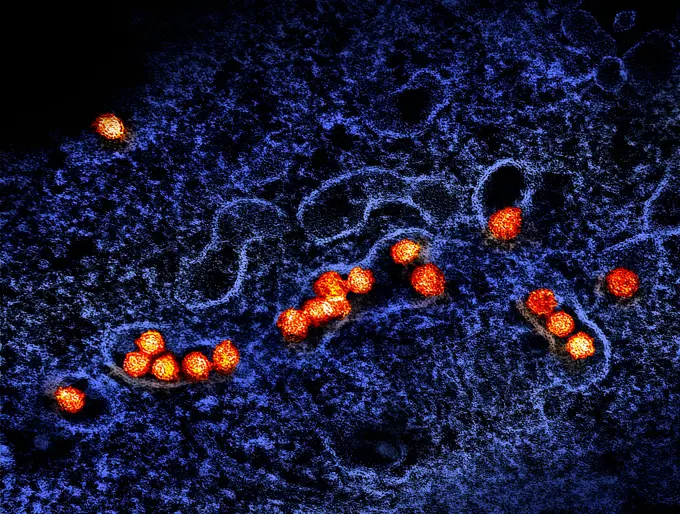
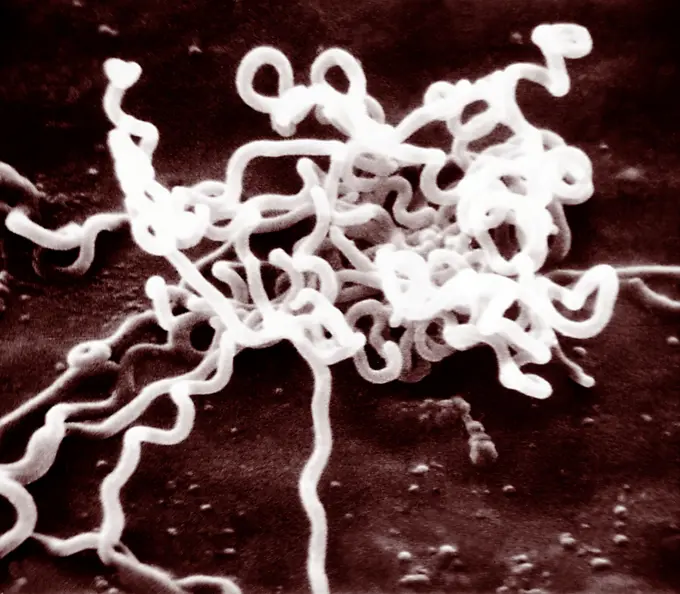
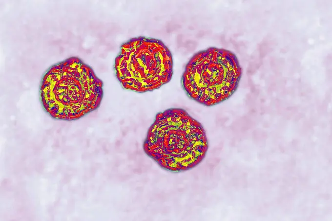
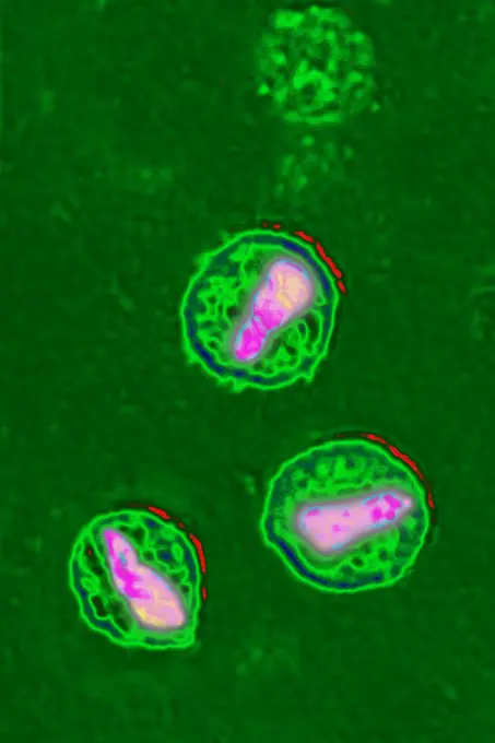
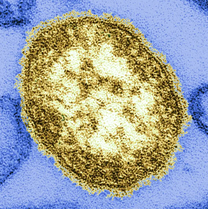
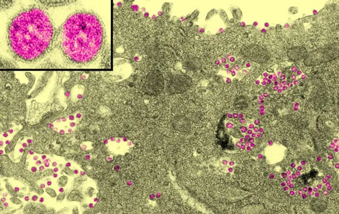
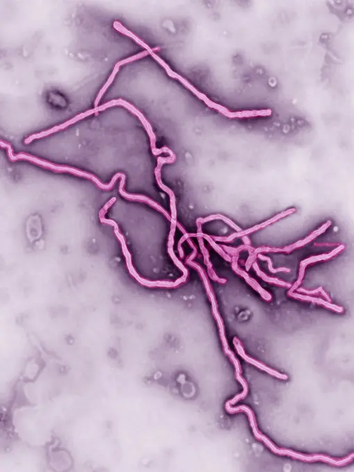
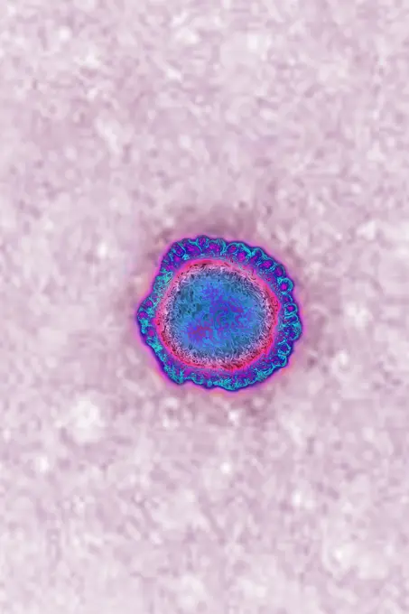
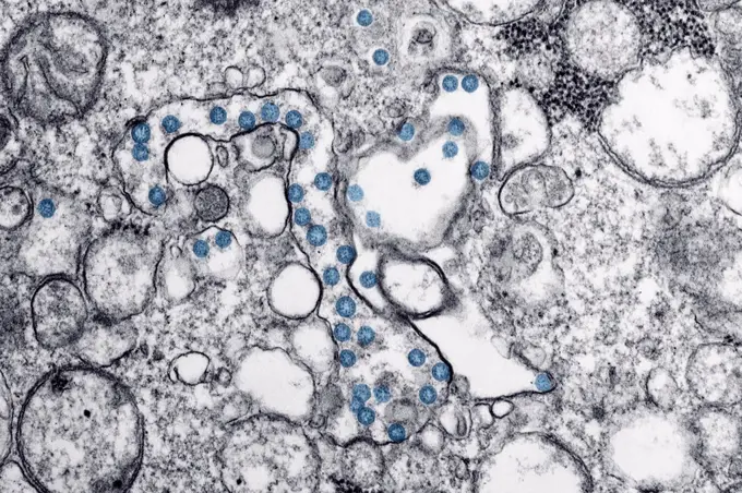
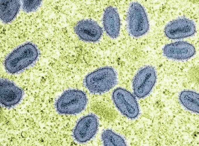
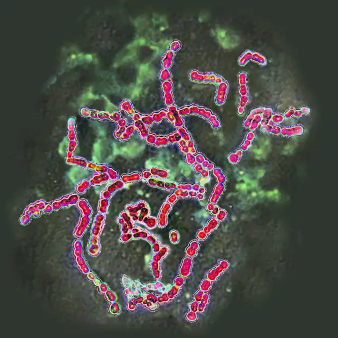
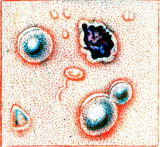
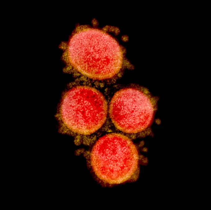
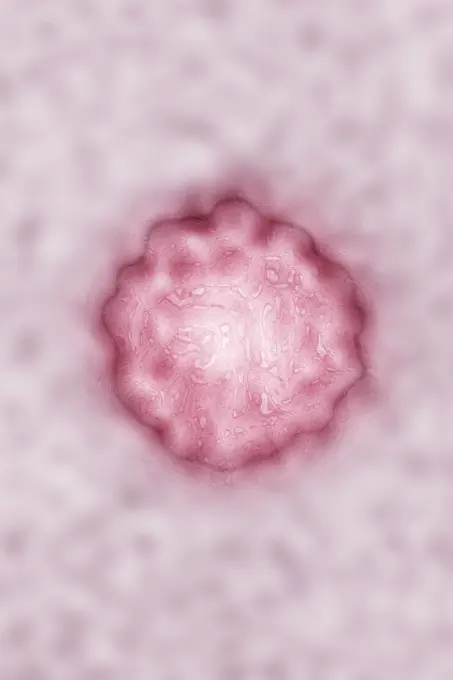
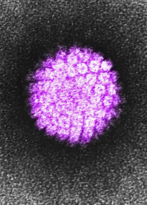
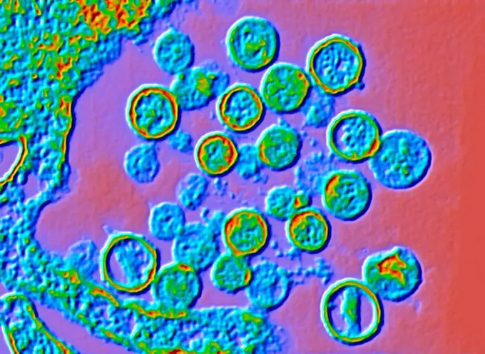
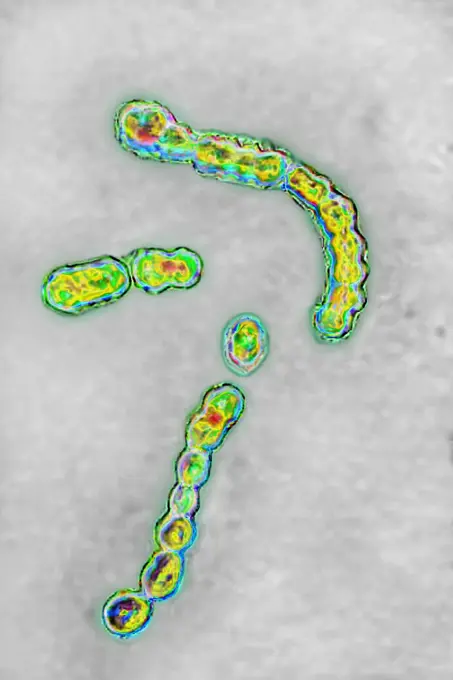
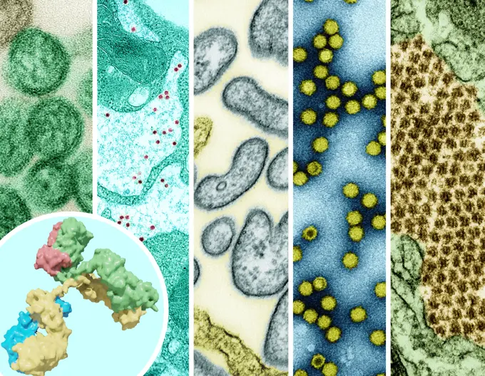
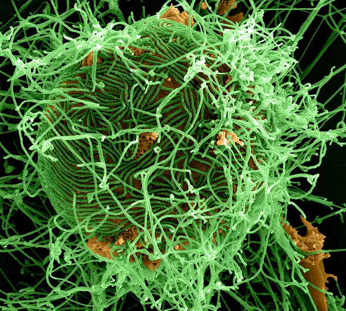
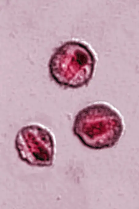
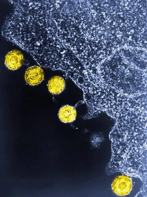
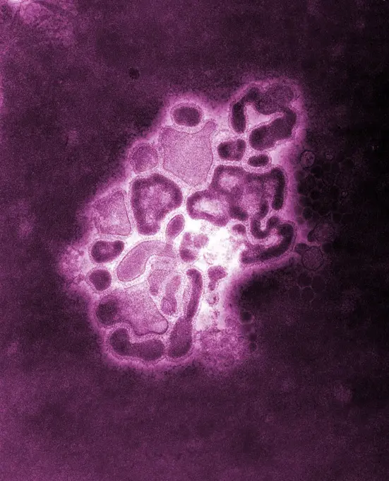
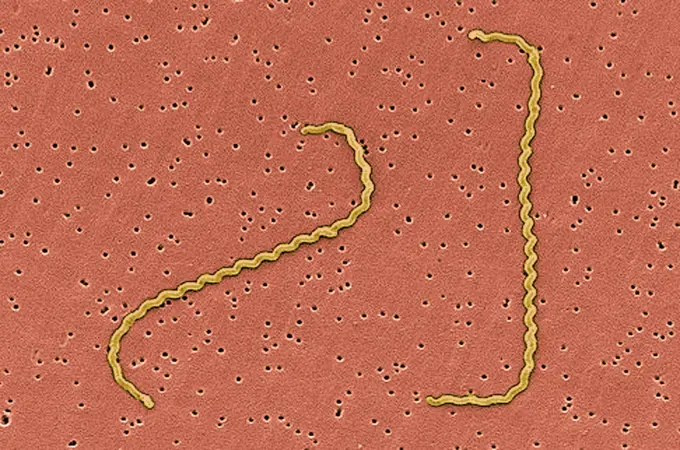
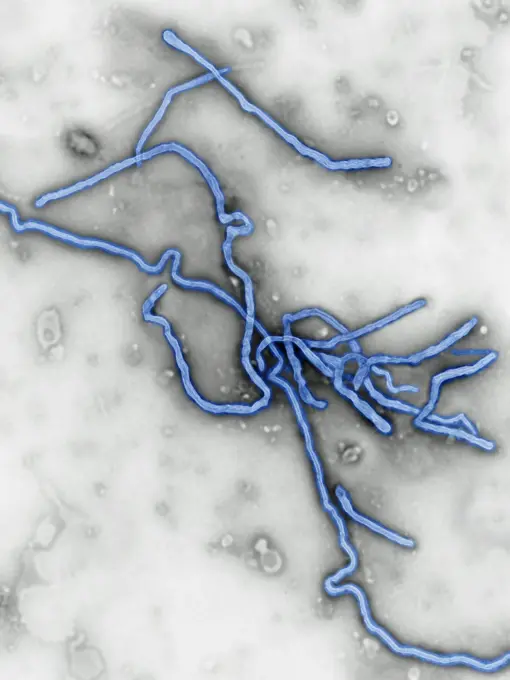
Colorized electron micrographs of various viruses including Tacaribe, influenza, herpes, and SARS-CoV-2, showing intricate details in shades of green, purple, and red.

This colorized transmission electron micrograph (TEM) depicts numbers of influenza A virions while in their late passage growth phase.
267 assets in this story
1899-53511307
Hepatitis B virus (Hepadnavirid family). HBV causes sexually transmitted or blood-borne hepatitis, cirrhosis occurs in 20% of cases, 15 to 20 years after the onset of infection. View produced from a transmission electron microscopy image. Viral diameter approximately: 22 nm.
824-63178999
Gonococcus (neisseria gonorrhoeae) is the bacterium responsable for gonorrhea. In men the symptoms are acute burning when urinating and some discharge (clap). In women the symptoms are burning when urinating, bartholin's cyst, cervisitis, salpingitis. Optical microscope x 1850.
824-63195694
This electron micrograph reveals both a paramyxovirus measles virus, and virions of the polyomavirus, simian virus SV40 (smaller circles). The envelope of the measles virus has broken, exposing the nucleocapsid filaments. Interest in SV40 has increased in the last several years because the virus was found in certain forms of cancer in humans, for instance mesotheliomas (rare tumors located in the lungs), brain, and bone tumors (Carbone et al. 1994, Jasani et al . 2001).
1899-65662411
Colorized transmission electron micrograph of avian influenza A H5N1 virus particles (gold), grown in Madin-Darby Canine Kidney (MDCK) epithelial cells.
824-65830657
Transmission electron micrograph of West Nile virus particles (orange/gold) replicating within the cytoplasm of an infected VERO E6 cell (blue).
824-63123715
Electron micrograph of Treponema pallidum on cultures of cotton-tail rabbit epithelium cells (Sf1Ep).Treponema pallidum is the causative agent of syphilis. In the United States, over 35,600 cases of syphilis were reported by health officials in 1999.
4297-1749
Transmission electron micrograph of Colorado tick fever virus (CTFV)
4269-25300
Ebola Virus. Color enhanced Transmission Electron Micrograph (TEM) of the ebola virus.
824-63188642
Osteoporosis of the lumbar vertebrae, seen on a side x-ray.
1746-111525033
Marburg virus is a hemorrhagic fever virus first described during small epidemics in the German cities Marburg and Frankfurt and the Yugoslavian capital Belgrade in the 1960s. Marburg virus (MARV) causes severe disease in humans and nonhuman primates in the form of viral hemorrhagic fever.
824-57657686
Colorized transmission electron micrograph of monkeypox virus particles (red) found within an infected cell (blue), cultured in the laboratory.
824-63198892
Human Papilloma virus (HPV). HPV causes STDs, condyloma acuminata warts that can lead to cervical cancer. It is also the cause of skin infections, warts. Image produced using high-dynamic-range imaging (HDRI) from an image taken with transmission electron microscopy. Viral diameter around 55 nm.
824-63194499
This colorized negative stained transmission electron micrograph (TEM) depicted some of the ultrastructural morphology of the A/CA/4/09 swine flu virus. Swine Influenza (swine flu) is a respiratory disease of pigs caused by type A influenza virus that regularly causes outbreaks of influenza in pigs. Swine flu viruses cause high levels of illness and low death rates in pigs. Swine influenza viruses may circulate among swine throughout the year, but most outbreaks occur during the late fall and winter months similar to outbreaks in humans. The classical swine flu virus (an influenza type A H1N1 virus) was first isolated from a pig in 1930.
824-63224490
Hepatitis C virus (close to the Flavivirid family). HCV causes blood-borne hepatitis, cirrhosis occurs in 25% of cases, 10 to 20 years after the onset of infection, with a risk of hepatic carcinoma. View produced from a transmission electron microscopy image. Viral diameter approximately: 50 to 60 nm.
1899-54027723
Human immunodeficiency virus (HIV), the retrovirus responsible for AIDS. Image produced with transmission electron microscope (viral diameter ranges from around 110 to 125 nm).
824-63217278
Human immunodeficiency virus (HIV), the retrovirus responsible for AIDS. Image produced with transmission electron microscope (viral diameter ranges from around 110 to 125 nm).
824-63195689
This thin-section transmission electron micrograph (TEM) revealed the ultrastructural appearance of a single virus particle, or virion, of measles virus. The measles virus is a paramyxovirus, of the genus Morbillivirus. It is 100-200 nm in diameter, with a core of single-stranded RNA, and is closely related to the Rinderpest and canine distemper viruses. Two membrane envelope proteins are important in pathogenesis. They are the F (fusion) protein, which is responsible for fusion of virus and host cell membranes, viral penetration, and hemolysis, and the H (hemagglutinin) protein, which is responsible for adsorption of virus to cells. There is only one antigenic type of measles virus. Although studies have documented changes in the H glycoprotein, these changes do not appear to be epidemiologically important (ie no change in vaccine efficacy has been observed).
4197-V65369289
4128-20038493
Cholesterol molecule, illustration
4297-1859
Scanning electron microscopic image of the coronaviruses contained within cytoplasmic membrane-bound vacuoles and cisternae of the rough endoplasmic reticulum
4297-1216
Scanning Electron micrograph of infected Vero E6 cell showing coronavirus particles within cytoplasmic membrane-bound vacuoles and the cisternae of the rough endoplasmic reticulum
4128-30420575
Marburg viruses, illustration
824-63190225
Ebola virus responsible for lightning hemorrhagic fever, this virus is part of the filoviridae family. Image made from a transmission electron microscope view.
824-65830683
Transmission electron micrograph of vesicular stomatitis virus (VSV) particles (teal) found in extracellular space and budding from the plasma membrane of infected cells (purple).
824-63222231
Coronavirus (CoV). viruses of the Coronaviridae family and the Orthocoronavirinae subfamily. It is a pathogen of respiratory syndromes. Viewed from a transmission electron microscopy (TEM) image. Viral diameter 80nm to 100nm.
4269-27642
Herpes simplex virus. Electron micrograph of the Herpes simplex virus.
824-63183642
Pox virus. Image produced using high-dynamic-range imaging (HDRI) from an image taken with transmission electron microscopy. Viral diameter ranges from around 100 to 300 nm.
1899-65662410
Colorized transmission electron micrograph of avian influenza A H5N1 virus particles (yellow), grown in Madin-Darby Canine Kidney (MDCK) epithelial cells (blue).
4048-16067045
Novel Coronavirus SARS-CoV-2, also called, 'severe acute respiratory syndrome coronavirus 2', causes 'coronavirus disease-2019', abbreviated as COVID-19. Transmission electron microscopic image of an isolate from the first US case of COVID-19. The spherical viral particles, colorized blue, contain cross-section through the viral genome, seen as black dots. Viruses are to small to be seen through an optical microscope. Image by Centers for Disease Control and Prevention (CDC)  (BSLOC_2020_2_250)
4269-2986
Transmission electron micrograph (TEM) of the avian influenza A H5N1 virus. Magnification x 150,000.
824-63194497
This colorized negative stained transmission electron micrograph (TEM) depicted some of the ultrastructural morphology of the A/CA/4/09 swine flu virus. Swine Influenza (swine flu) is a respiratory disease of pigs caused by type A influenza virus that regularly causes outbreaks of influenza in pigs. Swine flu viruses cause high levels of illness and low death rates in pigs. Swine influenza viruses may circulate among swine throughout the year, but most outbreaks occur during the late fall and winter months similar to outbreaks in humans. The classical swine flu virus (an influenza type A H1N1 virus) was first isolated from a pig in 1930.
1899-65662343
Colorized transmission electron micrograph of mpox virus particles (blue) found within an infected cell (green), cultured in the laboratory.
824-65830828
Transmission electron micrograph of HIV-1 virus particles (purple) from infected H9 cells, produced in cell culture. The particles exhibit two stages of replication: the two arcs are immature particles budding from the plasma membrane of the cell, and the center spherical particle is a mature form in extracellular space.
824-63222228
Coronavirus (CoV). viruses of the Coronaviridae family and the Orthocoronavirinae subfamily. It is a pathogen of respiratory syndromes. Viewed from a transmission electron microscopy (TEM) image. Viral diameter 80nm to 100nm.
1899-61460468
Transmission electron micrograph of West Nile virus particles (gold) replicating within the cytoplasm of an infected VERO E6 cell (pink)
824-63197035
Pox virus. Image produced using high-dynamic-range imaging (HDRI) from an image taken with transmission electron microscopy. Viral diameter ranges from around 100 to 300 nm.
824-63222970
Pneumococcal bacteria (streptococcus pneumoniae). Pneumococcus is an important pathogen in humans. It is responsible for many infections (pneumonia). He was responsible for pneumonia during the Spanish flu pandemic). Visualized by optical microscopy.
824-63190244
Meningococcus or Neisseria meningitidis, is a bacterium that can cause meningitis. The bacteria are germs that can be found in the rhinopharynx, where it can cause benign rhinopharyngitis or an asymptomatic state. Only one in 400 carriers become victim to a serious meningococcal disease, mostly commonly acute purulent meningitis. Seen through an optical microscope X 1000.
824-57660057
Group A streptococci pyogenes. It is the cause of skin infections (impetigo), abscesses, bronchopulmonary infections, a form of sore throat which can develop into complications such as acute rheumatic fever. Image produced from optical microscopy.
1525-28047888
Pustules of scabies, vintage engraved illustration.
824-63223221
Transmission electron micrograph of SARS-CoV-2 virus particles, isolated from a patient. Image captured and color-enhanced at the NIAID Integrated Research Facility (IRF) in Fort Detrick, Maryland. Credit: NIAID.
4297-1748
Electron micrograph of Varicella (Chickenpox) virus
4128R-13818128
Streptococcus mutans bacteria, computer illustration. These are Gram-positive, non-motile, spherical bacteria (cocci) which typically form chains of c...
824-63218221
Hepatitis A virus (picornavirus). HAV causes a hepatitis that is almost always benign. Image made from transmission electron microscopy. Approximate viral diameter: 30 nm.
824-57657659
Variola virus (poxviruses are large viruses of 300/200/100 nm. They are pathogenic for humans such as smallpox and vaccinia viruses as well as animal viruses that infect humans such as viruses monkeypox, cowpox, milker's nodule, pustular dermatitis in sheep Transmission electron microscopy.
4297-1425
Electron micrograph of a negatively stained human papilloma virus (HPV) which occurs in human warts. Warts on the hands and feet have never been known to progress to cancer. However, after many years cervical warts can become cancerous
4128R-13928397
Color enhanced transmission electron micrograph (TEM) of Hantavirus particles. Hantavirus is a respiratory disease carried in wild rodents such as dee...
824-63222960
Bacteria streptococcus pyogenes, responsible for infections, including angina, bronchopulmonary infections pneumonia, infection of wounds and skin. Optical microscopy view.
824-66066022
A 3D rendering of an antibody (foreground left), and examples of high-priority &quotprototype&quot pathogens that have the potential to threaten human health/are the focus of pandemic preparedness research efforts (left to right): hantavirus, yellow fever virus, Nipah virus, picornavirus, and Chikungunya. As of September 2024, The National Institutes of Health (NIH) has established a pandemic preparedness research network known as ReVAMPP to conduct research on high-priority pathogens with the goal of developing effective vaccines and monoclonal antibodies. Credit: NIAID See related September 13, 2024 NIAID news release, &quotNIH Awards Establish Pandemic Preparedness Research Network,&quot at.
824-63194378
Colorized scanning electron micrograph of filamentous Ebola virus particles (green) attached and budding from a chronically infected VERO E6 cell (orange) (25,000x magnification). Produced by the National Institute of Allergy and Infectious Diseases (NIAID), under a magnification of 25,000X, this digitally-colorized scanning electron micrograph (SEM) depicts numerous filamentous Ebola virus particles (green) budding from a chronically-infected VERO E6 cell ( orange). Ebola hemorrhagic fever (Ebola HF) is one of numerous Viral Hemorrhagic Fevers. It is a severe, often fatal disease in humans and nonhuman primates (such as monkeys, gorillas, and chimpanzees). Ebola HF is caused by infection with a virus of the family Filoviridae, genus Ebolavirus. When infection occurs, symptoms usually begin abruptly. The first Ebolavirus species was discovered in 1976 in what is now the Democratic Republic of the Congo near the Ebola River. Since then, outbreaks have appeared sporadically.
824-57657698
Transmission electron micrograph of West Nile virus particles (red) replicating within the cytoplasm of an infected VERO E6 cell (blue).
4128R-13575734
Illustration showing the structure of an antibody, or immunoglobulin, molecule. These Y-shaped molecules have two arms that can bind to specific antig...
1525-28047899
Cowpox from cows, vintage engraved illustration.
1899-54027721
Human immunodeficiency virus (HIV), the retrovirus responsible for AIDS. Image produced with transmission electron microscope (viral diameter ranges from around 110 to 125 nm).
824-63217275
Human immunodeficiency virus (HIV), the retrovirus responsible for AIDS. Image produced with transmission electron microscope (viral diameter ranges from around 110 to 125 nm).
824-63223262
Colorized transmission electron micrograph of negatively stained SW31 (swine strain) influenza virus particles. Credit: NIAID.
4269-7072
Colored transmission electron micrograph (TEM) of a virion (virus particles, stained brown) of the parainfluenza virus, a member of the paramyxovirus class causing minor respiratory infections.
6145-44620489
'Blueberries' and 'Popcorn'
824-63123774
Scanning electron micrograph of Leptospira interrogans strain RGA. Two spirochetes bound to a 0.2 µm filter. Strain RGA was isolated in 1915 by Uhlenhuth and Fromme from the blood of a soldier in Belgium. Leptospira is known to cause the infectious disea
4297-1412
colorized electron micrograph image of HHV-6, or the human herpes virus-6, was thought to infect b-cells and was at one time called HBLV, human b-lymphotropic virus
4269-27808
Hantavirus Pulmonary Syndrome. Ligh micrograph of an atypical enlarged lymphocyte found in the blood smear from a patientwith Hantavirus Pulmonary Syndrome (HPS), an acute respiratory infection caused by the Virus Sin Nombre.
824-63194486
Under a plate magnification of 37,800X, this colorized transmission electron micrograph (TEM) depicted a strain of swine flu, the A/New Jersey/76 (Hsw1N1) virus, while in the virus' first developmental passage through a chicken egg. Swine Influenza (swine flu) is a respiratory disease of pigs caused by type A influenza that regularly cause outbreaks of influenza among pigs. Swine flu viruses cause high levels of illness and low death rates among pigs. Swine influenza viruses may circulate in swine throughout the year, but most outbreaks among swine herds occur during the late fall and winter months similar to humans. The classical swine flu virus (an influenza type A H1N1 virus) was first isolated from a pig in 1930.
4269-5517
Color enhanced Transmission Electron Micrograph (TEM) of the ebola virus, an RNA virus (filovirus) causing hemorrhagic fever.
4128R-14324195
Threadworms in the gut, illustration
4269-7084
Color enhanced scanning electron micrograph (SEM) of Leptospira interrogans strain RGA. Leptospira is a type of Gram negative spirochete. It is know to cause the infectious disease leptospirosis, and other diseases with similar symptoms attacking the liver (jaundice) or the meninges of the brain (meningitis).
824-63190226
Ebola virus responsible for lightning hemorrhagic fever, this virus is part of the filoviridae family. Image made from a transmission electron microscope view.
1848-53286860
Graphic representation of the Ebola virus on blue background
PREVIOUS
of 3
NEXT
2401 S. Ervay, Suite 206
Dallas, Texas 75215
United States
Browse
CategoriesGalleriesFree Research
For Creators
License Your ContentSuperStock For StudentsFrame of Mind BlogContributor PortalNew Image SpotlightLicense AI Imagery
Resources
About UsLicensingTerms & ConditionsAPI AccessPrivacyPricing
Contact
+1 866 236 0087help@superstock.com Contact form
©2026 SuperStock. All Rights Reserved. -A
This site is protected by reCAPTCHA and Google Privacy Policy and Terms of Service apply.